


Download PDF
Abstract
The Mediterranean diet is associated with reduced cardiometabolic risk, yet its physiological effects during pregnancy and its impact on placental metabolism remain incompletely understood. This study aimed to determine whether maternal adherence to a Mediterranean diet during pregnancy influences placental lipid metabolism and signalling pathways involved in nutrient handling, tissue remodelling, and inflammation, and to assess their relationship with pregnancy outcomes. Placental samples and clinical outcome data were analysed from pregnant women participating in an unblinded randomized clinical trial of a Mediterranean diet intervention. Placental fatty acid composition was quantified, and the expression of genes and signalling pathways involved in lipid metabolism, nutrient transport, inflammation, and tissue remodelling was evaluated. Maternal adherence to a Mediterranean diet during pregnancy was associated with significant alterations in placental fatty acid composition, including reduced C18:0 and C24:0 and increased C18:1n9c, C20:3n6, and C22:0, with lower total saturated fatty acids and higher monounsaturated fatty acids. Placental expression of lipid metabolism regulators ALOX15 and PPARγ was reduced, alongside downregulation of AKT and p38 MAPK signalling pathways. Placentas from mothers adhering to the Mediterranean diet also showed lower expression of amino acid and glucose transporters SLC3A2 and SLC2A1, as well as altered inflammatory and extracellular matrix remodelling markers, including decreased SOCS3 and GHR and increased PAI1 and MMP3. Maternal adherence to a Mediterranean diet during pregnancy modifies placental fatty acid composition and regulates pathways involved in lipid handling, nutrient transport, inflammation, and tissue remodelling, providing insight into mechanisms linking maternal diet with placental metabolic function.
Subjects
Introduction
Diet during pregnancy is a critical determinant of fetal growth1,2,3. Studies in humans and animal models have shown that poor maternal nutrition (both excess and deficiency) from before or during pregnancy can compromise pregnancy outcomes, with both short- and long-term consequences for the offspring4,5,6,7,8. Such consequences include fetal growth restriction, hypoglycemia, and an increased risk of cardiometabolic disorders, including diabetes and hypertension, as well as increased risk of neuropsychiatric conditions, including schizophrenia9.
The Mediterranean diet is characterized by a high intake of vegetables, fruits, legumes, nuts, dairy products (principally cheese and yogurt), and extra-virgin olive oil (EVOO); a moderate intake of white meat and fish (particularly oily fish); and limited intake of red and processed meats10. It is rich in mono- and polyunsaturated fatty acids and emphasizes foods with antioxidant and anti-inflammatory properties, which protect against oxidative stress, DNA damage, and inflammation while promoting cell proliferation, and angiogenesis11,12. Accordingly, adherence to a Mediterranean diet has been associated with a reduced risk of cardiovascular disease, cancer, diabetes, and neurodegenerative disorders13,14,15,16,17,18,19,20.
In pregnant populations, adherence to a Mediterranean diet has also yielded promising outcomes. Several studies have shown that key components of this diet, such as EVOO, and nuts, are associated with a reduced risk of gestational diabetes mellitus21,22,23. Recently, a randomised clinical trial (Improving Mothers for a Better PrenAtal Care Trial BarCeloNa, IMPACT BCN) investigated the effects of structured lifestyle interventions, including a Mediterranean diet, in pregnant women at high risk for small-for-gestational-age (SGA) newborns and reported a significant reduction in its prevalence24. Consistently, adherence to a Mediterranean diet in other human cohorts has been associated with lower odds of preeclampsia25 and reduced risk of preterm birth among overweight and obese women26. Moreover, Mediterranean diet intake correlates positively with maternal plasma folate and serum vitamin B12 concentrations27, key micronutrients required for DNA synthesis, methylation, and normal fetal development. Indeed, within the same IMPACT BCN trial, offspring of mothers with higher adherence to a Mediterranean diet showed greater total fetal brain volume at magnetic resonance evaluation and higher scores on the Neonatal Neurobehavioral Assessment Scale28. The benefits of the Mediterranean diet during pregnancy extend beyond the perinatal period, with improvements in neurodevelopment also observed at 2 years of age29. Similar studies have demonstrated that maternal adherence to this dietary pattern is associated with fetal cardiac structure and function, including increased right ventricular fractional area change and reduced myocardial wall thickness30. At the placental level, it has been shown that combining maternal exercise with Mediterranean diet can prevent placental telomere shortening31.
Consequently, in recent years, modifying maternal diet to incorporate Mediterranean diet products before or during pregnancy has been considered a promising strategy to prevent or mitigate pregnancy-related complications. Nonetheless, further characterization of the metabolic effects of a Mediterranean diet on placental function, the organ that mediates nutrient and signalling exchange between the mother and fetus, remains to be elucidated. The aim of this study was to investigate the impact of maternal adherence to a Mediterranean diet on placental lipid metabolism and signalling pathways involved in nutrient handling, tissue remodelling, and inflammation, and to examine their association with pregnancy outcomes. To this end, we utilized clinical outcome data and placentas from women with and without adherence to a Mediterranean diet enrolled in the IMPACT BCN trial24,32,33.
Materials and methods
Participants and ethics
Placentas were selected from biobank samples collected during the IMPACT BCN trial. This was a parallel, unblinded randomized clinical trial conducted at BCNatal (Hospital Clínic and Hospital Sant Joan de Déu), a large referral center for maternal–fetal and neonatal medicine in Barcelona, Spain. Participants were enrolled after eligibility screening during routine second-trimester ultrasound examinations (19.0–23.6 weeks of gestation) and randomized in a 1:1:1 ratio into three groups: a nutritional intervention based on a Mediterranean diet with supplementation of EVOO and walnuts; a Stress reduction program; or a control group without any intervention (usual care). More details are reported in the study protocol elsewhere34.
All methods were carried out in accordance with relevant guidelines and regulations. The study was approved by the Institutional Review Board (HCB-2016-0830). The trial was registered at ClinicalTrials.gov (identifier: NCT03166332) on 19/04/2017. The Institutional Review Board of the Hospital Clínic of Barcelona approved the study (HCB-2016-0830) on16/12/2016 and the trial was registered at ClinicalTrials.gov (identifier: NCT03166332) on 19/04/2017. Written informed consent was obtained from all participants.
In all participants of the trial at enrolment (19–23.6 weeks’ gestation) and at final assessment (34–36 weeks’ gestation), a 151-item Food Frequency Questionnaire (FFQ)35, and a 17-item Mediterranean Diet Adherence Screener (preg-MEDAS) score, both validated for this study population, were provided36. For the present analysis, we considered participants from Mediterranean diet and Usual care groups, non-obese (pre-pregnancy BMI < 30 kg/m2) and with uncomplicated pregnancies, defined as those not affected by SGA, preterm birth, preeclampsia, or gestational diabetes mellitus (n = 14 per group). In addition, among women allocated to the Mediterranean diet intervention, only those with high adherence were included, defined as an improvement of ≥ 3 points in the final preg-MEDAS score36.
Mediterranean diet program
The dietary intervention consisted of monthly individual and group assessments, performed by trained nutritionists, from recruitment (19.0−23.6 weeks’ gestation) until the conclusion of the program (34−36 weeks’ gestation). In each assessment, the goal was to change the overall Mediterranean dietary pattern rather than focusing on single nutrients, including whole-grain cereals (≥ 5 servings per day); vegetables and dairy products (≥ 3 servings per day); fresh fruit (≥ 2 servings per day); and legumes, nuts, fish, and white meat (≥ 3 servings per week), together with the free provision of EVOO (2 L per month) and walnuts (15 g per day; 450 g per month). Further details are provided in the trial protocol34.
Placental sampling
Placental tissue samples were collected immediately after delivery. Briefly, soon after placental expulsion, four full-thickness biopsies of macroscopically normal placental parenchyma (one from each placental quadrant) were collected and placed in cryogenic tubes on dry ice within minutes of delivery. Samples were maintained on dry ice for 1–4 h depending on the day of collection, then stored at − 80 °C until further processing for molecular analysis. Subsequently, placentas were cleared of fetal membranes, the umbilical cord, and adherent blood clots. Trimmed placentas were weighed, and their length, width, and thickness were measured using a manual caliper. Placental efficiency was calculated as the fetal-to-placental weight ratio.
Placental RNA extraction
Placental RNA was extracted from frozen placental tissues (7 placentas per group and sex, total n = 28) using the RNeasy Fibrous Tissue Mini Kit (Qiagen, Hilden, Germany) following the manufacturer’s protocol and as described elsewhere37. The quantity of RNA extracted was determined using a NanoDrop spectrophotometer (NanoDrop Technologies, Inc., Auburn, AL), and the RNA was reverse transcribed using a high-capacity cDNA reverse transcription kit (Applied Biosystems, Foster City, USA) according to the manufacturer’s instructions. Quantitative real-time PCR was performed in duplicates using the primer sequences specified in Table S1. The relative mRNA expression levels were normalized to the geometric mean of two AGCCACATCGCTCAGACAC eping genes (GAPDH and HPRT1, which remained stably expressed across the two groups) and calculated using the 2−ΔΔCT method38.
Placental protein extraction
Approximately 50 mg of frozen placental samples were used to extract protein. Briefly, placental samples were previously washed in RIPA buffer to remove excess blood. Samples were then homogenized with fresh RIPA buffer containing a protease inhibitor cocktail (Complete Mini Protease Inhibitor Cocktail, 1 tablet/10 ml), 1mM β-glycerophosphate, and 1mM sodium orthovanadate (Na3VO4) using a bead-based method. omogenization was performed using a MagNA Lyser (Roche) for three cycles of 20 s at 6000 rpm, with 5-min incubations on ice between cycles. Samples were then placed on ice for approximately one hour to allow protein dissociation, centrifuged to remove placental debris, and the resulting protein lysates were stored at − 80 °C. Total placental protein concentration was determined using the Pierce™ BCA Protein Assay Kit (Thermo Fisher Scientific).
Proteins were separated by electrophoresis and transferred onto 0.2 μm nitrocellulose membranes (Bio-Rad Laboratories Inc., Hercules, CA, USA). Membranes were blocked with fetal bovine serum and/or skimmed milk, depending on whether phosphorylated or total proteins were analyzed, respectively, and incubated overnight at 4 °C with primary antibodies (Table S2). The following day, membranes were washed several times with TBS-Tween and incubated with HRP-conjugated secondary antibodies (1:10,000 dilution; NA931 or NA934; Amersham ECL Mouse/Rabbit IgG). Immunocomplexes were detected using SuperSignal™ West Femto Maximum Sensitivity Substrate (Thermo Fisher Scientific) and imaged with the iBright 1500 Imaging System. To control for variations in protein loading, membranes were stained with Ponceau S, and signal intensities were normalized to Ponceau S staining or to total protein levels, as appropriate.
Placental fatty acid analysis
For fatty acid analysis, placental samples were converted into fatty acid methyl esters (FAMEs) using the method described elsewhere39,40. Briefly, FAMEs were derived from freeze-dried placentas by employing a solution of 10% acetyl chloride in methanol and toluene, heated at 70 °C for 120 min. Post-heating, 6% potassium carbonate and toluene were introduced, followed by centrifugation at 1500 g for 5 min. The resulting organic layers containing FAMEs were segregated into individual vials and stored at − 20 °C for subsequent analysis.
The FAMEs were analyzed using a gas chromatograph (Varian CP-3800; Agilent, USA) equipped with a flame ionization detector and an SP-2560 column (Supelco, Bellefonte, PA, USA). The analytical process involved injecting 1.0 µL of the sample in split mode at a 1:30 split ratio. The detector and injector oven temperatures were maintained at 260 °C. The oven temperature profile began at 140 °C for 5 min, increased by 4 °C per minute to 240 °C, then increased by 20 °C within 1 min to 260 °C, where it remained constant for 15 min. Identification of FAMEs was accomplished by comparison with a standard FAME mixture (Supelco® 37-component FAME Mix; Sigma Aldrich). Results were presented as a percentage of the total identified FAMEs.
Statistical analysis
Maternal data were analyzed using Student’s t-test or Chi square test. Newborn weight and placental morphometric parameters, as well as all placental lipid profiling and gene expression data, were analyzed by two-way ANOVA (diet and newborn sex), followed by Tukey’s multiple-comparisons test. Assumptions of normality and homogeneity of variances were verified using the Shapiro–Wilk test and Bartlett’s test, respectively. Western blot data were analysed by Student’s t-test for each newborn sex separately. A P value < 0.05 was considered statistically significant.
Results
Maternal characteristics, newborn weights and placental macroscopic phenotype
No differences were observed in maternal baseline characteristics or perinatal results among the study groups (Table 1)
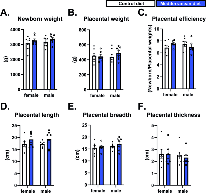
Regarding newborn and placental macroscopic phenotypes, we observed no differences in birth weight, placental weight, placental efficiency), or placental length, breadth, or thickness between women in the usual care and Mediterranean diet groups (Fig. 1A–F)
Newborn biometric parameters and placental characteristics. Newborn biometric data, placental weight, placental efficiency (defined as the ratio of fetal to placental weight), and placental macroscopic parameters. Data were analysed by two-way ANOVA followed by Tukey’s post hoc test. Data are shown as individual values, with columns representing the mean ± SEM
Mediterranean diet induced changes in placental fatty-acid lipid species
The total content of 16 lipid species within the placenta were determined. Principal component analysis showed modest clustering of placentas from usual care and Mediterranean diet groups, indicating diet-associated variation in placental lipid composition (Fig. 2A). To identify placental fatty-acid species contributing most strongly to dietary discrimination, we performed a supervised Random Forest analysis (Fig. 2B). Among the highest-ranking lipids were the long-chain saturated fatty acid C24:0, total saturated fatty acid (SFA), and C18:0, suggesting that saturated lipid species were major contributors to the separation between dietary groups.
Mediterranean diet alters placental lipid composition. (A,B) Principal component analysis (PCA) and random forest analysis of placental lipid profiles. (C–G) Placental lipid species significantly altered by maternal adherence to the Mediterranean diet. Lipid species are expressed as the proportion (%) of total measured placental lipids. Saturated fatty acids (SFA) were defined as the sum of C14:0, C16:0, C18:0, C20:0, C22:0 and C24:0; monounsaturated fatty acids (MUFA) as the sum of C16:1, C18:1n9c, C20:1 and C22:1n9; and polyunsaturated fatty acids (PUFA) as the sum of C18:2n6c, C20:2, C20:3n6, C20:3n3, C20:4n6, C20:5n3 and C22:6n3. Data were analysed by two-way ANOVA followed by Tukey’s post hoc test. *P < 0.05. Data are shown as individual values, with columns representing the mean ± SEM.
Of the 16 lipid species detected (Fig. 2 and Fig. S1), five were significantly altered by a maternal Mediterranean diet. Specifically, placentas from the Mediterranean diet group exhibited reduced levels of the saturated fatty acids C18:0 and C24:0 (Fig. 2C,G). In contrast, the monounsaturated fatty acid (MUFA) C18:1n9c and the polyunsaturated fatty acid (PUFA) C20:3n6, as well as the saturated fatty acid C22:0, were significantly increased in placentas exposed to the Mediterranean diet (Fig. 2D–F). Notably, the increase in C20:3n6 was particularly pronounced in male placentas, indicating a sex-specific dietary effect.
When lipid species were grouped by class, maternal adherence to a Mediterranean diet was associated with a significant reduction in total saturated fatty acid (SFA), driven predominantly by changes in male placentas (Fig. 2H). Conversely, total MUFA were significantly elevated in male placentas (Fig. 2I), whereas no significant differences were observed in total PUFA levels (Fig. 2J). Taken together, these data suggest that a Mediterranean diet affects the placental lipid profile
Mediterranean diet alters select cellular signalling pathways in the placenta
Exposure to fatty acids can alter the phosphorylation status of key metabolic signalling pathways, including AKT and MAPKs41,42,43. In Mediterranean diet-exposed placentas, we observed a significant reduction in phosphorylated AKT levels in male, but not female, placentas, without changes in total AKT protein abundance (Fig. 3A–C). Moreover, the phosphorylated levels of p38 MAPK, a signalling pathway involved in multiple other cellular processes including vascular growth44,45,46, were significantly reduced in both male and female placentas. Notably, total p38 MAPK protein levels were significantly increased in male placentas (Fig. 3D–F). Other key components of other metabolic and growth-related pathways, namely PI3K and mTOR (PI3K-p85α, mTOR-GbL, and RAPTOR), were not changed by a maternal Mediterranean diet (Fig. 3). Taken together, these data suggest that a maternal Mediterranean diet modulates select metabolic and growth signalling pathways in the human placenta.
Mediterranean diet alters placental signaling pathways. Data were analyzed separately using Student’s t-test. *P < 0.05 was considered statistically significant. Data are shown as individual values, with columns representing the mean ± SEM. Levels of phosphorylated proteins were normalized to the corresponding total protein abundance, and total protein levels were normalized to Ponceau S staining
Mediterranean diet reduces placental expression of nutrient transporters and lipid metabolism-related and regulatory genes
We next analysed the mRNA expression of three key nutrient transporters: SLC3A2, which is involved in amino acid transport and trophoblast function47; SLC2A1, a major glucose transporter48; and SLC2A8, a class III facilitative glucose/fructose transporter49. This revealed that a maternal Mediterranean diet significantly downregulated SLC3A2 and SLC2A1 mRNA expression, whereas SLC2A8 expression remained unchanged (Fig. 4A–C)
Mediterranean diet reduces mRNA levels of key nutrient transporters and decreases the abundance of lipid-handling genes and proteins (ALOX15 and PPARγ). Gene expression data were analyzed by two-way ANOVA followed by Tukey’s post hoc test. PPARγ protein levels were analyzed using Student’s t-test and normalized to Ponceau S staining. *P < 0.05 was considered statistically significant. Data are shown as individual values, with columns representing the mean ± SEM
We also measured the mRNA levels of genes involved in mitochondrial fatty acid β-oxidation (ACADL, ACADM, ACADVL), fatty acid synthesis (FASN), and lipid transport (APOE). However, none of these genes were altered by a maternal Mediterranean diet (Fig. S2A–E). In contrast, ALOX15, a gene involved in lipid peroxidation, was significantly reduced in placentas from the Mediterranean diet group (Fig. 4D). Furthermore, although mRNA levels were unchanged, the abundance of PPARG, a conserved regulator of placentation50 and PUFA metabolism, was significantly reduced in both male and female placentas from the Mediterranean diet group (Fig. 4E,F). Taken together, these findings indicate that Mediterranean diet selectively modulates nutrient transport and lipid peroxidation pathways in the placenta.
Mediterranean diet modulates placental inflammation and extracellular matrix remodelling genes and proteins
Inflammation and extracellular matrix (ECM) remodelling are tightly coupled processes, as inflammatory signalling induces ECM degradation and reorganization through the regulation of proteases and matrix components, while changes in ECM composition and structure feedback to modulate inflammatory responses and tissue function. In light of this interplay, and given that consumption of Mediterranean diet components has been linked to reduced inflammation51, we next examined the expression of genes involved in inflammatory signalling and ECM remodelling. Proteins encoded by the RAGE, PTGS2, and SOCS3 genes are known to coordinate cytokine-driven responses52,53,54, while those encoding GHR, PAI1, and MMP3 contribute to ECM remodelling55,56,57. While no differences were observed in the mRNA levels of RAGE or PTGS2, SOCS3 expression was significantly reduced in female placentas of mothers adhering to the Mediterranean diet (Fig. 5A–C). In contrast, we found a significant overall reduction in the GHR mRNA levels in the placentas of the Mediterranean diet group (Fig. 5D). At the protein level, PAI1 and MMP3 were significantly increased in female placentas from the Mediterranean diet group, whereas in males, only PAI1 abundance was elevated, with no change observed for MMP3 (Fig. 5E,F). Taken together, these data indicate that Mediterranean diet modulates the expression of genes and the abundance of proteins involved in inflammation and extracellular matrix remodelling in the placenta.
Mediterranean diet reduces mRNA levels of genes involved in placental inflammation and growth signaling, while increasing proteins associated with extracellular matrix remodeling. (A–D) Relative mRNA expression of RAGE, PTGS2, SOCS3, and GHR in placental tissue. (E,F) Protein abundance of PAI1 and MMP3 with corresponding immunoblots. Protein levels were normalized to Ponceau S staining. Gene expression data were analyzed by two-way ANOVA followed by Tukey’s post hoc test. Protein data were analyzed using Student’s t-test. P < 0.05 was considered statistically significant. Data are shown as individual values, with columns representing the mean ± SEM.
Discussion
In this study, we show that maternal adherence to a Mediterranean diet alters placental lipid composition and modulates signalling pathways, nutrient transporter expression, and inflammatory and extracellular matrix–related pathways. Several of these effects were sex-specific, suggesting differential responses of male and female placentas to maternal dietary exposure. Notably, these molecular changes occurred in the absence of differences in placental macroscopic phenotype or neonatal weight
Consistent with these observations, we observed significant alterations in five placental lipid species, along with key genes and proteins involved in lipid handling, including ALOX1558 and its downstream target, PPAR gamma59(both reduced in the Mediterranean diet group). Among these, the saturated long-chain fatty acid C18:0 (stearic acid) was significantly reduced in placentas from women consuming a Mediterranean diet. This finding is of interest given that accumulation of saturated fatty acids has been shown to activate pro-inflammatory pathways, including increased cytokine production60. In line with previous reports linking elevated C18:0 to metabolic dysfunction, women with GDM exhibit higher circulating levels of C18:0 in maternal plasma61, and studies in primary human trophoblasts have demonstrated that C18:0 stimulates the synthesis and release of pro-inflammatory cytokines, including TNF-α, IL-6, and IL-862.
In addition, the proportion of C24:0 (lignoceric acid) was significantly reduced relative to other detected lipid species in the placenta of the Mediterranean diet group. Although C24:0 is present at low abundance in the placenta, its levels are responsive to maternal dietary exposures, as demonstrated in animal models subjected to obesogenic diets during gestation63. Studies in both human cohorts and animal models have identified alterations in lipid species containing C24:0 in obese and diabetic states, and animal models using high-energy diets during gestation have shown that such exposures can lead to metabolic dysfunction in the offspring63,64. While accumulation of specific lipid species containing C18:0 and C24:0, including ceramides, has been associated with mitochondrial dysfunction, reduced oxidative capacity, increased fatty acid accumulation, and insulin resistance63,64, our analysis measured total fatty acid abundance and did not assess ceramide species. Therefore, the biological implications of the reduced abundance of C18:0 and C24:0 observed in the present study should be interpreted with caution. Nevertheless, these changes may reflect alterations in placental lipid metabolism that could influence mitochondrial function and metabolic efficiency, although further studies are required to confirm these mechanisms. Even in the absence of differences in birthweight, such placental adaptations could influence the intrauterine environment and may have implications for long-term offspring health.
Among the lipid species increased in placentas from the Mediterranean diet group, C22:0 (behenic acid) warrants particular attention. Studies in animal models have shown that C22:0 can alleviate inflammation and insulin resistance in GDM, at least in part by modulating the TLR4/NF-κB signalling pathway. In addition, in vitro studies using placental tissues have demonstrated that this fatty acid reduces the secretion of pro-inflammatory cytokines, including IL-6, IL-17, and TNF-α, as well as chemokines such as CCL3, CCL8, CXCL2, and CXCL65.
We also observed a sexually dimorphic response in the proportion of C20:3n-6 (dihomo-γ-linolenic acid), a precursor for eicosanoid synthesis, which was significantly increased in male but not female placentas in the Mediterranean diet group. In the control group, male placentas, on average, had lower levels of this fatty acid than female placentas, but this difference was not statistically significant. Moreover, information regarding the role of this lipid species in placental function remains limited. Future studies are needed to determine whether changes in placental lipid species handling with the maternal Mediterranean diet reflect increased peroxisomal fatty acid oxidation. Alternatively, these changes may indicate a shift away from lipid storage pathways toward enhanced lipid catabolism in the placenta, or increased transplacental transfer of these fatty acids to the developing fetus.
Both AKT and p38MAPK are critical signalling pathways important for metabolism, inflammation, and cell growth66,67,68. Phosphorylation of both pathways was significantly reduced in the placentas of mothers following a Mediterranean diet. Notably, these changes occurred in the absence of alterations in the upstream regulatory unit of PI3K-p85 or in the downstream mediators growth and nutrient-sensing mediators, mTOR, and RAPTOR. Although further investigation is required, these findings suggest that the observed changes may reflect alterations in specific input signals, such as lipid species, membrane-associated receptors, or hormonal cues, rather than changes in the expression of core components of the PI3K–mTOR signalling axis.
In support of this interpretation, p38MAPK activity has been shown to promote the expression of suppressor of cytokine signalling 3 (SOCS3)69, and we observed significantly reduced SOCS3 mRNA levels in female placentas exposed to the Mediterranean diet. Moreover, studies in non-placental systems have demonstrated that growth hormone receptor (GHR) deficiency inhibits activation of the PI3K–AKT signalling pathway70. Consistent with this, we detected an overall reduction in GHR mRNA levels in the Mediterranean diet group, which may contribute to the attenuation of AKT signalling observed in these placentas. In addition, we observed a significant reduction in SLC3A2 expression in the Mediterranean diet placentas. SLC3A2 is an integrin-associated protein that forms the heavy chain of heteromeric amino-acid transporters and modulates integrin-dependent signalling in the placenta. Knockdown of SLC3A2 has been shown to reduce AKT phosphorylation71,72, suggesting that decreased SLC3A2 expression may further contribute to reduced AKT activity. Notably, we also detected reduced mRNA levels of SLC2A1 (GLUT1), the primary placental glucose transporter. While SLC2A1 expression is regulated by multiple signalling and metabolic cues, including growth factor and insulin-related pathways73,74, its reduction may reflect broader alterations in nutrient-sensing and transport mechanisms associated with attenuated AKT signalling, although a direct causal relationship cannot be inferred from the present data and will require further work.
Finally, we detected increased protein levels of plasminogen activator inhibitor-1 (PAI1; SERPINE1) in both male and female placentas exposed to the Mediterranean diet. Although elevated PAI1 levels have been associated with pregnancy complications such as GDM, preeclampsia, and fetal growth restriction56, PAI1 also plays an essential physiological role in placental development. This protein is expressed in extravillous interstitial and vascular trophoblasts, where it contributes to the regulation of extracellular matrix degradation, thereby controlling trophoblast invasion and ensuring appropriate remodelling of maternal spiral arteries while preventing excessive invasion56. Interestingly, in female placentas exposed to the Mediterranean diet, we also observed increased levels of matrix metalloproteinase-3 (MMP3). Under physiological conditions, PAI1 participates in the fine-tuning of matrix metalloproteinase activity by regulating the plasminogen–plasmin system. The concomitant increase in PAI1 and MMP3 in female placentas may therefore reflect a balanced remodelling programme rather than a pathological phenotype, although further studies will be required to define the functional consequences of this coordinated regulation.
Importantly, despite the observed molecular and biochemical adaptations in the placenta, neither newborn birth weight nor gross placental phenotype differed between the Mediterranean diet and control groups, irrespective of fetal sex. These findings suggest that maternal adherence to the Mediterranean diet may promote subtle functional and molecular placental adaptations without altering overall fetal growth at birth. However, whether these placental changes confer long-term benefits to the offspring remains unknown. Longitudinal follow-up studies will be essential to determine whether such adaptations influence susceptibility to metabolic, neurological, or cardiovascular disorders later in life.
Limitations and future research
This study has some limitations that should be considered when interpreting the findings. Although we identified changes in placental lipid composition as well as gene and protein expression, further functional studies will be required to determine the extent to which these molecular adaptations translate into changes in placental metabolism and nutrient transporter expression. For example, future experiments could involve treating trophoblast organoids or placental explants with the fatty acid species altered by maternal diet to better define their functional effects.
The sample size was relatively modest, and validation in larger cohorts will be important to confirm these findings and to further explore potential subgroup differences, including sex-specific effects
In addition, our placental fatty acid analysis assessed the relative proportion of specific fatty acid species rather than their absolute concentrations. Future studies incorporating quantitative and integrative lipidomics approaches would provide a more comprehensive understanding of placental lipid metabolism
While we measured placental protein expression of key extracellular matrix regulators such as PAI1 and MMP3, circulating levels in maternal and fetal compartments were not evaluated. Assessing these parameters could provide additional insight into their potential role in maternal-fetal signalling
Conclusions
In conclusion, this study provides evidence that maternal adherence to the Mediterranean diet modulates placental lipid composition, nutrient transport, and signalling pathways controlling growth and inflammation, highlighting the placenta as a key mediator of maternal diet-fetal interactions. However, larger and well-powered studies integrating state-of-the-art approaches, including transcriptomics, proteomics, metabolomics, and detailed histological analyses, will be required to fully characterize the impact of maternal dietary patterns on placental phenotype and to assess the long-term benefits of these changes for maternal and offspring health.
Data availability
The datasets are available from the corresponding author upon reasonable request
References
Moreno-Fernandez, J., Ochoa, J. J., Lopez-Frias, M. & Diaz-Castro, J. Impact of early nutrition, physical activity and sleep on the fetal programming of disease in the pregnancy: A narrative review. Nutrients12, 3900. https://doi.org/10.3390/nu12123900 (2020)
Parrettini, S., Caroli, A. & Torlone, E. Nutrition and metabolic adaptations in physiological and complicated pregnancy: Focus on obesity and gestational diabetes. Front. Endocrinol. (Lausanne). 11, 611929. https://doi.org/10.3389/fendo.2020.611929 (2020)
Yajnik, C. S. & Deshmukh, U. S. Fetal programming: maternal nutrition and role of one-carbon metabolism. Rev. Endocr. Metab. Disord. 13, 121–127. https://doi.org/10.1007/s11154-012-9214-8 (2012)
Luca, A-C. et al. The first 1000 days: Maternal nutrient intake-a window of opportunity for pulmonary hypertension-a narrative review. Nutrients18, 424. https://doi.org/10.3390/nu18030424 (2026)
Lopez-Tello, J. et al. Competition for materno-fetal rePLoS One. 12, e0169194. https://doi.org/10.1371/journal.pone.0169194 (2017)
Parenti, M. & Paquette, A. G. Periconceptional and prenatal nutrition shapes the fetal epigenomic landscape. Epigenomics17, 1621–1639. https://doi.org/10.1080/17501911.2025.2605942 (2025)
Alum, E. U. et al. Maternal nutrition, toxicants, and epigenetic programming of obesity across generations. Diabetes Metab. Syndr. Obes.18, 4873–4911. https://doi.org/10.2147/DMSO.S579409 (2025)
Cui, S. et al. Maternal nutritional imbalance during pregnancy and the development of fetal-origin cardiovascular diseases. Front. Nutr.13, 1717069. https://doi.org/10.3389/fnut.2026.1717069 (2026)
Kirkbride, J. B. et al. Prenatal nutrition, epigenetics and schizophrenia risk: can we test causal effects? Epigenomics. 4, 303–315. https://doi.org/10.2217/epi.12.20 (2012)
Willett, W. C. et al. Mediterranean diet pyramid: a cultural model for healthy eating. Am. J. Clin. Nutr.61, 1402S–1406S. https://doi.org/10.1093/ajcn/61.6.1402S (1995)
Aleksandrova, K., Koelman, L. & Rodrigues, C. E. Dietary patterns and biomarkers of oxidative stress and inflammation: A systematic review of observational and intervention studies. Redox Biol.42, 101869. https://doi.org/10.1016/j.redox.2021.101869 (2021)
González, C. M., Martínez, L., Ros, G. & Nieto, G. Evaluation of nutritional profile and total antioxidant capacity of the Mediterranean diet of southern Spain. Food Sci. Nutr.7, 3853–3862. https://doi.org/10.1002/fsn3.1211 (2019)
Nucci, D. et al. Mediterranean diet in cancer patients’ survival: A systematic review and meta-analysis for tertiary prevention featured in the Italian National Guidelines La Dieta Mediterranea. Nutrition145, 113071. https://doi.org/10.1016/j.nut.2025.113071 (2025)
Volpe, R. et al. Efficacy of Mediterranean diet for the prevention of cardiovascular disease in patients: A systematic review and meta-analysis featured in the Italian National Guidelines La Dieta Mediterranea. Nutrition144, 113053. https://doi.org/10.1016/j.nut.2025.113053 (2026)
Zuliani, G. et al. Efficacy of Mediterranean diet for the prevention of neurological diseases: A systematic review and meta-analysis featured in the Italian National Guidelines La Dieta Mediterranea. Nutrition142, 112990. https://doi.org/10.1016/j.nut.2025.112990 (2026)
Muscogiuri, G. et al. Mediterranean diet for the management of pre-existing metabolic diseases: Evidence from a systematic review and meta-analysis featured in the Italian national guidelines La Dieta Mediterranea. Nutrition142, 112975. https://doi.org/10.1016/j.nut.2025.112975 (2026)
Estruch, R. et al. Primary prevention of cardiovascular disease with a mediterranean diet supplemented with extra-virgin olive oil or nuts. N. Engl. J. Med.378, e34. https://doi.org/10.1056/NEJMoa1800389 (2018)
Martínez-Lapiscina, E. H. et al. Mediterranean diet improves cognition: the PREDIMED-NAVARRA randomised trial. J. Neurol. Neurosurg. Psychiatry. 84, 1318–1325. https://doi.org/10.1136/jnnp-2012-304792 (2013)
Martínez-González, M. A. et al. Benefits of the mediterranean diet: Insights from the PREDIMED study. Prog. Cardiovasc. Dis.58, 50–60. https://doi.org/10.1016/j.pcad.2015.04.003 (2015)
Protocol derial. JAMA Intern. Med. ;178:1731–1732. https://doi.org/10.1001/jamainternmed.2018.6460. (2018)
Assaf-Balut, C. et al. A Mediterranean diet with additional extra virgin olive oil and pistachios reduces the incidence of gestational diabetes mellitus (GDM): A randomized controlled trial: The St. Carlos GDM prevention study. PLoS One. 12, e0185873. https://doi.org/10.1371/journal.pone.0185873 (2017)
Zhao, L. et al. Does a MediDiet with additional extra virgin olive oil and pistachios reduce the incidence of gestational diabetes? Endocr. Pract.28, 135–141. https://doi.org/10.1016/j.eprac.2021.08.010 (2022)
Amati, F., Hassounah, S. & Swaka, A. The impact of mediterranean dietary patterns during pregnancy on maternal and offspring health. Nutrients11, 1098. https://doi.org/10.3390/nu11051098 (2019)
Crovetto, F. et al. Effects of mediterranean diet or mindfulness-based stress reduction on prevention of small-for-gestational age birth weights in newborns born to at-risk pregnant individuals: The IMPACT BCN randomized clinical trial. JAMA326, 2150–2160. https://doi.org/10.1001/jama.2021.20178 (2021)
Minhas, A. S. et al. Mediterranean-style diet and risk of preeclampsia by race in the boston birth cohort. J. Am. Heart Assoc.11, e022589. https://doi.org/10.1161/JAHA.121.022589 (2022)
Saunders, L. et al. Effect of a Mediterranean diet during pregnancy on fetal growth and preterm delivery: results from a French Caribbean Mother-Child Cohort Study (TIMOUN). Paediatr. Perinat. Epidemiol.28, 235–244. https://doi.org/10.1111/ppe.12113 (2014)
Timmermans, S. et al. The Mediterranean diet and fetal size parameters: The Generation R Study 2014. https://doi.org/10.1159/000356110
Nakaki, A. et al. Effects of Mediterranean diet or mindfulness-based stress reduction on fetal and neonatal brain development: a secondary analysis of a randomized clinical trial. Am. J. Obstet. Gynecol. MFM. 5, 101188. https://doi.org/10.1016/j.ajogmf.2023.101188 (2023)
Crovetto, F. et al. Effect of a mediterranean diet or mindfulness-based stress reduction during pregnancy on child neurodevelopment: a prespecified analysis of the IMPACT BCN randomized clinical trial. JAMA Netw. Open.6, e2330255. https://doi.org/10.1001/jamanetworkopen.2023.30255 (2023)
Youssef, L. et al. Effects of maternal Mediterranean diet on fetal cardiac function in high-risk pregnancies: a secondary analysis of the IMPACT BCN randomized trial. Am. J. Obstet. Gynecol. MFM. https://doi.org/10.1016/j.ajogmf.2025.101875 (2026)
Flor-Alemany, M. et al. Influence of an exercise intervention plus an optimal Mediterranean diet adherence during pregnancy on the telomere length of the placenta. GESTAFIT Project Placenta. 136, 42–45. https://doi.org/10.1016/j.placenta.2023.04.002 (2023)
Nakaki, A. et al. Effects of mediterranean diet or mindfulness-based stress reduction during pregnancy on fetal brain development detected by neurosonography: a secondary analysis of a randomized clinical trial (IMPACT BCN). Fetal Diagn. Ther.52, 46–58. https://doi.org/10.1159/000540580 (2025)
Nakaki, A. et al. The mediterranean diet in pregnancy: Implications for maternal brain morphometry in a secondary analysis of the IMPACT BCN randomized clinical trial. Nutrients16, 1604. https://doi.org/10.3390/nu16111604 (2024)
Crovetto, F. et al. Mediterranean diet, mindfulness-based stress reduction and usual care during pregnancy for reducing fetal growth restriction and adverse perinatal outcomes: IMPACT BCN (Improving Mothers for a better PrenAtal Care Trial BarCeloNa): a study protocol for a randomized controlled trial. Trials22, 362. https://doi.org/10.1186/s13063-021-05309-2 (2021)
Juton, C. et al. Reliability and concurrent and construct validity of a food frequency questionnaire for pregnant women at high risk to develop fetal growth restriction. Nutrients13, 1629. https://doi.org/10.3390/nu13051629 (2021)
Castro-Barquero, S. et al. Validation of a pregnancy-adapted Mediterranean Diet Adherence Screener (preg-MEDAS): a validation study nested in the Improving Mothers for a better PrenAtal Care Trial BarCeloNa (IMPACT BCN) trial. Am. J. Clin. Nutr.120, 449–458. https://doi.org/10.1016/j.ajcnut.2024.05.025 (2024)
Yong, H. E. J. et al. Integrated placental modelling of histology with gene expression to identify functional impact on fetal growth. Cells12, 1093. https://doi.org/10.3390/cells12071093 (2023)
Livak, K. J. & Schmittgen, T. D. Analysis of relative gene expression data using real-time quantitative PCR and the 2(-Delta Delta C(T)) Method. Methods25, 402–408. https://doi.org/10.1006/meth.2001.1262 (2001)
Cabezas, A. et al. Effect of the inclusion of rumen-protected amino acids in the diet of growing beef cattle on animal performance and meat quality. Front. Anim. Sci.https://doi.org/10.3389/fanim.2023.1269775 (2023)
Lee, M. R. F., Tweed, J. K. S., Kim, E. J. & Scollan, N. D. Beef, chicken and lamb fatty acid analysis–a simplified direct bimethylation procedure using freeze-dried material. Meat Sci.92, 863–866. https://doi.org/10.1016/j.meatsci.2012.06.013 (2012)
Aggarwal, R. et al. Chronic exposure to palmitic acid down-regulates AKT in beta-cells through activation of mTOR. Am. J. Pathol.192, 130–145. https://doi.org/10.1016/j.ajpath.2021.09.008 (2022)
Simon, M. N., Azevedo-Martins, A. K., Amanso, A. M., Carvalho, C. R. O. & Curi, R. Persistent activation of Akt or ERK prevents the toxicity induced by saturated and polyunsaturated fatty acids in RINm5F β-cells. Toxicol. Vitro. 22, 1018–1024. https://doi.org/10.1016/j.tiv.2008.02.012 (2008)
Chai, W. & Liu, Z. p38 mitogen-activated protein kinase mediates palmitate-induced apoptosis but not inhibitor of nuclear factor-kappaB degradation in human coronary artery endothelial cells. Endocrinology148, 1622–1628. https://doi.org/10.1210/en.2006-1068 (2007)
Romero-Becerra, R., Santamans, A. M., Folgueira, C. & Sabio, G. p38 MAPK pathway in the heart: New insights in health and disease. Int. J. Mol. Sci.21, 7412. https://doi.org/10.3390/ijms21197412 (2020)
Cuadrado, A. & Nebreda, A. R. Mechanisms and functions of p38 MAPK signalling. Biochem. J.429, 403–417. https://doi.org/10.1042/BJ20100323 (2010)
Shi, Y. & Gaestel, M. In the cellular garden of forking paths: how p38 MAPKs signal for downstream assistance. Biol. Chem.383, 1519–1536. https://doi.org/10.1515/BC.2002.173 (2002)
Simner, C. et al. DNA methylation of amino acid transporter genes in the human placenta. Placenta60, 64–73. https://doi.org/10.1016/j.placenta.2017.10.010 (2017)
Kramer, A. C. et al. Steroids regulate SLC2A1 and SLC2A3 to deliver glucose into trophectoderm for metabolism10/endocr/bqaa098 (2020)
Lipka, A. et al. The impact of SLC2A8 RNA interference on glucose uptake and the transcriptome of human trophoblast cells. Cells13, 391. https://doi.org/10.3390/cells13050391 (2024)
Dominguez, E. M. et al. PPARG directs trophoblast cell fate and establishment of the uterine-placental interface. bioRxiv 2025:2025.10.08.681177. https://doi.org/10.1101/2025.10.08.681177
Keshani, M. et al. Mediterranean diet reduces inflammation in adults: A systematic review and meta-analysis of randomized controlled trials. Nutr. Rev.https://doi.org/10.1093/nutrit/nuaf213 (2025)
Chandra, N. et al. Inflammatory mediators and the RAGE pathway in placental tissues of pregnancies complicated by severe preeclampsia. Front. Reprod. Health. 7, 1587699. https://doi.org/10.3389/frph.2025.1587699 (2025)
Li, R. et al. LPS-induced PTGS2 manipulates the inflammatory response through trophoblast invasion in preeclampsia016/j.repbio.2022.100696 (2022)
Justina, V. D. et al. Increased expression of STAT3 and SOCS3 in placenta from hyperglycemic rats. Eur. J. Histochem.63, 3054. https://doi.org/10.4081/ejh.2019.3054 (2019)
Silva, C. M., Kloth, M. T., Lyons, C. E., Dunn, C. R. & Kirk, S. E. Intracellular signaling by growth hormone variant (GH-V). Growth Horm. IGF Res.12, 374–380. https://doi.org/10.1016/s1096-6374(02)00048-5 (2002)
Ye, Y. et al. Role of plasminogen activator inhibitor type 1 in pathologies of female reproductive diseases. Int. J. Mol. Sci.18, 1651. https://doi.org/10.3390/ijms18081651 (2017)
Husslein, H. et al. Expression, regulation and functional characterization of matrix metalloproteinase-3 of human trophoblast. Placenta30, 284–291. https://doi.org/10.1016/j.placenta.2008.12.002 (2009)
Kwon, H-J., Kim, S-N., Kim, Y-A. & Lee, Y-H. The contribution of arachidonate 15-lipoxygenase in tissue macrophages to adipose tissue remodeling. Cell. Death Dis.7, e2285–e2285. https://doi.org/10.1038/cddis.2016.190 (2016)
Singh, N. K. & Rao, G. N. Emerging role of 12/15-Lipoxygenase (ALOX15) in human pathologies. Prog. Lipid Res.73, 28–45. https://doi.org/10.1016/j.plipres.2018.11.001 (2019)
Pathmaperuma, A. N. et al. Fatty acids alter glycerolipid metabolism and induce lipid droplet formation, syncytialisation and cytokine production in human trophoblasts with minimal glucose effect or interaction. Placenta31, 230–239. https://doi.org/10.1016/j.placenta.2009.12.013 (2010)
Furse, S. et al. Altered lipid metabolism in obese women with gestational diabetes and associations with offspring adiposity. J. Clin. Endocrinol. Metab.107, e2825–e2832. https://doi.org/10.1210/clinem/dgac206 (2022)
Yang, X. et al. Saturated fatty acids enhance TLR4 immune pathways in human trophoblasts. Hum. Reprod.30, 2152–2159. https://doi.org/10.1093/humrep/dev173 (2015)
Álvarez, D. et al. Effects of a high-fat diet and docosahexaenoic acid during pregnancy on fatty acid composition in the fetal livers of mice. Nutrients15, 4696. https://doi.org/10.3390/nu15214696 (2023)
Camacho-Morales, A. et al. Plasma C24:0 ceramide impairs adipose tissue remodeling and promotes liver steatosis and glucose imbalance in offspring of rats. Heliyon10, e39206. https://doi.org/10.1016/j.heliyon.2024.e39206 (2024)
Liu, K. et al. Behenic acid alletes mellitus by regulating TLR4/NF-κB signaling pathway. iScience27, 111019. https://doi.org/10.1016/j.isci.2024.111019 (2024)
Fontana, F., Giannitti, G., Marchesi, S. & Limonta, P. The PI3K/Akt pathway and glucose metabolism: a dangerous liaison in cancer. Int. J. Biol. Sci.20, 3113–3125. https://doi.org/10.7150/ijbs.89942 (2024)
Acosta-Martinez, M. & Cabail, M. Z. The PI3K/Akt pathway in meta-inflammation. Int. J. Mol. Sci.23, 15330. https://doi.org/10.3390/ijms232315330 (2022)
Bengal, E. & Aviram, S. p38α MAPK Regulation of Energy Metabolism in Skeletal Muscle Offers a Therapeutic Path for Type 2 Diabetes. Cells14, 1277. https://doi.org/10.3390/cells14161277 (2025)
Chaves de Souza, J. A. et al. SOCS3 expression correlates with severity of inflammation, expression of proinflammatory cytokines, and activation of STAT3 and p38 MAPK in LPS-induced inflammation in vivo. Mediat. Inflamm.2013, 650812. https://doi.org/10.1155/2013/650812 (2013)
Yan, H-Z. et al. GHR is involved in gastric cell growth and apoptosis//doi.org/10.1111/jcmm.16160 (2021)
Zhu, B. et al. SLC3A2 is upregulated in human osteosarcoma and promotes tumor growth through the PI3K/Akt signaling pathway. Oncol. Rep.37, 2575–2582. https://doi.org/10.3892/or.2017.5530 (2017)
Feral, C. C. et al. CD98hc (SLC3A2) mediates integrin signaling. Proc. Natl. Acad. Sci.102, 355–360. https://doi.org/10.1073/pnas.0404852102 (2005)
Illsley, N. P. & Baumann, M. U. Human placental glucose transport in fetoplacental growth and metabolism. Biochim. Biophys. Acta Mol. Basis Dis.1866, 165359. https://doi.org/10.1016/j.bbadis.2018.12.010 (2020)
Hauguel-de Mouzon, S. et al. Developmental expression of Glut1 glucose transporter and c-fos genes in human placental cells. Placenta15, 35–46. https://doi.org/10.1016/s0143-4004(05)80234-6 (1994)
Acknowledgements
We thank the Clinic-IDIBAPS Biobank and the “Biobanc de l’Hospital Infantil Sant Joan de Déu per a la Investigació” integrated in the Spanish Biobank Network of ISCIII for the sample and data procurement
Funding
JL-T is supported by an Attraction of Talent Grant from the Community of Madrid (grant No. 2023-T1/SAL-GL-28960, CESAR NOMBELA fellowship) and a Sir Henry Wellcome Postdoctoral Fellowship (220456/Z/20/Z). LY was supported by the grant FJC2021-048123-I, funded by MCIN/AEI/https://doi.org/10.13039/501100011033 and by the European Union “NextGenerationEU”/PRT, and the BITRECS fellowship programme “Biomedicine International training research programme for excellent clinician-scientist” by Barcelona Clinic Hospital and funded by the “la Caixa” Foundation (code num. LCF/PR/SP23/52950012). F Crovetto was supported by the Departament de Salut de la Generalitat de Catalunya (SLT042/25/000050). F Crispi was supported by Instituto de Salud Carlos III (INT25/00064). ANS-P is supported by a Lister Institute of Preventive Medicine Research Prize (RG93692). Additionally, the project was partially funded by a grant from Fundación Mutua Madrileña en la financiación y desarrollo del proyecto AP16002/2024; with the support of Fundació La Marató de TV3 Projecte 202415-30; and Instituto de Salud Carlos III (ISCIII), (PI24/00127, PI22/00684, PI22/00109), co-funded by the European Union. Funders had no role in the design and conduct of the study; collection, management, analysis, and interpretation of the data; preparation, review, or approval of the manuscript; and decision to submit the manuscript for publication.
Author information
Author notes
F. Crispi and A. N. Sferruzzi-Perri contributed equally to this work
Authors and Affiliations
Department of Physiology, Development, and Neuroscience, The Loke Centre for Trophoblast Research, University of Cambridge, Cambridge, UK
Jorge Lopez-Tello & A. N. Sferruzzi-Perri
Metabolic and Immune Diseases Department, Biomedical Research Institute Sols-Morreale (IIBM), Spanish National Research Council (CSIC) – Autonoma University of Madrid (UAM), Madrid, Spain
Jorge Lopez-Tello
Department of Physiology, Faculty of Medicine, Autonomous University of Madrid, Madrid, Spain
Jorge Lopez-Tello
BCNatal Fetal Medicine Research Center (Hospital Clínic and HospitalSant Joan de Déu), University of Barcelona, Barcelona, Spain
L. Youssef, F. Crovetto, E. Gratacos & F. Crispi
Institut d’Investigacions Biomèdiques August Pi i Sunyer (IDIBAPS), Barcelona, Spain
L. Youssef & F. Crispi
Animal Production Department, Veterinary Faculty, Complutense University of Madrid, Avenida Puerta de Hierro s/n, Madrid, 28040, Spain
R. Bermejo-Poza, A. Cabezas & J. De la Fuente
Institut de Recerca Sant Joan de Déu (IRSJD), Barcelona, Spain
F. Crovetto & E. Gratacos
Spanish Network in Maternal, Neonatal, Child and developmental Health Research, RICORS-SAMID, RD24/0013/0004Instituto de Salud Carlos III, Madrid, Spain
F. Crovetto & E. Gratacos
Centre for Biomedical Research on Rare Diseases (CIBER-ER), Madrid, Spain
E. Gratacos & F. Crispi
Authors
- Jorge Lopez-TelloView author publications
Search author on:PubMed Google Scholar
- L. YoussefView author publications
Search author on:PubMed Google Scholar
- R. Bermejo-PozaView author publications
Search author on:PubMed Google Scholar
- A. CabezasView author publications
Search author on:PubMed Google Scholar
- J. De la FuenteView author publications
Search author on:PubMed Google Scholar
- F. CrovettoView author publications
Search author on:PubMed Google Scholar
- E. GratacosView author publications
Search author on:PubMed Google Scholar
- F. CrispiView author publications
Search author on:PubMed Google Scholar
- A. N. Sferruzzi-PerriView author publications
Search author on:PubMed Google Scholar
Contributions
Acquired human placental samples (L.Y., A.C., F.C., E.G., F.C.), fatty acid analysis (R.B.-P., A.C., J.D.L.F.), laboratory work (J.L.-T.), acquired funding (J.L.-T., A.N.S.-P., L.Y., A.C., F.C., E.G., F.C.), prepared the figures (J.L.-T.), designed laboratory experiments (J.L.-T., A.N.S.-P.), data analysis (J.L.-T., L.Y., R.B.-P.), drafted original version of the article (J.L.-T.), all authors proofread the final version of the manuscript
Ethics declarations
Competing interests
The authors declare no competing interests
Additional information
Publisher’s note
Springer Nature remains neutral with regard to jurisdictional claims in published maps and institutional affiliations
Supplementary Information
Below is the link to the electronic supplementary material
Supplementary Material 1 (download PDF )
Supplementary Material 2 (download DOCX )
Supplementary Material 3 (download TIF )
Supplementary Material 4 (download TIF )
Rights and permissions
Open Access This article is licensed under a Creative Commons Attribution 4.0 International License, which permits use, sharing, adaptation, distribution and reproduction in any medium or format, as long as you give appropriate credit to the original author(s) and the source, provide a link to the Creative Commons licence, and indicate if changes were made. The images or other third party material in this article are included in the article’s Creative Commons licence, unless indicated otherwise in a credit line to the material. If material is not included in the article’s Creative Commons licence and your intended use is not permitted by statutory regulation or exceeds the permitted use, you will need to obtain permission directly from the copyright holder. To view a copy of this licence, visit http://creativecommons.org/licenses/by/4.0/.
About this article
Cite this article
Lopez-Tello, J., Youssef, L., Bermejo-Poza, R. et al. Placental lipid handling, growth and inflammatory pathways are modified by a maternal Mediterranean diet.
Sci Rep16, 21820 (2026). https://doi.org/10.1038/s41598-026-60877-0
Received:27 March 2026
Accepted:30 June 2026
Published:28 July 2026
Version of record:28 July 2026
DOI
:https://doi.org/10.1038/s41598-026-60877-0
