


Download PDF
Abstract
Mitochondria are essential for cellular homeostasis, integrating various signals to control key cellular functions such as metabolism, apoptosis, inflammation, cell proliferation and redox balance. Given their multifaceted functions, it is not surprising that mitochondrial dysfunction has been implicated as a key contributor to the pathogenesis of numerous human diseases. Consequently, preserving mitochondrial integrity and functionality is vital for overall organismal health. Mitochondrial health is safeguarded by a sophisticated and tightly regulated network of quality control systems. These include mitochondrial proteostasis, which ensures proper protein folding and degradation; mitochondrial biogenesis, which governs the synthesis of new mitochondria; mitochondrial dynamics, encompassing fusion and fission processes; and mitophagy, the selective autophagic removal of damaged mitochondria. Additionally, these core systems are intricately connected to other crucial mitochondrial processes, such as the maintenance of mitochondrial DNA integrity, the regulation of cristae architecture, and the control of mitochondrial permeability transition, all of which are indispensable for optimal mitochondrial performance. Preclinical and clinical studies consistently demonstrate a strong link between impairments in these quality control mechanisms and both aging and the development of a wide spectrum of diseases. These include cancer, metabolic disorders, cardiovascular conditions, neurodegenerative diseases and autoimmune pathologies. In this review, we explore the different facets of mitochondrial quality control and discuss their implications in disease progression and aging. Furthermore, we highlight recent advances in interventions and therapies aimed at modulating mitochondrial quality control, providing an overview of their potential to mitigate disease burden and promote healthy aging.
Subjects
Introduction
Mitochondria are indispensable and highly specialized organelles that perform a broad range of crucial roles in the cell. While best known for generating energy through oxidative phosphorylation (OXPHOS), their functions extend far beyond ATP generation. Mitochondria act as critical hubs for calcium buffering and signaling, integrate and regulate numerous metabolic pathways, govern intrinsic apoptosis, and participate actively in innate immune responses. These diverse activities underscore the complex, dynamic, and multifaceted nature of mitochondrial biology.
Given their centrality to cellular homeostasis, mitochondria are continuously exposed to metabolic, environmental, and physiological stresses that can compromise their function and ultimately threaten cellular health. To mitigate these risks, cells have evolved an intricate network of mitochondrial quality control (MQC) mechanisms tailored to monitor, repair, and, when necessary, eliminate damaged mitochondrial components. These MQC pathways operate at multiple levels, spanning molecular chaperones and proteases that maintain proteostasis, mechanisms of fusion and fission that segregate dysfunctional mitochondrial segments, mitophagy pathways that selectively remove damaged mitochondria and mitochondrial biogenesis programs that replenish the organellar population.
Mitochondrial dysfunction is a hallmark of aging and a major contributor to numerous age-related pathologies.1 Importantly, disruptions in MQC mechanisms are implicated in aging and in various human diseases.2,3,4 Consequently, unraveling the molecular mechanisms governing MQC has become a major focus in the fields of cellular biology, aging research, and disease pathogenesis. Defects in individual MQC components—ranging from dysregulated mitochondrial dynamics to impaired mitophagy—have been implicated in a wide spectrum of human diseases, including neurodegenerative disorders, metabolic and cardiovascular diseases, cancer, and autoimmune conditions. However, despite substantial progress in understanding individual MQC pathways, the full extent of their contributions to disease onset, progression and severity remains incompletely understood. Furthermore, the interplay among the various MQC mechanisms and how cells integrate these pathways in response to distinct stressors continue to be an area of active investigation.
The goal of this review is to provide a comprehensive and integrative overview of the current understanding of MQC mechanisms. We will summarize the major molecular pathways that preserve mitochondrial integrity, including proteostasis systems, organelle dynamics, mitophagy, and biogenesis. In addition, we will discuss the implications of MQC in health and various disease contexts, such as metabolic syndrome, cardiovascular dysfunction, malignant transformation, neurodegenerative diseases, autoimmune disorders, and the aging process. Finally, we will examine emerging therapeutic strategies aimed at modulating MQC to prevent or ameliorate disease. By consolidating knowledge across these areas, this review aims to illuminate the central importance of MQC in maintaining cellular and organismal health and to identify potential avenues for clinical intervention.
Mitochondrial quality control mechanisms
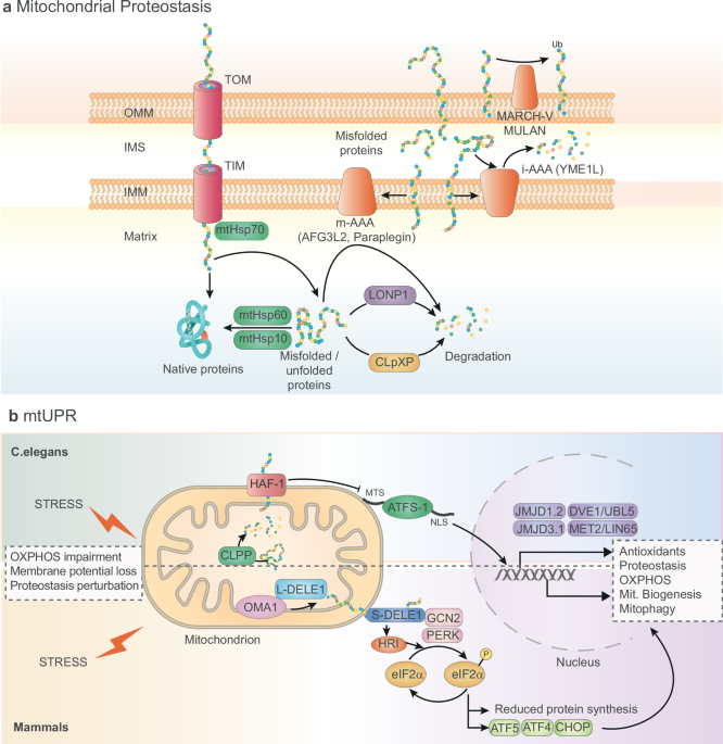
As essential organelles in maintaining cellular homeostasis, mitochondria require constant surveillance to ensure their proper functioning. MQC involves a diverse array of coordinated pathways operating at the molecular, organelle and cellular levels (Figs. 1–3). These pathways are activated depending on the type and severity of damage.5 The key components of the MQC system are detailed below
Mitochondrial quality control mechanisms at molecular level. a Mitochondrial proteostasis. Nuclear-encoded mitochondrial proteins enter mitochondrial transported by TOM and TIM complexes, and mtHSP70 assists in their folding upon transportation. The mtHSP60/mtHSP10 chaperonin complex encapsulates unfolded or partially folded proteins and promotes their proper folding. Misfolded proteins that cannot be refolded are degraded by mitochondrial proteases such as LONP1 and ClpXP in the matrix, and m-AAA or i-AAA proteases are located in the inner mitochondrial membrane. Ubiquitin ligases such as MARCH-V and MULAN target outer mitochondrial membrane proteins for proteasomal degradation. b Mitochondrial UPR (mtUPR). In C. elegans, CLPP detects and degrades misfolded or damaged proteins, and the peptides generated are transported to the cytosol by HAF-1, activating the nuclear localization of ATFS-1, which, together with the action of the histone demethylases JMJD1.2 and JMJD3.1 and the transcription factors DVE-1/UBL-5 and MET-2/LIN-65, activates the transcription of a subset of genes to restore mitochondrial health. In mammals, mtUPR is activated by the phosphorylation of eIF2α by the action of different kinases (HRI, GCN2 and PERK). This leads to increased translation of ATF5, ATF4 and CHOP, which triggers the expression of genes involved in mitochondrial homeostasis
Mitochondrial quality control mechanisms at the organellar level. a Mitochondrial biogenesis is regulated by PGC1α, which coactivates the transcription factors NRF1/2 to induce the expression of nuclear-encoded mitochondrial genes. TFAM controls mtDNA transcription and replication. b Mitochondrial dynamics is regulated by mitochondrial fusion and fission proteins. MFN1, MFN2 and OPA1 drive mitochondrial fusion, while MTFP1 inhibits it. On the other hand, DRP1, FIS1, MFF, and MID49/51 regulate mitochondrial fission. c Mitophagy can follow a Ub-dependent pathway or a receptor-mediated pathway. Ub-dependent mitophagy requires the ubiquitination of different mitochondrial proteins by PINK1-activated Parkin. Ubiquitinated proteins are recognized by different mitophagy adapters (p62, OPTN, NBR1, and NPD52), which bind to LC3 in the nascent autophagosome. Other Ub ligases, such as MUL1 and SIAH-1, have been described to participate in this type of mitophagy. Receptor-mediated mitophagy depends on the activation of mitophagy receptors (BNIP3, BNIP3L/NIX, BCL2L13, FUNDC1 and FKBP8), which interact with LC3 in the autophagosomal membrane. d Mitochondrial-derived vesicles (MDVs) originate from the action of PINK1 and Parkin or RAB9/SNX9 and are excised from mitochondria by the mitochondrial fission machinery. Once formed, they are delivered to the endosome or lysosome by the participation of STX17, RAB7 and VPS39/41
Mitochondrial quality control at the cellular level. Mitochondrial transfer between cells occurs through extracellular vesicles (mitoEVs) or tunneling nanotubes (TNTs) as a novel mechanism to secrete and eliminate damaged mitochondria. Mitochondrial components can also be transferred through the generation of MDVs
Mitochondrial protein quality control
Mitochondrial proteostasis (protein homeostasis) is essential for cellular function, energy production and signaling.6 Given their central role in bioenergetics and metabolism, mitochondria require a tightly regulated balance of protein synthesis, folding and degradation to prevent dysfunction.7 To achieve this proteostatic balance, mitochondria rely on specialized import and folding mechanisms (Fig. 1a). As nearly all mitochondrial proteins are encoded by the nuclear genome, they must be correctly imported into the mitochondria and then folded and assembled into functional complexes. The complex architecture of the mitochondrion, comprising the outer membrane, intermembrane space, inner membrane and matrix, necessitates precisely regulated mechanisms for protein import and assembly. Specialized translocase complexes, including the translocase of the outer membrane (TOM) and translocase of the inner membrane (TIM) complexes, manage protein import and assembly. The TOM complex serves as the primary entry point for nearly all nuclear-encoded mitochondrial proteins, whereas the TIM complex transports them to the mitochondrial matrix or inserts them into the inner membrane. For a more detailed view of mitochondrial protein import mechanisms and regulation, we refer to excellent reviews in the field.8,9 Once inside the mitochondria, proteins are folded, assembled or degraded. At this step, MQC involves chaperones to assist with protein folding and proteases to degrade misfolded proteins.10
Mitochondrial chaperones play a vital role in maintaining proper mitochondrial function by ensuring correct protein folding, preventing aggregation and refolding misfolded proteins (Fig. 1a). Key players include the heat shock protein Hsp60/Hsp10 chaperonin complex in the matrix, which encapsulates unfolded or partially folded proteins and promotes their proper folding within a protected environment through an ATP-dependent mechanism.11 Other chaperones, such as mitochondrial Hsp70 (mtHsp70), assist in protein translocation and folding upon import. mtHsp70 binds newly imported polypeptides, stabilizing and guiding them to their final destinations within the matrix or inner membrane, working in concert with the presequence translocase-associated motor (PAM).12 In yeast, Hsp78 assists in refolding aggregated proteins under heat stress, working alongside Hsp70.13 In the intermembrane space, small TIM chaperones ferry hydrophobic proteins with internal targeting sequences through the aqueous environment, preventing their release into the aqueous phase.9 Small TIM chaperones are heterohexameric complexes that can be soluble (Tim9-Tim10 complex) or bound to the IMM (Tim9-Tim10-Tim12 complex). Critically, under conditions of cellular stress, mitochondrial chaperones not only support proper folding but also cooperate with MQC systems, directing irreversibly misfolded proteins for degradation. This prevents aggregation and preserves mitochondrial health.10
Mitochondrial protein quality control relies heavily on ATPases associated with diverse cellular activities (AAA+ proteases). These proteases reside in various mitochondrial compartments (Fig. 1a). Within the inner mitochondrial membrane (IMM), two permanently anchored AAA+ proteases, intermembrane space-AAA (i-AAA) and matrix-AAA (m-AAA), oversee protein turnover. The i-AAA protease, facing the intermembrane space, degrades unstable proteins at this location, whereas the m-AAA protease, with its active site facing the matrix, assists in assembling mitochondrial ribosomes and degrading nonassembled proteins.14 i-AAA proteases form homo-oligomeric assemblies of Yme1 in yeast or Yme1-like ATPase (YME1L) in humans, whereas m-AAA proteases are hetero-oligomers of two subunits, Yta10 and Yta12 in yeast, or AFG3 ATPase family gene 3-like 2 (AFG3L2) and paraplegin in humans.15 In the mitochondrial matrix, two other AAA+ proteases, LON peptidase 1 (LONP1) and caseinolytic mitochondrial matrix peptidase (ClpXP), primarily degrade oxidatively damaged proteins.16,17 While the outer mitochondrial membrane (OMM) lacks dedicated proteases, it utilizes the ubiquitin‒proteasome system to regulate its protein landscape. Outer membrane-bound ubiquitin ligases, such as membrane-associated ring-CH-type finger 5 (MARCH-V/MITOL) and mitochondrial ubiquitin ligase activator of NF-κB (MULAN), tag specific proteins for degradation, maintaining proteome stability.18 The ubiquitin‒proteasome system also influences protein management across mitochondrial compartments by controlling precursor stability in the cytosol,19 highlighting its broader role in mitochondrial protein homeostasis.
Mitochondrial proteases not only degrade proteins and maintain quality control but also coordinate various MQC mechanisms. For example, YME1L works with the metalloprotease overlapping proteolytic activity with m-AAA protease 1 (OMA1) to cleave the mitochondrial dynamin-like GTPase, optic atrophy 1 (OPA1), a protein crucial for mitochondrial fusion, leading to mitochondrial fragmentation.20 Importantly, stress can activate OMA1, which then cleaves the signaling peptide of DAP3-binding cell death enhancer 1 (DELE1), producing a short form. This truncated form accumulates in the cytosol and triggers the integrated stress response.21,22 Additionally, two mitochondrial proteases, mitochondrial processing peptidase (MPP) and presenilin-associated rhomboid-like protease (PARL), are involved in mitophagy by processing and further degrading PTEN-induced kinase 1 (PINK1).23
Mitochondrial unfolded protein response
The mitochondrial unfolded protein response (mtUPR) is a transcriptional pathway activated in response to a wide range of mitochondrial stresses, including reactive oxygen species (ROS), protein import failure, blocked mitochondrial translation, mitochondrial DNA (mtDNA) depletion, electron transport chain impairment, and mito-nuclear protein imbalance (Fig. 1b). The mtUPR restores mitochondrial function by increasing the expression of nuclear-encoded chaperones and proteases involved in protein folding and degradation.24 While initially characterized in mammalian cells,25 the molecular components of the mtUPR have been extensively studied in Caenorhabditis elegans. In worms, the protease caseinolytic protease P (CLPP-1) is thought to detect and degrade misfolded or damaged mitochondrial proteins, generating oligopeptides that are then exported to the cytosol by the matrix peptide exporter HAF-1, activating the mtUPR.26,27 Activating transcription factor associated with stress-1 (ATFS-1) is the primary messenger of mitochondrial stress to the nucleus in worms.28 ATFS-1 contains both a nuclear localization sequence and a mitochondrial-targeting sequence. Under basal conditions, the mitochondrial-targeting sequence directs ATFS-1 to the mitochondria, where it is quickly degraded by LONP1. However, mitochondrial stress impairs protein import, causing ATFS-1 to accumulate in the cytosol. The nuclear localization sequence then directs ATFS-1 to the nucleus, where it activates genes that restore mitochondrial health (mitochondrial chaperones and proteases, protein import machinery and ROS detoxification).29 While ATFS-1 is the key messenger, other factors also contribute to the nuclear response. For example, the histone demethylases JMJD-1.2 and JMJD-3.1 facilitate access to mtUPR gene promoters.30 Additionally, the transcription factor defective proventriculus in Drosophila homolog 1 (DVE-1), its co-factor ubiquitin-like 5 (UBL-5), histone methyltransferase-like 2 (MET-2) and its co-factor abnormal cell lineage 65 (LIN-65) regulate the expression of mitochondrial chaperones.26,31,32
In mammals, the mtUPR relies on key transcription factors, including the mammalian ortholog of ATFS-1, activating transcription factor 5 (ATF5), ATF4 and C/EBP homologous protein (CHOP)25,33 (Fig. 1b). The mammalian mtUPR is activated by the phosphorylation of eukaryotic translation initiation factor 2 subunit 1 (eIF2α) by eIF2α kinases 3 and 4 (EIF2AK3/PERK and EIF2AK4/GCN2). These kinases are activated by ROS, amino acid depletion and endoplasmic reticulum (ER) stress, suggesting that the mtUPR could also work alongside the integrated stress response.34,35 Phosphorylation of eIF2α reduces global protein synthesis while promoting the translation of specific mRNAs, including those encoding CHOP, ATF4 and ATF5.36,37 As previously mentioned, the mtUPR can also be triggered by the processing of the long form of DELE1 by OMA1, leading to the cytosolic accumulation of truncated DELE1, which activates EIF2AK1 (also known as HRI), which phosphorylates eIF2α.21 In addition to activation of these mtUPR mechanisms by proteotoxic stress in the mitochondrial matrix, a separate mtUPR pathway responds to unfolded protein stress within the IMS. This IMS-specific pathway involves AKT phosphorylation and activation of the nuclear hormone receptor estrogen receptor alpha (ERα), which in turn increases the expression of the IMS protease HtrA2 and the transcription factor nuclear respiratory factor 1 (NRF1), a key regulator of mitochondrial biogenesis.38
Mitochondrial biogenesis
Mitochondrial biogenesis––the creation of new mitochondria––is a tightly regulated process essential for tissue development, adapting to energy stress and recovering from mitochondrial damage. This complex process requires coordinated gene expression from both the nucleus and the mitochondria, together with mitochondrial protein import, lipid synthesis and mitochondrial DNA (mtDNA) replication (Fig. 2a). A key regulator of this process is peroxisome proliferator-activated receptor gamma coactivator 1-alpha (PGC-1α), often considered the “master regulator” of mitochondrial biogenesis. PGC-1α coactivates transcription factors such as NRF1 and NRF2,39 which then promote the expression of nuclear-encoded mitochondrial genes. This, in turn, drives the transcription of genes involved in OXPHOS, the machinery for importing proteins into the mitochondria and mtDNA replication.40
Another critical player in mitochondrial biogenesis is mitochondrial transcription factor A (TFAM), which controls mtDNA transcription and replication. Maintaining the mitochondrial genome and proper organelle function depends on TFAM activity.41 Mitochondrial biogenesis is also tightly regulated by energy-sensing pathways. For example, AMP-activated protein kinase (AMPK) activates PGC-1α when energy levels are low.42 Similarly, sirtuin 1 (SIRT1) activates PGC-1α (by deacetylation) in response to nicotinamide adenine dinucleotide (NAD+) levels, linking mitochondrial biogenesis to cellular metabolic states.43 Finally, the mechanistic target of rapamycin (mTOR) supports mitochondrial biogenesis in nutrient-rich conditions by regulating protein synthesis and mitochondrial translation.44
Mitochondrial dynamics
When molecular MQC fails or mitochondrial damage exceeds a certain threshold, a second line of defense is activated––organellar quality control. This involves mitochondrial dynamics, continuous mitochondrial fusion and fission processes crucial for mitochondrial and cellular homeostasis (Fig. 2b). These dynamics allow mitochondria to adapt to cellular stress and are crucial for quality control.45 Mitochondrial fusion merges individual mitochondria into larger, interconnected networks, distributing mitochondrial contents and mitigating localized damage while ensuring optimal mtDNA integrity. Fusion occurs in two steps: OMM fusion and IMM fusion. In mammals, OMM fusion is mediated by mitofusin 1 (MFN1) and mitofusin 2 (MFN2) GTPases located on the OMM. These proteins tether adjacent mitochondria and promote membrane merging by forming homotypic (MFN1-MFN1) or heterotypic (MFN1-MFN2) complexes.46 MFN2 also tethers mitochondria to the ER, facilitating crucial interorganelle communication for lipid transfer and calcium homeostasis.47,48 IMM fusion requires the activity of OPA1, which is located in the inner membrane. OPA1 exists in long (L-OPA1) and short (S-OPA1) forms. L-OPA1 anchors to the inner membrane, while s-OPA1 is generated by proteolytic cleavage of L-OPA1. This cleavage is mediated by OMA1 in response to mitochondrial depolarization, ATP depletion and other stresses or constitutively by YME1L, which is upregulated during OXPHOS stimulation.49 The coordinated activity of both long and short OPA1 isoforms is critical for fusion. The specific functions of both isoforms are still under debate; however, L-OPA seems to be more important for mitochondrial fusion and s-OPA1 for sustaining energetic efficiency.50 Beyond fusion, OPA1 maintains cristae architecture, preserving mitochondrial function and preventing cytochrome c release during apoptosis.51 In addition to mitofusins and OPA1, phosphatidic acid (generated by mitochondrion-localized phospholipase D) and cardiolipin also play important roles in mitochondrial fusion.52,53 Conversely, it has recently been demonstrated that the IMM protein mitochondrial fission process 1 (MTFP1) inhibits mitochondrial fusion, creating IMM subdomains targeted for fission and subsequent autophagic degradation.54
Mitochondrial fission, which involves cytosolic and mitochondrial proteins, cytoskeletal elements and regulatory pathways, is orchestrated by the large GTPase dynamin-related protein 1 (DRP1).55 Inactive DRP1 resides in the cytosol, but mitochondrial stress or division signals trigger its recruitment to the OMM, mediated by adapter proteins, including mitochondrial fission protein 1 (FIS1), mitochondrial fission factor (MFF) and mitochondrial dynamics proteins of 49 and 51 kDa (MiD49 and MiD51).56 Mitochondrial fission enables organelle redistribution during mitosis, facilitates apoptosis by segregating damaged mitochondria and supports mitophagy by preparing dysfunctional segments for removal.57 Furthermore, DRP1 drives two different types of mitochondrial fission: midzone division, which promotes mitochondrial biogenesis, and peripheral division, which is associated with mitophagy.58
The mechanisms governing mitochondrial fusion and fission are complex and involve the coordinated action of the core mitochondrial dynamics machinery. This includes their structural characteristics and posttranslational modifications, mitochondrial membrane lipids and interactions with other organelles such as the ER. For a comprehensive review of the molecular mechanisms controlling mitochondrial dynamics, we refer the reader to a recent excellent review.46
Mitophagy
Mitophagy, the selective degradation of mitochondria via autophagy, involves a complex interplay of molecules and pathways that identify, isolate and breakdown defective mitochondria. This process begins with the identification of damaged mitochondria, which are then sequestered within autophagosomes and delivered to lysosomes for degradation. Mitophagy is broadly classified into two types based on the “eat me” signals that tag damaged mitochondria: ubiquitin (Ub)-mediated mitophagy and receptor-mediated mitophagy59 (Fig. 2c).
Ubiquitin-mediated mitophagy involves the attachment of Ub chains onto damaged mitochondria by E3-ubiquitin ligases, which are recognized by autophagy adapter proteins, triggering the formation of the mitophagosome. One of the best-understood Ub-mediated mitophagy mechanisms is the PINK1-Parkin pathway. Under normal conditions, PINK1 is imported into healthy mitochondria via the TOM complex, where it is degraded by proteases such as PARL. However, when mitochondria lose membrane potential (a sign of dysfunction), PINK1 import is impaired, causing PINK1 to accumulate on the OMM.60 This stabilized PINK1 then dimerizes and is activated by autophosphorylation. Active PINK1 phosphorylates serine 65 of Ub and Parkin, an E3 ubiquitin ligase, at specific residues, activating its ubiquitin ligase activity.61 Interestingly, PINK1 can also phosphorylate MFN2, further promoting Parkin binding and ubiquitination.62 Activated Parkin ubiquitinates numerous OMM proteins, including voltage-dependent anion channel 1 (VDAC1), creating docking sites for Ub-binding autophagy adapter proteins or autophagy receptors, such as p62/sequestosome 1 (SQSTM1), neighbor of BRCA1 gene 1 (NBR1), optineurin (OPTN), and calcium-binding and coiled-coil domain-containing protein 2 (CALCOCO2/NDP52).63 These receptors, possessing both ub-binding domains and LC3-interacting regions, bridge damaged mitochondria to LC3-positive autophagosomal membranes. LC3-II, the lipidated form of LC3, localizes to the autophagosomal membrane and participates in autophagosome formation and elongation. Therefore, the interaction of adapter proteins with LC3 ultimately leads to the engulfment of damaged mitochondria within autophagosomes, which then fuse with lysosomes for degradation.63 In addition to Parkin, other E3-Ub ligases, such as seven in absentia homolog (SIAH)-1 and mitochondrial Ub ligase 1/mitochondrial-anchored protein ligase (MUL1/MAPL), can drive mitophagy under specific contexts.59
Receptor-mediated mitophagy is driven by receptor proteins residing on the OMM, such as BCL2/adenovirus E1B 19 kDa protein-interacting protein 3 (BNIP3), BCL2/adenovirus E1B 19 kDa protein-interacting protein 3-like (BNIP3L/NIX), BCL2-like 13 (BCL2L13/Bcl-rambo), FUN14 domain containing 1 (FUNDC1) and FK506-binding protein 8 (FKBP8). Each of these receptors contains an LC3-interacting region, enabling them to bind LC3-positive autophagosomal membranes and recruit damaged mitochondria for engulfment.59 Because these receptors are constitutively present on the OMM, the key regulatory step in mitophagy is the upregulation or activation of the receptors themselves. For instance, BNIP3, NIX and FUNDC1 levels are regulated transcriptionally and posttranslationally through ubiquitination and phosphorylation.64,65,66,67 Receptor-mediated mitophagy contributes to both basal mitochondrial turnover and induced mitophagy in response to various cellular stresses and conditions, such as hypoxia, cellular differentiation, tissue remodeling and metabolic adaptation.67,68,69 For a more detailed discussion of the molecular regulation and physiological functions of mitophagy, readers are directed to other comprehensive reviews.59,70
Mitochondrial-derived vesicles and mitochondrial transfer
Finally, other MQC systems have been described, including mitochondrial-derived vesicles (MDVs, Fig. 2d), the release of mitochondria in extracellular vesicles (EVs) and the transfer of mitochondria between cells (Fig. 3)
Mitochondria-derived vesicles are small, single- or double-membrane vesicles that bud from mitochondria and transport damaged mitochondrial components, such as oxidized proteins and lipids, to lysosomes for degradation or to peroxisomes for detoxification. While MDV formation occurs at a basal level under physiological conditions, it increases during mitochondrial stress.71 This process begins with the identification of damaged mitochondrial regions, triggered by oxidative stress or mitochondrial depolarization. MDV formation and function involve several key molecules and complex processes that ensure proper cargo targeting and delivery.4 For example, the retromer complex components vacuolar protein sorting ortholog 35 (VPS35) and VPS26A are involved in cargo selection.72 PINK1 and Parkin initiate MDV biogenesis in response to mitochondrial stress,73 while sortin nexin 9 (SNX9) and the small GTPAse RAB9 participate in inflammation-induced MDV formation.74 The mitochondrial fission machinery, including DRP1 and its adapter proteins MFF, MiD49 and MiD51, regulates final MDV fission.75 Finally, the SNARE protein syntaxin 17 (STX17) together with the small GTPase RAB7 and VPS39/VPS41 are required for delivering PINK1/Parkin-generated MDVs to the late endosome/lysosome.76
Mitochondrial transfer, the exchange of mitochondria between cells, occurs through various mechanisms, including EVs, tunneling nanotubes (TNTs) and direct engulfment. Cells can also secrete mitochondria as a (novel) quality control mechanism to eliminate damaged organelles. Although progress has been made in recent years, there is still relatively little knowledge about the molecular mechanisms that underlie the different intercellular mitochondria transfer axes. For a detailed view of the different mitochondrial transfer mechanisms, we direct the reader to another excellent review.77 Recent studies have shown that diverse cell types, such as cardiomyocytes, adipocytes, astrocytes and mesenchymal stromal cells, release vesicles containing defective mitochondria.78,79,80,81 These EVs, termed mitoEVs, can contain whole mitochondria or mitochondrial components. mitoEVs can originate from MDVs, representing a quality control pathway when lysosomal function is compromised.82,83 Beyond mitochondrial homeostasis, mitoEVs also participate in intercellular communication, cell development, immune modulation and bioenergetic remodeling.84 TNTs are actin-based bridges between cells, ranging from 50 to 1000 nm in diameter. These structures facilitate the direct transfer of intact mitochondria, allowing healthy organelles to be redistributed to recipient cells with dysfunctional mitochondria. This transfer can restore metabolic function and reduce oxidative damage.85 While TNTs can form in healthy cells under normal conditions, their formation is also triggered by various stressors, including ultraviolet radiation, oxidative stress, DNA damage, nutrient restriction and senescence.86 For example, TNT-mediated mitochondrial transfer has been observed between astrocytes and neurons, demonstrating neuroprotective effects in mice,80 and between healthy and prostate cancer cells.87
Other aspects related to MQC: mtDNA integrity, cristae dynamics, and mitochondrial permeability transition pore (mPTP)
In addition to the main mechanisms of the MQC system, there are other aspects important for mitochondrial quality that need to be considered (Fig. 4). As described above, nuclear-encoded proteins such as TFAM, PGC1α, and NRF1/2 regulate mitochondrial biogenesis and are important for mtDNA replication under both basal and stress conditions.39,40,41 In addition to these proteins, the core machinery of mtDNA replication includes DNA polymerase γ (POLG), TWINKLE helicase and mitochondrial RNA polymerase (POLRMT)88 (Fig. 4a). Importantly, intrinsic errors of mtDNA replication caused by mutations in these genes can induce alterations in mtDNA, such as mutations or deletions, which are associated with mitochondrial dysfunction, metabolic alterations and the development of a number of different human diseases and aging.89,90,91 In addition to alterations in mtDNA replication, defects in mtDNA repair are also associated with disease. For instance, mtDNA mutator mice, which have a POLG deficient in proofreading activity, accumulate high levels of point mutations, leading to multisystemic disease and premature aging.92 To avoid the accumulation of mtDNA damage, mitochondria have different mtDNA repair mechanisms, such as base excision repair (BER), mismatch repair and double-strand break repair systems.93 When the damage cannot be repaired or is persistent, mtDNA can be degraded by the action of endonuclease G (EndoG) or POLG.94,95 Importantly, MQC mechanisms can be activated in response to mtDNA damage, such as mitochondrial fragmentation followed by mitophagy,96 and the induction of the ISR via the OMA1-DELE1-HRI pathway in response to mtDNA double-strand breaks.97 Interestingly, defects in some MQC mechanisms, such as mitophagy and mitochondrial dynamics, may promote the mislocation of mtDNA outside mitochondria, where it is a key signal triggering sterile inflammation.98,99 In this regard, cytosolic mtDNA can engage with either cyclic GAMP synthase (cGAS), triggering the type I interferon response and the nuclear factor kappa B (NFκB)-dependent pathway,100 or inflammasomes, promoting the secretion of interleukin 1β and 18.101 In addition, endosomal localization of mtDNA can induce Toll-like receptor 9 (TLR9)-dependent activation of the NFκB inflammatory pathway98 (Fig. 4a). In addition to mtDNA, other molecules released from damaged mitochondria, including mitochondrial RNA (mtRNA) and cardiolipins, have been shown to trigger inflammatory responses.102
Other aspects related to mitochondrial quality control. a Mitochondrial DNA (mtDNA) maintenance and integrity are preserved by the control of mtDNA replication by TFAM, POLG and TWINKLE; mtDNA repair systems (base excision repair (BER), mismatch repair and double-strand break repair systems); and mtDNA degradation carried out by POLG or EndoG. In addition, mtDNA mislocation outside mitochondria can trigger inflammatory responses through activation of the cGAS/STING pathway (type I interferon response), NLRP3 inflammasome (IL-1β and IL-18 secretion), or TLR9 (NFκB-induced inflammation). b Mitochondrial crista structure and organization. The MICOs complex and OPA1 protein are responsible for cristae structure and dynamics, which play an essential role in mitochondrial health. c Mitochondrial permeability transition pore (mPTP). The major components of mPTP are VDAC, ANT, PiC, ATP synthase and CypD
The organization and structure of cristae (deep invaginations of the IMM) is essential for proper mitochondrial function. In this regard, crista shape is key for the activity of some Krebs cycle enzymes, the formation and elimination of ROS, OXPHOS supercomplex (SC) stability and assembly, and mitochondrial calcium sensing and signaling.103 Different proteins have been described to be involved in the regulation of cristae structure and remodeling, such as the mitochondrial contact site and cristae organizing system (MICOS) and OPA1104 (Fig. 4b). For a detailed view of the different IMM-shaping machineries and how they regulate cristae dynamics, readers are directed to another excellent review.104 Alterations in these IMM-shaping machineries are associated with mitochondrial dysfunction and the development of disease. In this regard, different studies have shown that cristae disorganization induced by the absence of different MICOS subunits is associated with alterations in OXPHOS complexes, mitochondrial fragmentation and dysfunction,105,106 and the development of mitochondrial encephalopathy with liver disease.107 Regarding OPA1, mutations causing dominant optic atrophy (DOA) lead to cristae morphology and OXPHOS defects,108 and OPA1-dependent cristae remodeling is associated with the correct functionality of the OXPHOS system and protection from a range of pathological conditions in mice, such as muscular atrophy, heart and brain ischemia and reperfusion, and mitochondrial diseases.109,110 In addition, OPA1 has been described to regulate SC formation and stabilization and therefore respiratory efficiency.111
The mPTP is a protein complex located in the IMM that, when activated, forms a mega-channel permeable to ions and low molecular weight solutes (up to 1500 Da in size). The molecular nature of mPTP is still under debate, but a group of proteins have been proposed to be responsible for its formation, including adenine nucleotide translocator (ANT), voltage-dependent anion channels (VDACs), mitochondrial inorganic phosphate carrier (PiC) and F1/F0 ATP synthase112 (Fig. 4c). The channel is voltage-gated, activated by low mitochondrial membrane potential (Δψm) and inhibited by high Δψm, and it is physiologically activated by mitochondrial matrix calcium overload or excessive mitochondrial ROS production.113 In addition, cyclophilin D (CypD), the most important mPTP-associated protein, binds to and activates the channel, and posttranslational modifications of this protein, such as phosphorylation and acetylation, have been described to modulate mPTP activity.113 In addition, some mitochondrial chaperones, including HSP60 and HSP90, have been shown to interact with and inhibit CypD, conferring protection against mPTP opening and cell death.114,115 Physiological and transient opening of the mPTP is important for calcium efflux from the mitochondrial matrix and therefore for the regulation of calcium-related metabolism, as well as for the extrusion of endogenous ROS into the cytoplasm. In contrast, persistent mPTP opening has a strong impact on mitochondrial activity since it causes the depletion of NADH and the disassembly of respiratory SCs, and prolonged pore opening in a large number of mitochondria in the cell can finally lead to cell death.112,116,117 Interestingly, oxidized mtDNA can also be released from mitochondria via mPTP and trigger inflammatory responses.118 Therefore, alterations in mPTP or its regulators have a profound impact on cellular metabolism and have been described to be linked with aging and disease.119
Mitochondrial quality control and disease
Given the key role of mitochondria in cellular and tissue homeostasis, mitochondrial dysfunction has been associated with several diseases, many of which, as discussed below, are age-related conditions (Fig. 5). Consequently, we propose that disrupted MQC mechanisms contribute to the development of these diseases
Role of MQC in the development of disease. Alterations in different MQC systems are associated with the development of various diseases, including metabolic disorders, cardiovascular conditions, cancer, neurodegenerative diseases and autoimmune pathologies
MQC and metabolic diseases
Role of mtUPR and proteostasis in metabolic disease
Several preclinical studies have demonstrated a role for mtUPR and proteostasis in metabolic disease (Table 1). Indeed, various mitochondrial chaperones have been implicated in insulin resistance. For example, lower levels of GRP75/mtHSP70 in the liver and white adipose tissue (WAT) are observed in insulin-resistant mice, while inducing GRP75/mtHSP70 expression prevents both obesity and insulin resistance.120 Similarly, insulin-resistant humans show reduced HSP72 protein expression and increased c-Jun terminal kinase (JNK) phosphorylation in skeletal muscle,121 and mice lacking HSP72 develop insulin resistance and obesity.122 Conversely, overexpressing HSP72 in rodent skeletal muscle improves insulin sensitivity by increasing mitochondrial number and oxidative capacity.123 Interestingly, heterozygous whole-body HSP60 knockout mice fed a high-fat diet (HFD) exhibit lower body weight with reduced fat mass, along with improved insulin sensitivity and glucose tolerance. However, these same mice also display dysfunctional adipose tissue, characterized by altered metabolism, elevated insulin-independent glucose uptake, adipocyte hyperplasia and local insulin resistance.124
Mitochondrial proteases are also associated with metabolic disturbances. While some might be expected to support healthy metabolic function, their disruption can paradoxically improve certain metabolic parameters. For example, mice lacking the protease ClpP exhibit reduced adiposity, improved insulin sensitivity and protection against diet-induced obesity, glucose intolerance, insulin resistance and hepatic steatosis.125 This protection is likely due to a compensatory increase in mitochondrial biogenesis and respiration in WAT. Similarly, deleting Lonp1 specifically in muscle leads to an imbalance in mitochondrial proteostasis, triggering the mtUPR, which acts distally to activate thermogenesis in adipose tissue and decrease lipogenesis in the liver, ultimately conferring resistance to diet-induced obesity.126 Conversely, mice lacking Oma1, a protease involved in OPA1 processing, develop obesity, hepatic steatosis and reduced thermogenesis. These effects are exacerbated by metabolic stress, such as HFD, demonstrating that a functional OMA1-OPA1 system is essential for appropriate metabolic adaptation to stress.127
Role of mitochondrial biogenesis in metabolic disease
Obesity and type 2 diabetes mellitus (T2DM) are often associated with decreased mitochondrial mass and/or function in skeletal muscle.128 This is thought to be caused by impaired mitochondrial biogenesis, as evidenced by reduced expression of PGC1α and related genes. However, whether these alterations are linked with insulin resistance is still under debate.129
Role of mitochondrial dynamics in metabolic disease
Mitochondrial dynamics, which is closely linked to metabolism, adapt to metabolic needs130 and is a key pathophysiological component of metabolic diseases.131 In both humans and experimental models of obesity, insulin resistance is associated with impaired mitochondrial dynamics and altered levels of proteins that regulate fusion and fission in metabolically important tissues132 (Table 1)
With regard to mitochondrial fusion, MFN2 expression in skeletal muscle is reduced in obesity and T2DM in humans and mouse models, and Mfn2 mRNA levels in muscle correlate with insulin sensitivity in both nondiabetic individuals and those with T2DM.133 Consistent with this, Mfn2 deficiency in mouse skeletal muscle leads to insulin resistance in response to HFD and aging.134,135 Similarly, human metabolic dysfunction-associated fatty liver disease is linked to decreased MFN2 expression, and Mfn2 deficiency in mouse liver causes hepatic steatosis, insulin resistance and glucose intolerance early in life, progressing to fibrosis and liver cancer with age.47,134 In human adipose tissue, biallelic MFN2 mutations cause mitochondrial dysfunction, adipose tissue hyperplasia and impaired leptin expression,136 and adult mice lacking MFN2 in white adipocytes showed increased food intake, adiposity and impaired glucose metabolism regardless of diet.137 Conversely, Mfn2 ablation in brown adipose tissue (BAT) blunts thermogenesis but protects against HFD-induced glucose intolerance, insulin resistance and hepatic steatosis.138,139 Mitochondrial fusion is also important for central energy balance in the hypothalamus. In mice, specific ablation of Mfn2 or Opa1 in anorexigenic proopiomelanocortin neurons increases food intake and induces obesity and energy imbalance,140,141 whereas Mfn1 ablation impairs glucose homeostasis due to defective insulin secretion.142 In contrast, Mfn2 or Mfn1 ablation in orexigenic agouti-related peptide neurons protects against diet-induced obesity.143 Interestingly, deficiency of hepatic MFN1 also protects against diet-induced obesity and insulin resistance in mice,144 possibly via compensatory improvements in mitochondrial function. Similarly, Opa1 ablation in muscle or BAT protects against diet-induced obesity and insulin resistance through an FGF21-mediated compensatory mechanism.145,146 In individuals with obesity, OPA1 gene expression is significantly reduced in visceral adipose tissue, and ubiquitous mild induction of Opa1 in diet-induced obese mice improves glucose tolerance and insulin sensitivity by increasing adipose tissue expandability and browning.147 Finally, loss of function of Opa1 in mouse white adipocytes reduces WAT mass and alters lipid handling, leading to hepatic steatosis and glucose intolerance, but surprisingly protects against diet-induced obesity.148
Mitochondrial fission is also associated with metabolic homeostasis. Studies in obese mice have shown that pharmacologically inhibiting DRP1 or specifically deleting Drp1 in muscle improves insulin sensitivity.149,150 In the liver, while one study found increased DRP1 expression in rats fed a high-fructose diet but not a HFD,151 another found that liver-specific ablation of Drp1 in mice actually protected against HFD-induced obesity by increasing energy expenditure, driven by ER stress and subsequent FGF21 production.152 In the hypothalamus, deleting Drp1 in proopiomelanocortin neurons enhanced their activity and improved systemic glucose metabolism.153 Consistent with these findings, DRP1 expression in human adipose tissue correlates positively with obesity and insulin resistance.154 Furthermore, DRP1 activity is regulated by posttranscriptional modifications. For example, whole-body and skeletal muscle-specific ablation of calcineurin, which dephosphorylates DRP1, increases phosphorylation of DRP1 at S637, leading to DRP1 inactivation, mitochondrial elongation and protection against HFD-induced obesity.155 Similarly, mice with DRP1 S600A knock-in, which prevents DRP1 activation by phosphorylation at S600, show improved glucose tolerance and thermogenesis.156 Interestingly, HFD-fed mice show increased expression and activity of the small GTPase RalA in WAT, which counteracts the inhibitory S637 phosphorylation of DRP1, promoting mitochondrial fission and contributing to weight gain and metabolic dysregulation.154
Role of mitophagy in metabolic disease
Mitophagy appears to be impaired in individuals with T2DM. This is supported by several observations, including decreased expression of mitophagy-related markers in peripheral blood mononuclear cells,157 lower Parkin expression in myotubes158 and reduced PINK1 expression in both muscle and adipose tissue.159 While these findings suggest a link between impaired mitophagy and the development of metabolic disease, studies performed in different model organisms have yielded conflicting results about whether altering mitophagy is beneficial or harmful in the development of metabolic diseases (Table 1). These effects appear to depend on the specific tissue and the presence or absence of complementary mechanisms. For example, in mice, muscle-specific deletion of Fundc1 protects against diet-induced obesity and insulin resistance by triggering muscle-adipose crosstalk to promote fat burning in adipose tissue through FGF21.160 Conversely, whole-body ablation of Fundc1 worsened diet-induced metabolic dysfunction by inducing WAT remodeling and inflammation.161 Notably, overexpressing Fundc1 specifically in the pancreas improved HFD-induced insulin resistance and obesity.162
The impact of PINK1/Parkin-induced mitophagy on metabolism has been investigated in various animal models. In skeletal muscle, activating the PINK1-Parkin pathway with brain-derived neurotrophic factor (BDNF) improves mitochondrial quality, whereas muscle-specific Bdnf deletion exacerbates HFD-induced metabolic dysfunction.163 Similarly, Hsp72 ablation impairs Parkin activity, leading to damaged mitochondrial accumulation and muscle insulin resistance.122 Hepatic deletion of Parkin increases susceptibility to diet-induced steatosis, inflammation, fibrosis and insulin resistance.164 Conversely, preserving Parkin-mediated mitophagy improves β-cell function in diabetes models.165 In BAT, increased Parkin levels correlate with thermogenic inactivation, and Parkin knockout mice exhibit enhanced thermogenesis and improved metabolic parameters under HFD,166 potentially due to its role in thermogenic beige adipocyte retention.167 However, other studies suggest that activation of the PINK1/Parkin pathway promotes thermogenesis,168 as Pink1 global or BAT-specific deletion in mice induces BAT dysfunction and an obesity-prone phenotype.169 BNIP3-driven mitophagy also plays a role in metabolic dysfunction. Global Bnip3 deletion increases inflammation and steatohepatitis-like features in mice fed a standard chow diet170 and reduces adiposity but worsens liver steatosis and systemic insulin resistance in HFD-fed mice.171 Interestingly, the mitophagy activator urolithin A ameliorates insulin resistance in obese rodents,172 suggesting mitophagy as a potential therapeutic target for metabolic diseases.
Mitochondrial transfer and metabolic disease
Mitochondrial transfer can have complex metabolic effects. Transfer from a dysfunctional cell to a healthy cell can induce metabolic decline in the recipient, while transfer from a healthy cell to a dysfunctional cell can improve the metabolic state of the recipient. The role of this transfer in the context of metabolic diseases is now being explored. For example, both white and brown adipocytes can transfer mitochondria to macrophages, a process important for thermogenesis,83 that is impaired in obesity.173 In the heart, defective macrophage phagocytosis leads to impaired clearance of extracellular mitochondria and metabolic dysfunction.79 Interestingly, mitochondria released in EVs from stressed adipocytes can signal to the heart,78 demonstrating that mitochondrial transfer facilitates both intercellular and interorgan communication. While human data on mitochondrial transfer in metabolic disorders are limited, studies suggest a link between the release of mitochondrial components and T2DM. Lower circulating mtDNA levels are associated with T2DM,174 and low blood mtDNA content correlates with impaired glucose-stimulated islet β cell function, HbA1c levels, fasting plasma glucose and age of onset in T2DM.174,175
Other aspects related to MQC and metabolic diseases
Loss of mtDNA integrity is associated with the development of mitochondrial diseases, a diverse group of inherited metabolic disorders that ultimately impair mitochondrial bioenergetics. Some of those are mitochondrial encephalopathy, lactic acidosis, stroke-like episodes (MELAS), Kearns-Sayre syndrome (KSS), Pearson syndrome, some cases of Leigh syndrome and chronic progressive external ophthalmoplegia (CPEO). These diseases can be caused by mtDNA mutations or deletions or by alterations in proteins involved in mtDNA maintenance, such as POLG, TWINKLE, and TFAM. For a comprehensive view of mitochondrial diseases, we direct the reader to a specific review on this topic.91 Regarding metabolic diseases, different studies have shown that mice lacking different components of the mtDNA damage repair machinery are more prone to obesity, glucose intolerance, and insulin resistance.176,177 In humans, a common polymorphism in the BER enzyme 8-oxoguanine DNA glycosylase OGG1 correlates with increased body mass index, total cholesterol, fasting blood glucose and incidence of T2DM.178,179 In mice, alterations in cristae organization induced by dysregulation of MICOS components are associated with metabolic alterations, such as liver inflammation and liver fibrosis,180 and mPTP opening has been found to contribute to insulin resistance in muscle.181
In summary, while alterations in different MQC mechanisms are clearly linked to metabolic disorders (Table 1), it remains unclear whether these anomalies are a cause or a consequence of mitochondrial dysfunction and whether they can trigger metabolic disease. The relationship between these alterations and metabolic disturbances likely depends on the activation of adaptive pathways, which are probably triggered by the degree of mitochondrial damage. Finally, the specific tissue- or cell-dependent signals and/or events that contribute to impaired MQC are not yet fully understood.
MQC and cardiovascular disease (CVD)
Role of mtUPR and proteostasis in CVD
Cardiovascular diseases, such as heart failure, cardiomyopathy, myocardial ischemia‒reperfusion (I/R) injury and atherosclerosis, are characterized by mitochondrial damage and stress, such as unfolded and misfolded protein accumulation, calcium overload and oxidative stress. The mtUPR is known to play an important role in CVD development (Table 2). While mtUPR components can be down- or upregulated depending on the specific CVD, activating the mtUPR has shown promise as a therapeutic strategy. In rodent models of chronic pressure overload, pharmacologically boosting the mtUPR enhances cardiomyocyte survival and ventricular function.182 Similarly, upregulating the mtUPR through dietary supplements, drugs or genetic manipulation protects against cardiac hypertrophy in cardiomyopathies and spontaneous hypertension183,184 and cardiac injury in cardiometabolic syndromes.185,186 Interestingly, the HSP60/HSP10 ratio has been proposed as a potential biomarker of CVD,187 and enhancing HSP70 in the heart—through genetic or pharmacological approaches—protects against acute but not chronic cardiac stress in mice.188 Regarding mitochondrial proteases, while loss of Clpp can alleviate mitochondrial cardiomyopathy, it does not appear to affect the mtUPR.189 Ablation of the OMA1 protease has been shown to be protective in different mouse models of heart failure by protecting against cristae remodeling and cardiomyocyte death.190 However, in a genetic mouse model of mitochondrial cardiomyopathy induced by cardiac-specific ablation of Cox10, activation of OMA1 results in activation of the DELE1-ATF4 signaling axis of the ISR, which confers protection from cardiomyopathy.191
Role of mitochondrial biogenesis in CVD
Mitochondrial biogenesis is disrupted early in the progression of heart failure.192 Although increasing PGC-1α expression does not guarantee restored heart function, studies in different rodent models of heart failure have shown that stimulating mitochondrial biogenesis through different signaling pathways can improve cardiac performance and myocardial dysfunction.193,194 Consistent with this, diabetic cardiomyopathy is characterized by reduced PGC-1α levels and decreased mitochondrial activity in cardiomyocytes, together with increased myocardial injury. In these cases, boosting the activity of SIRT1 and SIRT6 may offer a potential strategy to restore mitochondrial biogenesis.195
Role of mitochondrial dynamics in CVD
Cardiac tissue is highly oxidative, and proper mitochondrial function, dependent on a balanced fusion-fission process, is crucial for maintaining a healthy cardiovascular system and responding to stress (Table 2). Loss of MFN1/2, OPA1 and DRP1 in cardiomyocytes causes a profound shift in this fusion-fission balance that impacts cardiac performance. For example, reduced Mfn2 expression is observed in experimental models of heart failure (induced by pressure overload) and diabetic cardiomyopathy and correlates with worsening cardiac abnormalities.196,197 Studies using loss-of-function approaches have underscored the importance of fusion-related proteins for cardiovascular health. Mice with conditional, combined Mfn1/2 knockout develop lethal dilated cardiomyopathy as adults.198 Similarly, studies in cardiomyocytes specifically lacking MFN2 have demonstrated its essential role in maintaining heart function.62,199 However, while combined Mfn1/Mfn2 ablation impairs myocardial contractile function, it paradoxically protects mice from acute I/R injury.200 This suggests that mitofusins play complex and context-dependent roles in heart physiology, potentially differing in response to short-term versus long-term stressors. A newly discovered pathway involving ubiquitin-specific protease 28 (USP28)-dependent activation of PPARα-MFN2 was found to restore mitochondrial function and improve cardiac structure and performance in diabetic hearts.201 In humans, MFN2 is downregulated in diabetic cardiomyopathy,202 and a rare Mfn2 mutation causing perinatal cardiomyopathy in mice is overrepresented in clinical cardiomyopathy.203 The expression of OPA1 is decreased in failing human hearts (explanted at transplant) and rodent heart failure models,204 and mice with a mutant version of Opa1, leading to a 50% reduction in OPA1 protein, develop late-onset cardiomyopathy.205 Additionally, imbalanced OPA1 processing leading to accelerated OPA1 proteolysis and mitochondrial fragmentation has a causal role in heart failure development in mice.206 Conversely, restoring OPA1 processing and reducing mitochondrial fragmentation in mice improves cardiac function,207 and increased OPA1 levels in mice protect against I/R injury.109 During I/R, left ventricle dysfunction is associated with DRP1 activation,208 and acutely inhibiting DRP1 activity with Mdivi-1 protects against heart hypertrophy and preserves function after I/R injury209 or myocardial infarction210 in mice. However, chronic DRP1 dysfunction, in both mutant and cardiac-specific Drp1 knockout mice, induces cardiac dysfunction, likely due to impaired mitophagy.211 Some effects of MFN2 deficiency on the heart also stem from disrupted mitophagy,62 reflecting the link between mitochondrial dynamics and mitophagy. Collectively, these findings indicate that maintaining the mitochondrial fusion-fission balance is essential for cardiomyocyte survival and heart function.
Role of mitophagy in CVD
In cardiac muscle, removal of damaged mitochondria is crucial for maintaining cardiac structure and function under normal conditions, and impaired mitophagy is associated with aging-induced cardiac abnormalities.212 Human heart failure is associated with a downregulation of mitophagic genes in cardiac tissue.213 Despite promising cardioprotective results with the mitophagy activator urolithin A in animal models, a recent randomized, double-blind, placebo-controlled crossover trial failed to find a positive effect of urothilin A supplementation in improving cardiac function in patients with heart failure.214 However, activating mitophagy by pharmacological and/or genetic strategies has been shown to be effective against cardiac dysfunction in experimental models of cardiometabolic syndrome, heart failure and diabetic cardiomyopathy.185,215,216 In the latter, the specific mitophagy mechanisms involved vary depending on the disease stage. In early phases, upregulation of Atg7-mediated mitophagy plays a key role in maintaining cardiac performance, while activation of Rab9-dependent alternative mitophagy becomes more important in later stages.217,218 In pressure overload-induced cardiac hypertrophy, ULK1-mediated alternative mitophagy is the primary protective mechanism against cardiac dysfunction.219 During I/R injury, clearing defective mitochondria is beneficial. In this regard, Parkin knockout mice have exacerbated infarct expansion,220 and FUNDC1-dependent mitophagy offers protection.221 Mitophagy also plays a role in age-related cardiac dysfunction. Mitophagy decreases with age in mouse hearts, and promoting Parkin-induced mitophagy alleviates aging-related decline in cardiac function.222 One possible explanation for this decline in mitophagy is increased Shank3 levels in aging hearts. Shank3, a heart-enriched protein, inhibits Parkin-mediated mitophagy by binding to CaMKII and preventing its mitochondrial translocation.223 Collectively, these findings suggest that mitophagy is essential for maintaining cardiac function and heart homeostasis under pathological or stressful conditions.
Mitochondrial transfer and CVD
Extracellular mitochondria are emerging as important regulators in CVD progression.224 Cardiomyocytes can eject damaged mitochondria, which are then engulfed and eliminated by resident macrophages, a process amplified during cardiac stress and crucial for cardiac function.79 Conversely, cardiomyocytes can also absorb mitochondria released by macrophages, triggering ferroptosis and cellular injury.225 This bidirectional mitochondrial exchange between cardiomyocytes and macrophages highlights its importance for heart homeostasis and function. Consequently, mitochondrial transfer and EVs are being explored as potential CVD therapies. For example, intramyocardial injection of mitochondria-rich EVs from autologous stem cell-derived cardiomyocytes improves postmyocardial infarction cardiac function in animal models.226 Similarly, cardiac-derived EVs have shown promise in mitigating I/R injury by delivering ATP synthase F1 subunit alpha (ATP5a1).227
Other aspects related to MQC and CVD
Alterations in mtDNA integrity are associated with the development of CVDs. Experiments using mDNA mutator mice demonstrated that mutations in mtDNA led to cardiac hypertrophy at the age of 6 months.92 Indeed, cardiomyopathy has been shown to be present in 20% of patients with mitochondrial disease harboring different mutations in mtDNA.228 Interestingly, circulating levels of mtDNA (possibly released from damaged mitochondria) have been described to be increased after myocardial infarction and to be associated with increased mortality in heart failure.229,230 Regarding mitochondrial crista organization, transgenic mice overexpressing the MICOS subunit MIC60/mitofilin show reduced type 1 diabetes-associated cardiomyopathy symptoms.231 Finally, numerous studies have demonstrated a key role of mPTP opening in cardiomyocyte cell death during I/R.232 In this regard, genetic ablation or pharmacological inhibition of CypD and ANT protects against heart damage during I/R.233,234
In summary, given the dependence of the heart on mitochondrial oxidative metabolism, disruption in MQC processes is believed to be a major driver of CVD and cardiac aging235 (Table 2). However, further research is necessary to fully elucidate the mechanisms underlying these mitochondrial quality changes in CVD and to confirm their presence and relevance in humans
MQC and neurodegenerative diseases
Role of mtUPR and proteostasis in neurodegenerative diseases
Neurons, with their high metabolic demand and complex morphology, rely heavily on intact mtUPR signaling and mitochondrial proteostasis. Accordingly, neurodegenerative diseases often exhibit evidence of either insufficient or maladaptive activation of these pathways. Mutations in the mitochondrial protease HTRA2/OMI have been found in sporadic cases of Parkinson’s disease (PD), and Htra2-deficient mice show neurodegeneration and Parkinson-like phenotypes.236 Mutations in paraplegin, a subunit of the m-AAA protease, cause hereditary spastic paraplegia (HSP),237 and paraplegin-deficient mice recapitulate the symptoms of the disease.238 Mutations in Hsp60 and decreased expression of LONP1 and CLPP have also been documented in HSP patients,239,240 and downregulation of Hsp60 and Clpp mRNA has been found in fibroblasts derived from Huntington’s Disease (HD) patients.241 The mitochondrial protease Pitrilysin Metallopeptidase 1 (PITRM1), a matrix peptidase that clears amyloid-β (Aβ) fragments, is deficient in Alzheimer’s Disease (AD) brains and model systems, leading to intramitochondrial Aβ accumulation and synaptic degeneration.242 In addition, reduced expression of LONP1 has been found in experimental models of AD and in the brains of AD patients.243 Consequently, LONP1 knockdown caused neuronal dysfunction, while restored neuronal expression of LONP1 in in vitro and in vivo models of AD rescued Aβ-induced mitochondrial alterations and cognitive deficits.243 Interestingly, other studies have demonstrated an increase in mtUPR-associated genes in AD patients and mouse models, including mitochondrial chaperones and proteases, suggesting a protective response during disease progression.244,245 Activation of mtUPR has also been described in PD models, which confers protection from neurotoxicity and neuronal death.246 However, in HD patients and mouse models, mtUPR has been shown to be impaired due to the sequestration of ATF5 into mutant huntingtin (mHTT) aggregates.247 In amyotrophic lateral sclerosis (ALS), the aberrant accumulation of the RNA-binding protein transactive response DNA-binding protein-43 kDa (TDP-43) or FUS leads to energy failure and proteostatic stress that activates mtUPR signaling,248,249 and in models of ALS carrying SOD1 mutations, mtUPR has also been shown to be activated in a sex-specific manner.250 In addition, ALS-linked mutations in the coiled-coil-helix-coiled-coil-helix domain containing 10 (CHCHD10) gene, which encodes a mitochondrial protein involved in cristae morphology, activate OMA1-dependent cleavage of OPA1, which disorganizes cristae and triggers the DELE1-HRI-eIF2α-dependent mtUPR.251 Regarding protein import stress, in PD, misfolded α-synuclein binds the TOM20 receptor and interferes with TOM20-TOM22 cooperation, leading to protein import failure and mitochondrial dysfunction.252 In HD, mHTT has been reported to engage the TIM23 import machinery and to produce a measurable defect in translocation of nuclear-encoded preproteins into the matrix/inner membrane, depleting imported respiratory subunits and contributing to OXPHOS compromise and neuronal vulnerability. However, the submitochondrial localization of mHTT and the universality of direct TIM23 blockade remain contested across models.253,254
Role of mitochondrial biogenesis in neurodegenerative diseases
Neurodegenerative disorders consistently display suppression of the mitochondrial biogenesis program, which is associated with a repression of PGC-1α signaling. Studies in AD patient brains and transgenic mouse models have documented reduced PGC-1α expression, accompanied by diminished mtDNA content and elevated Aβ levels. Experimental restoration of PGC-1α expression in these models was associated with decreased amyloid burden and improved mitochondrial function.255,256 Genome-wide transcriptomic profiling of dopaminergic neurons and the substantia nigra of PD patients has also revealed coordinated downregulation of PGC-1α-responsive genes.257 In HD, mHTT interferes with the coactivator function of PGC-1α and its downstream partners TFAM and NRF-1, leading to the repression of mitochondrial gene expression and increased vulnerability of striatal neurons.258,259,260 Likewise, in ALS, reduced expression of PGC-1α and PGC-1α-regulated factors has been observed in both patient tissue and SOD1 mutant mice, and experimental restoration of PGC-1α in ALS models prolongs survival and improves motor performance.261,262
Role of mitochondrial dynamics in neurodegenerative diseases
Disruption of mitochondrial dynamics is a consistent feature of neurodegeneration and directly interfaces with other axes of MQC. Mutations in OPA1 and MFN2 are responsible for Charcot-Marie-Tooth 2 A and dominant optic atrophy neuropathies, respectively.263,264,265 In addition, a mutation in DRP1 has been associated with a severe type of infantile neurodegeneration, characterized by microcephaly, optic atrophy and abnormal brain development.266 KO models of these genes in various organisms, including Drosophila, zebrafish, and mice, have yielded similar phenotypes.267 In mice, specific ablation of Mfn2 in the brain leads to degeneration of the cerebellum,268 and neural cell-specific ablation of Drp1 leads to premature death as a result of brain hypoplasia.269
Regarding neurodegenerative diseases, analyses of AD patient brains and neuronal models report increased levels of fission proteins (DRP1, FIS1) concurrent with decreased fusion proteins (MFN1/2, OPA1).270 In addition, an increased interaction between DRP1 and Aβ or TAU has been observed in neurons from patients with AD,270 together with nitrosylation of DRP1, induced by nitric oxide released from Aβ, which leads to excessive mitochondrial fission and neuronal synaptic damage, aggravating AD progression.271 In HD, mHTT interacts with DRP1, enhancing its enzymatic activity and producing excessive mitochondrial fragmentation and defects in axonal mitochondrial transport and neuronal cell death.272,273 Finally, in ALS, increased mitochondrial fission by hyperactivation of DRP1 in spinal cord motor neurons exacerbates the progression of the disease, whereas its negative modulation rescues neuronal cell death.274
Role of mitophagy in neurodegenerative diseases
Failures at multiple steps of mitophagy have been observed across several neurodegenerative disorders. Thus, loss-of-function mutations in PINK1 or PRKN genes are responsible for early-onset hereditary PD, as they impair mitophagy, leading to the accumulation of damaged mitochondria.275,276 Interestingly, Parkin inactivation results in the accumulation of PARIS, a Parkin-interacting substrate, which represses PGC-1α promoter activity, thereby leading to impaired mitochondrial renewal and contributing to neurodegeneration in PD.277 Moreover, a recent study revealed that under conditions associated with PD, inactivated Parkin leads to NLRP3 inflammasome activation and promotes neuroinflammation.278 In AD models, the TAU protein has been shown to inhibit mitophagy by binding to Parkin and blocking its translocation to damaged mitochondria.279 However, Parkin-mediated mitophagy has been found to be increased in neurons with TAU lesions in the early stages of AD and to impair synaptic function,280 suggesting that mitophagy could be beneficial or detrimental depending on the stage of the disease. In HD, mHTT has been shown to impair Parkin translocation to mitochondria in a Drosophila model of the disease, and PINK1 overexpression counteracts the neurotoxicity of mHTT.281 In ALS, mitophagy has been described to be activated in the spinal cord of SOD1 mutant mice, and Parkin genetic ablation delays disease progression, suggesting that Parkin-induced mitophagy is associated with the progression of ALS.282 However, some ALS-linked mutations in TANK-binding kinase (TBK1), a kinase with an essential role in mitophagy, have been shown to inhibit mitophagy and contribute to disease pathogenesis.283 Finally, cytoplasmic accumulation of TDP-43, a disease hallmark for many cases of ALS, is linked to Parkin reduction and cleaved PINK1 cytosolic accumulation in mouse primary neurons and an ALS mouse model.284
Mitochondrial transfer and neurodegenerative diseases
A few studies have described associations between intercellular mitochondrial transfer and neurodegenerative diseases. In PD, microglia have been shown to share healthy mitochondria with overburdened neurons via TNTs, restoring mitochondrial function and energy metabolism and alleviating cell death in neurons and PD-like symptoms in mice.285 In addition, under PD-related stress, decreased FGF13 levels have been described to induce the transfer of damaged mitochondria from neurons to glia in part via EVs, promoting neuroinflammation.286 In vitro studies have revealed that stressed astrocytes with accumulated α-synuclein oligomers extend TNTs to transfer them to healthy astrocytes, which, in turn, transfer mitochondria to assist with cellular function.287 Finally, astrocytes can also rescue the degeneration of dopaminergic neurons through intercellular mitochondrial transfer.288
Other aspects related to MQC and neurodegenerative diseases
Alterations in mtDNA integrity have been linked to neurodegeneration. In this regard, mice lacking the mtDNA repair proteins MTH1 and/or OGG1 exhibited severe striatal neurodegeneration.289 In humans, defects in POLG, higher levels of mtDNA deletions, or some polymorphisms in mtDNA repair enzymes have been reported in some cases of PD.290,291,292 In addition, both mtDNA and mtRNA mislocation have been shown to play a role in the development of neurodegenerative diseases. Thus, in mouse models of HD, the release of mtRNA induced by mHTT is associated with innate immune signaling activation in spiny projection neurons,293 and in ALS patient cells and disease models, TDP-43-induced release of mtDNA to the cytosol has been shown to drive neuroinflammation.294
Abnormal mitochondrial crista morphology is also observed in some neurodegenerative diseases. Several MIC60/mitofilin coding variants leading to altered crista morphology have been found in PD patients, and expression of this protein in PINK1-null flies rescues their behavioral defects and dopaminergic neurodegeneration.295 Mitochondrial structure and cristae organization alterations have been described in fibroblasts from HD patients,296 and in ALS, mutant variants of CHCHD10 have been shown to induce cristae disorganization by activating OPA1 cleavage.251,297 Finally, alterations in mPTP have also been linked to neurodegenerative pathophysiology. In this regard, mPTP activation has been documented in mouse models of AD, PD and HD.298,299,300,301 In AD, CypD deficiency has been shown to ameliorate neuronal alterations, learning and memory,300 and reduced stability of F1/F0-ATP synthase leads to mPTP activation, mitochondrial dysfunction and synaptic injury in neurons.301 Similarly, in PD, CypD ablation in mice delayed disease onset and extended lifespan.299
In summary, MQC in neurodegeneration is disrupted at multiple levels, from proteostasis and biogenesis to dynamics, mitophagy, and auxiliary pathways such as mitochondrial transfer. Together, these defects converge on the persistence of dysfunctional mitochondria and insufficient bioenergetic support in vulnerable neuronal populations, reinforcing the concept that MQC failure is a unifying mechanism of neurodegenerative disease (Table 3)
MQC and cancer
Role of mtUPR and proteostasis in cancer
Activation of the mtUPR has been frequently associated with tumor progression, although its effects may vary depending on tumor type and context. Several mtUPR effectors, including ATF4, ATF5, ERα, and the HSF1–SSBP1 axis, have been implicated in cancer progression or survival responses. For instance, ATF5 has been shown to suppress apoptosis and support invasive behavior, while HSF1 contributes to angiogenesis and tumor adaptation to stress.302 Recently, several novel molecular players activated by mtUPR have been identified to play a role in cancer progression. For instance, in thyroid cancer, mtUPR activation induced by mitochondrial stress drives the release of growth differentiation factor 15 (GDF15), a well-characterized mitokine, inducing STAT3 activation and determining tumor progression.303 Upregulation of β-catenin signaling by mtUPR promotes neuroendocrine prostate cancer,304 and the ERα–SIRT3–mtUPR axis has been proposed as a modulator of breast cancer subtype–specific metabolic responses in an age-dependent manner.305 mtUPR has also been associated with therapy resistance, particularly in response to oxidative stress–inducing agents.302 Interestingly, a signature of mtUPR-related genes is able to predict therapy sensitivity and the prognosis of hepatocellular carcinoma (HCC).306 Regarding mitochondrial proteases, their dysregulation or overactivation has been documented to contribute to tumor progression. In this regard, knockdown of LONP1 decreased the proliferation, invasion, and migration of HepG2 cells, suggesting that upregulated LONP1 expression contributed to the malignant behaviors of HCC cells.306 In addition, experimental strategies targeting mitochondrial proteases—such as HSP60, ClpP and LONP1—have been shown to impair cancer cell proliferation and invasion.302,307
Role of mitochondrial biogenesis in cancer
While glycolysis predominates in many tumors, several aggressive cancers—such as melanoma, prostate cancer, and HER2-positive breast cancer—or specific cellular subtypes within tumors show increased levels of PGC1α and enhanced mitochondrial biogenesis to sustain OXPHOS and support metastatic dissemination.308,309 In this regard, a recent study has shown that the molecular chaperone TRAP1, which is overexpressed in most human colorectal cancers and correlates with poor clinical outcome, enhances mitochondrial biogenesis via PGC1α-TFAM signaling.310 Additional evidence suggests a dual and context-dependent role of PGC1α in cancer, acting as both a transcriptional coactivator and a downstream target of oncogenic pathways to integrate diverse signals. For instance, different tumor-specific signaling axes—such as PML–PGC1α–PPARα and PGC1α–ERRα in breast cancer, MITF–PGC1α in melanoma, MYC–PGC1α in pancreatic ductal adenocarcinoma, and AR–PGC1α in prostate cancer—have been shown to regulate fuel flexibility and mitochondrial capacity.311 Finally, stress conditions can also influence mitochondrial biogenesis in cancer cells. For instance, in HCC, hypoxia induces PGC1α and mitochondrial biogenesis, promoting tumor survival and proliferation.312
Role of mitochondrial dynamics in cancer
Several studies have pinpointed mitochondrial dynamics as a pivotal factor in cancer progression. Increased mitochondrial fission mediated by FIS1 or DRP1 has been reported in HCC and is associated with metabolic reprogramming, proliferation, and enhanced metastatic potential.313,314 In other types of tumors, such as glioblastoma, DRP1-induced mitochondrial fission has also been associated with increased proliferative capacity and aggressiveness.315 Succinylation of MFF, whose expression is increased in ovarian cancer cells and correlates with poor prognosis in patients, induces mitochondrial fission and enhances tumor growth.316 Other studies highlight mechanisms involving transcriptional regulation of fission-related genes. In liver cancer stem-like cells (CSCs), the tumor suppressor YY2 negatively regulates DRP1 transcription, maintaining the stem cell pool with tumor-initiating capacity,317 and alternative DRP1 splice variants identified in tumor samples have been shown to influence mitochondrial dynamics and impact tumor progression.318 Interestingly, a broader systems biology approach has revealed fission-associated gene signatures associated with high risk and poorer prognosis in HCC.319 Mitochondrial fusion proteins have been reported to be either upregulated or downregulated depending on the tumor type and cellular context. MFN2, initially known as hyperplasia suppressor gene (HSC), was shown to be reduced in hyperproliferative vascular smooth muscle cells (VSMCs) from arteries in different rodent models of hypertension, and its overexpression partially suppressed VSMC proliferation.320 Further studies have shown that MFN2 stands out as an important inhibitory factor of tumor development and cell proliferation in ovarian cancer models.321 Similar to MFN2, MFN1 has also been shown to exert tumor-suppressive functions across distinct tumor types. MFN1 has consistently been linked to the suppression of cancer cell dissemination, acting through both metabolic and structural mechanisms. In HCC, its downregulation favors epithelial–mesenchymal transition (EMT) and metabolic reprogramming, while in breast cancer, mitochondrial fragmentation increases metastatic capacity. In both contexts, MFN1 restoration impairs key steps of tumor progression, such as migration, invasion, and proliferation.322,323 Finally, elevated OPA1 gene expression has been documented to increase metastatic potential in breast cancer and to confer treatment resistance and increase CSC properties in lung adenocarcinoma.324,325
Role of mitophagy in cancer
Mitophagy plays a pivotal role in maintaining mitochondrial quality and supporting the metabolic demands of cancer cells. The functional impact of mitophagy alterations in cancer is largely determined by cancer type and microenvironmental context. Mitophagy induction driven by BNIP3 and BNIP3L/NIX has been found to be beneficial for tumor growth under hypoxia,326 and Parkin-mediated mitophagy facilitates breast cancer cell survival under glucose starvation conditions.327 Additional evidence further supports the tumor-promoting role of mitophagy in specific settings. In this regard, an increase in mitophagy and mitochondrial biogenesis has been shown to be important for oral cancer cells to adapt and survive,328 and extracellular vesicles (EVs) released by highly metastatic tumor cells activate cancer-associated fibroblasts in lung cancer, which in turn enhance mtDNA release and trigger mitophagy, ultimately promoting metastasis.329 However, a reduction in mitophagy has also been shown to be beneficial for tumor development and metastatic potential. Under normoxia, loss of BNIP3 in breast cancer cell lines impairs mitochondrial turnover, leading to mitochondrial damage, ROS accumulation, and a metabolic shift toward glycolysis, promoting metastasis.330 In addition, a reduction in mitophagy during hypoxia induces an invasive phenotype of breast cancer cells and increases osteolytic bone metastasis.331 Importantly, mitophagy has recently been recognized as a mechanism contributing to therapy resistance in cancer. Thus, increased mitophagy has been associated with chemotherapy resistance in human non-small cell lung cancer cells,332,333 and PINK1-mediated mitophagy has been identified as a key mechanism for drug-tolerant persister cells during cancer therapy, highlighting its inhibition as a promising strategy to prevent relapse.334 However, although mitophagy often favors cancer cell survival under therapeutic stress, some studies suggest that its activation may, in certain contexts, counteract chemoresistance. For example, the induction of PINK1-Parkin-mediated mitophagy suppresses chemoresistance in ovarian cancer.335 Together, these findings highlight the dual role of mitophagy in either promoting or overcoming therapy resistance, depending on the tumor context and molecular regulators involved. Beyond its mechanistic contributions, recent bioinformatic analyses in triple-negative breast cancer have shown that mitophagy-related gene expression profiles can serve as prognostic markers and predictors of treatment response, reinforcing the potential of mitophagy as a therapeutic and stratification target in oncology.336
Mitochondrial transfer and cancer
Emerging research underscores mitochondrial transfer as a potent driver of intercellular signaling in the tumor microenvironment, enabling recipient cancer cells to boost metabolic flexibility, self-renewal, and aggressiveness. Tumor cells can acquire functional mitochondria from neighboring stromal fibroblasts, immune cells (e.g., T cells), platelets, endothelial cells, astrocytes, and cancer-associated stem cells through TNTs, extracellular vesicles, or direct cell–cell contacts.77 Glioblastoma stem cells can receive astrocyte mitochondria via TNTs, substantially increasing tumor initiation capacity,337 and cancer-associated neurons boost tumor metabolic plasticity by donating mitochondria to cancer cells, thereby enhancing stemness and metastatic potential.338 In melanoma, the adapter protein Miro1 regulates horizontal mitochondrial transfer, whereas in hepatocellular carcinoma, hypoxia has been shown to induce TNT-mediated mitochondrial exchange.339,340 Mitochondrial transfer has also been described to promote chemoresistance in different types of cancer, including breast and gastric cancer and glioblastoma.341,342,343 In addition, the transfer of mitochondria with mtDNA mutations from cancer cells to T lymphocytes has been shown to promote T-cell dysfunction and thereby chemoresistance due to immune evasion.344 However, mitochondrial transfer is not always associated with benefits for tumor growth. In this regard, mitochondrial transfer from osteocytes to bone metastatic cancer cells has been shown to trigger a cGAS/STING-mediated antitumor response.345
Other aspects of MQC and cancer
Several mtDNA mutations and deletions have been reported in various types of cancers. For instance, a deletion in the region encoding NADH:ubiquinone oxidoreductase (complex I) has been demonstrated to be associated with renal cell carcinoma,346 and a mutation in the Atp6 mitochondrial gene has been reported in prostate cancer cells.347 However, it is difficult to establish whether these mtDNA alterations are drivers of the cancer phenotype or are secondary to cell transformation. In this regard, the accumulation of mtDNA mutations in intestine-specific mutator mice increased tumor burden, suggesting that mtDNA mutations could contribute to accelerated intestinal cancer development.348 Interestingly, TFAM ablation in dendritic cells within the tumor microenvironment leads to mtDNA release and activation of the cGAS-STING pathway, enhancing antitumor immunity.349 Release and transfer of mtDNA has also been observed in cancer cells. In colorectal cancer, the transfer of complete mitochondrial genomes to surrounding epithelial cells promotes tumor progression,350 and VDAC-dependent release of mtDNA within EVs by senescent tumor cells leads to immunosuppression in the tumor microenvironment and thereby tumor progression.351 Regarding mPTP, some studies have demonstrated that its opening status may influence cancer cell survival and death.352 Different mPTP components, such as VDAC and ANT-2, are upregulated in cancer cells,353,354 suggesting an enhanced probability of mPTP formation and cell death, which is counteracted by an increase in the expression of antiapoptotic proteins from the Bcl-2 family.355
In summary, the role of mitochondria in different key aspects of tumor biology, such as metabolic reprogramming, stress responses and intercellular communication, establishes them as key modulators of cancer development. However, the precise role of the different MQC mechanisms in tumor progression depends on the type of cancer cell, external conditions such as hypoxia or starvation, the tumor microenvironment and the intrinsic tumor heterogenicity
MQC and autoimmune diseases
Role of mitochondrial biogenesis in autoimmune diseases
Several studies have documented a disruption in mitochondrial biogenesis associated with some autoimmune diseases. For example, PGC-1α levels and mitochondrial biogenesis are significantly reduced in the pyramidal neurons of the cortex in multiple sclerosis (MS) patients,356 whereas PGC-1α overexpression in neurons increases mitochondrial mass and activity and protects against neurodegeneration in a mouse model of MS.357 Interestingly, a striking upregulation of PGC-1α has been observed in reactive astrocytes from active demyelinating MS lesions, suggesting an endogenous astrocyte-driven protective mechanism to dampen oxidative damage and inflammation, thereby reducing neurodegeneration.358 However, in PBMCs and T cells derived from rheumatoid arthritis (RA) patients, mitochondrial mass is maintained constant despite a decrease in respiratory activity,359,360 which may be explained by reduced AMPK activation,361 suggesting a coordinated reduction in mitochondrial biogenesis and mitophagy.
Role of mitochondrial dynamics in autoimmune diseases
Alterations in mitochondrial dynamics can contribute to the immune dysregulation, oxidative stress, and inflammation seen in autoimmune diseases. In RA, fibroblast-like synoviocytes (FLS) exhibit high invasiveness and proliferation, contributing to the progression of the disease. It has been found that the expression of DRP1, MiD49 and MiD51 is increased in FLS from RA patients, and their knockdown or inhibition can mitigate RA symptoms in mouse models of RA.362,363 A similar scenario has been described in Sjögren’s syndrome (SS), a common chronic inflammatory autoimmune disease affecting the salivary and lacrimal glands. The expression levels of DRP1 are increased in PBMCs from SS patients and submandibular glands from SS mice, and repression of mitochondrial fission alleviates the symptoms of the disease.364 Furthermore, an increase in MFF gene expression has been observed in PBMCs from SS patients, together with a decrease in MFN1, MFN2 and OPA1 genes in salivary glands.365,366 In MS, a disease characterized by demyelinating lesions in the central nervous system, overactivation of DRP1 has been observed in oligodendrocytes, the cells responsible for myelin production, while its inhibition improves disease progression in mice.367 In this line, inhibition of mitochondrial fission by Mdivi-1 in T cells has also been shown to reduce demyelination and the severity of the disease in a mouse model of MS.368 In contrast, in systemic lupus erythematosus (SLE), DRP1 expression has been found to be decreased in patient-derived peripheral blood lymphocytes, and an increase in DRP1 in T cells can delay the development of the disease in mice.369 Additionally, the expression of MFF has been shown to be reduced in PBMCs from SLE patients compared to healthy subjects.370
Role of mitophagy in autoimmune diseases
Alterations in mitophagy have been proposed to contribute to the development of some autoimmune diseases. For example, in RA, FUNDC1 expression is decreased in FLS, and knockdown of PINK1 and Parkin has been shown to reverse the aggressive phenotype of these cells,363,371 indicating that a decrease in mitophagy in FLS is associated with the progression of RA. In contrast, an increase in mitophagy in T cells has been observed in RA patients,372 suggesting a cell-specific role of mitophagy in the pathophysiology of the disease. Disrupted mitophagic activity has been found in astrocytes in samples from MS patients, and activation of mitophagy in mouse primary and human embryonic stem cell-derived astrocytes is associated with improvements in an MS mouse model.373 Supporting the protective role of mitophagy in MS, PINK1-deficient mice exhibit an earlier onset and more severe symptoms than control mice, although this effect is only observed in adult mice.374 However, other studies have documented an increase in PINK1-Parkin-driven mitophagy in in vitro and in vivo MS models.375,376 Interestingly, increased mitophagy-related elements such as Parkin, PINK1 and ATG5 have been found to be increased in serum and cerebrospinal fluid from MS patients.377,378 These data suggest that mitophagy could constitute a compensatory mechanism aimed at removing damaged mitochondria in MS. Impairments in mitophagy are also linked to SLE pathogenesis. For instance, a failure in the autophagic degradation of mitochondria during erythroid cell maturation leads to the accumulation of red blood cells carrying mitochondria, which has been observed in SLE patients and is correlated with disease severity.379 In addition, reduced mitophagy has also been described in other cell types from SLE patients, such as T CD8+ cells and PBMCs.369,380
Mitochondrial transfer and autoimmune diseases
Few studies have proposed that mitochondrial transfer, by modulating immune and metabolic functions in recipient cells, might play a role in autoimmune diseases. For instance, mitochondrial transfer from mesenchymal stem cells (MSCs) to Th17 cells has been found to be reduced in RA, which contributes to increased IL-17 production and inflammation in RA synovitis.381 However, the transfer of exogenous mitochondria to T CD4+ cells from RA patients reduced the inflammatory profile and the development of RA symptoms in mice.382 In SLE, mitochondrial transfer from MSCs to T cells has been shown to decrease apoptosis in these cells, which has been proposed to play an important role in SLE treatment.383
Other aspects of MQC and autoimmune diseases
A number of studies have reported an association between alterations in mtDNA and the development of some autoimmune diseases. A reduction in mtDNA copy number has been linked to an increased risk of developing SLE and RA,384,385 and several mtDNA polymorphisms are responsible for conferring susceptibility toward RA and SLE.386,387 mtDNA-induced inflammation has also been shown to promote autoimmune disorders. In this regard, oxidative damage to mtDNA triggers an inflammatory response that is implicated in the development of SLE.388 Interestingly, neutrophils in SLE undergo enhanced NETosis (neutrophil extracellular trap formation), a process during which oxidized mtDNA is extruded and further fuels IFN-I production, and provide a source of autoantigens for antibody generation.389 In RA, TNF-induced mtDNA release, which activates the type I interferon response, promotes inflammation and the progression of the disease in mice.390 Importantly, mtDNA levels are significantly elevated in the plasma of RA patients compared to healthy subjects, suggesting that plasma mtDNA can be used as a biomarker for autoimmune diseases.391 Finally, mPTP has also been associated with autoimmune disorders, as revealed by the protective role of its inhibition in the development of MS in mice.392,393
In summary, numerous studies have highlighted the pivotal role of MQC in the regulation of immune cell homeostasis, activation and differentiation, and inflammatory cytokine production. Therefore, dysregulation of the different MQC systems is associated with the development of autoimmune diseases and could be a potential target for their treatment
Mitochondrial quality control and its contribution to aging: from model organisms to humans
Proper mitochondrial function depends on several MQC mechanisms, as described above. In this section, we examine how these mechanisms relate to aging in organisms ranging from simple models to humans
Role of mtUPR and proteostasis in aging
Mitochondrial protein quality control, maintained by proteases and chaperones, declines with age, causing misfolded proteins to accumulate. Studies in the fungus Podospora anserine have shown that the absence of ClpP (catalytic subunit of the ClpXP complex) or i-AAA proteases leads to a reduction in lifespan, although the latter effect is temperature-sensitive.394,395 In rodents, aging affects Lonp1 expression differently in various tissues: it decreases in skeletal muscle but increases in the heart. This suggests a tissue-specific regulation of Lonp1 expression during aging.396 Regarding mitochondrial chaperones, overexpression or repression of HSP70 in C. elegans and HSP22 in Drosophila melanogaster leads to an increase or a decrease in lifespan, respectively.397,398,399,400 Mitochondrial stress, such as electron transport chain disruption or mitonuclear protein imbalance, has been linked to lifespan extension in worms, flies and mice.401,402,403,404 This longevity is largely attributed to the activation of the mtUPR.401,403 However, the role of mtUPR as a pro-longevity effector depends on both time and context. In C. elegans, mtUPR promotes longevity only when triggered by early-life damage,401,403,405 pointing to mtUPR activation as a surveillance mechanism, monitoring mitochondrial quality early in life and influencing the rate of aging during adulthood. Conversely, chronic mitochondrial dysfunction in mice is detrimental and shortens lifespan.92 While it is unclear if persistent mitochondrial damage chronically activates mtUPR, its ectopic activation in C. elegans neurons causes cell death.406 In addition, in C. elegans, activation of mtUPR only in some tissues but not in others has been shown to be associated with increased longevity,401 indicating a tissue-specific role of this pathway in lifespan extension. Intriguingly, in worms and flies, mitochondrial stress in one tissue can trigger mtUPR activation in a cell-nonautonomous manner in distant tissues.407,408 In mice, growth/differentiation factor 15 (GDF15) and fibroblast growth factor 21 (FGF21), two mtUPR-associated secreted factors, enhance healthspan and longevity.409 These factors are also elevated during human aging,410 indicating that mitochondrial stress and mtUPR can be propagated from a specific tissue to the rest of the organism, increasing stress resistance and promoting lifespan. In summary, while these adaptive responses to mitochondrial stress appear beneficial for lifespan in a time- and tissue-specific manner, much of the research relies on genetically modified lower organisms. It remains to be established whether age-related mitochondrial changes activate these pathways in higher organisms and whether their regulation plays a physiological role in aging and longevity.
Role of mitochondrial dynamics in aging
Mitochondrial dynamics, the balance between fusion and fission, changes with age, although this relationship is complex and varies across species and tissues.2,411 Pioneering studies in lower organisms suggested a link between preservation of mitochondrial elongation and increased lifespan: elongated mitochondria correlated with longer lifespan in yeast 412 and C. elegans,413 while fragmented mitochondria were associated with shorter lifespan in Drosophila.414 In line with the pro-longevity effect of mitochondrial elongation, aging in worms was associated with mitochondrial fragmentation,415 and different longevity-promoting pathways depended on mitochondrial fusion.416 However, subsequent studies have challenged this simple correlation. For example, a recent study showed that overexpression of both mitochondrial fusion and fission genes in C. elegans extends longevity by conferring stress resistance.417 In Drosophila, two independent studies showed that increased fission and fragmented mitochondria increased lifespan,418,419 and both fusion and fission were required for the pro-longevity effects of AMPK activation or dietary restriction in C. elegans.420 It has been proposed that maintaining mitochondrial network homeostasis through coordinated fusion and fission is more important for lifespan extension than simply favoring one process over the other.420 In mice, both mitochondrial fusion and fission decline with age in various tissues and are implicated in age-related diseases.135,411 Human aging also involves changes in the expression of proteins engaged in mitochondrial dynamics and morphological changes in muscle mitochondria.99,421 Despite these clues, the independent contribution of mitochondrial dynamics to lifespan regulation in mice and humans remains unclear, although it likely plays a role in healthspan.
Role of mitophagy in aging
A growing number of studies show that mitophagy plays a crucial role in aging and longevity in different organisms. In Drosophila, loss of Parkin shortens lifespan,422 whereas its overexpression extends it.419 Similarly, inducing BNIP3-induced mitophagy in the adult fly nervous system also promotes organismal longevity.423 In C. elegans, mitophagy is necessary for the extended lifespan observed in several long-lived mutants424 and in response to mild mitochondrial stress induced by iron starvation.425 In addition, activation of mitophagy by the natural compound urolithin A or distinct diamines also extends lifespan in worms.426,427 In mice, the role of mitophagy in aging is more complex. It is generally accepted that mitophagy declines with age in different tissues; for example, mitophagy is decreased in heart and skeletal muscle from old mice compared with young counterparts,135,222 correlating with the accumulation of dysfunctional mitochondria and tissue failure. Notably, however, inducing mitophagy by overexpressing the autophagy modulator TP53INP2 in mouse muscle mitigates sarcopenia and promotes healthy aging.428 Defective mitophagy has been described in muscle satellite cells from aged mice and humans, contributing to the loss of muscle regenerative capacity.429 However, studies using different mitophagy reporters have yielded conflicting results. One study observed reduced mitophagy associated with aging in the hippocampus of transgenic mice expressing the mitophagy reporter mt-Keima,430 while a recent study using transgenic mice expressing the mitoQC mitophagy reporter found that aging is associated with stable or even increased mitophagy in multiple organs,431 challenging the view of reduced mitophagy during aging and highlighting the potential influence of methodology on these findings. Human studies also present a mixed picture. While most studies report dysfunctional mitophagy in older individuals, often measured by the expression of mitophagy regulators, others find no significant differences.3 For example, some studies have found reduced expression of several mitophagy-related genes (PRKN, ATG7, BECN1 and BNIP3) in skeletal muscle from frail older women432 and reduced Parkin levels in muscle from physically active old men.433 However, other research has found no differences in the expression of Parkin, BNIP3 and p62 in muscle from sedentary older individuals.434 Intriguingly, increased BNIP3 protein expression in the muscle of older individuals with healthy aging markers suggests that BNIP3-driven mitophagy may be protective.435 Moreover, increased accumulation of p-Ub, a well-established marker of PINK1-Parkin-related mitophagy, in the hippocampus of aged individuals may suggest decreased mitophagy.436 Despite these inconsistencies, several mitophagy modulators, such as urolithin A, spermidine and NAD+ enhancers, have shown promise in models of aging and age-related diseases.437,438,439
Other aspects related to MQC and their role in aging
Genomic instability affecting mtDNA has been proposed to contribute to aging and age-related pathologies. The first proposed evidence for a causative role of mtDNA mutations in aging came from studies in the mtDNA mutator mouse model, which exhibited an accelerated aging phenotype and a 50% reduction in lifespan.92,440 Further studies have proposed that mtDNA deletions and not point mutations act as drivers of aging in this mouse model.441 However, the concept of loss of mtDNA integrity as a driver of aging has been challenged by different studies using mouse models deficient in proteins involved in mtDNA replication, which accumulate mtDNA mutations and deletions without significantly affecting lifespan.442,443 Therefore, although mtDNA mutations have been shown to increase during aging across human tissues, their functional implication in the aging process remains unclear.444 Another proposed role for mtDNA in aging is through its ability to induce inflammation. One of the hallmarks of aging is the development of a low-grade, chronic, sterile inflammatory state often called “inflammaging”. In this regard, as described above, defects in MQC systems can lead to mtDNA release from mitochondria and the induction of inflammation, which could participate in aging and age-related pathologies. Activation of either the NLRP3 inflammasome or cGAS-STING pathway, which can be activated by mtDNA, has been proposed as a major contributor to chronic inflammation and functional decline during aging.445,446 In humans, unhealthy aging is characterized by reduced levels of the mitophagic protein BNIP3 and increased NLRP3 and inflammation in muscle.435 Interestingly, in humans, circulating mtDNA has also been shown to increase with age and correlate with serum inflammatory markers.447 Alterations in mitochondrial dynamics also induce inflammation by mislocation of mtDNA and activation of the NLRP3 or cGAS-STING pathways, which may explain the link between dysregulation of mitochondrial dynamics and some aging-related phenotypes.98,99 Activation of mPTP is enhanced in aging and age-related diseases, suggesting a role of the pore as a driver of the aging process.232 The mPTP opening threshold is lowered in mitochondria from old mice compared to young mice,448 and spontaneous oscillations in mPTP activity are associated with aging in C. elegans.449 Interestingly, mild inhibition of mPTP in heterozygous CypD KO mice, but not in homozygous mice, increased lifespan, suggesting that complete inhibition and loss of physiological roles of the mPTP could be detrimental.450 Remarkably, low mPTP activity is also required for the beneficial role of autophagy in lifespan extension in C. elegans.451 Finally, in humans, an increased sensitivity to mPTP opening has been described in the atrophied muscle of older subjects.433
Overall, while manipulating MQC clearly impacts lifespan in lower model organisms such as flies and worms, its role in mice and humans appears more strongly associated with healthy aging and preventing age-related diseases. Further human studies are necessary to precisely define how these mechanisms influence aging and longevity
Therapeutic strategies targeting MQC
As described in the previous sections, mitochondrial quality is essential for cellular and organismal health, and its decline is strongly linked to aging and age-related diseases. Remarkably, studies in different experimental models have shown that improving mitochondrial health can extend lifespan and mitigate age-related pathologies. Consequently, MQC has become a promising therapeutic target to combat these diseases and promote healthy aging. Several mitochondrial health-promoting strategies have been identified, including lifestyle and dietary changes, as well as pharmacological therapies (Fig. 6).
Strategies targeting mitochondrial quality control. Summary of the different strategies and compounds described to target mitochondrial quality control mechanisms and to promote mitochondrial health
Physical exercise is a potent catalyst for mitochondrial health, employing multiple mechanisms to enhance mitochondrial quality. Treadmill running activates AMPK and inhibits mTOR, triggering ULK1 activation and mitophagy in mouse muscle.452 Similarly, acute exercise promotes Parkin translocation to mitochondria and increases mitophagic flux in mouse muscle,453 a process dependent on PGC1α454 and impaired with age.453 Exercise rescues age-related mitochondrial dysfunction in both mouse muscle 99,455 and C. elegans456 and boosts mitochondrial fission and mitophagy in mouse muscle via the PGC1α/FNDC5/irisin pathway.457 However, the type and duration of exercise influences its effects on mitochondrial quality. In humans, endurance exercise,434,458 but not resistance training459 or acute exercise,460 improves age-related alterations in mitophagy and mitochondrial dynamics protein expression in muscle. In mice, long-term endurance training increases muscle mitophagy marker expression and Parkin translocation but paradoxically reduces exercise-induced mitophagic flux,461 possibly because improved mitochondrial function lessens the need for mitophagy. Beyond muscle, exercise may also enhance mitochondrial quality in other tissues, such as the heart and liver,462,463 expanding the beneficial effects of exercise at the whole-body level.
Caloric restriction (CR)––reducing food intake without causing malnutrition––is considered a highly effective strategy for delaying age-related diseases across species.464 In mice, CR promotes the expression and activation of PGC1α, sirtuins and AMPK, potentially contributing to the maintenance of mitochondrial biogenesis and mitophagy during aging.420,465 Human studies on CR and mitochondrial function in muscle have yielded mixed results. One study found that CR increased mitochondrial function, linked to increased mitochondrial biogenesis466; however, another trial found improved mitochondrial function without changes in mitochondrial mass.467
While increased physical activity and reduced calorie intake are known to improve mitochondrial quality and delay age-related diseases, their effectiveness varies depending on factors such as individual adherence, age and genetic predisposition. Biomedical research is heavily invested in developing drugs that mimic these interventions, but such drugs face challenges such as tissue specificity, long-term safety and inconsistent human efficacy. Resveratrol, a natural polyphenol and CR mimetic, improves mitochondrial function by influencing mitochondrial biogenesis, mitophagy and mitochondrial dynamics in several aging and/or age-related disease models.468 Similarly, the CR mimetic rapamycin promotes mitophagy in vitro by inducing PINK1, Parkin and BECN1.469 Other natural compounds, including berberine, quercetin, curcumin and spermidine, have also shown promise in promoting mitochondrial quality and mitigating age-related changes in preclinical models.470 However, human clinical evidence supporting their effects on mitochondrial health is limited. Urolithin A, a polyphenol metabolite derived from ellagic acid by gut bacteria, was found to increase longevity and reduce age-related disease by inducing mitophagy in preclinical models.437 It also improved muscle performance and increased the mitophagy marker phosphorylated Parkin in the muscle of middle-aged overweight adults in a randomized trial.471
Several synthetic compounds, described as mitochondrial health boosters, have demonstrated potential benefits in age-related diseases. For example, metformin, a common hypoglycemic drug, promotes mitophagy through AMPK activation in peripheral blood mononuclear cells from patients with T2DM, both in vitro and in vivo.472 Nicotinamide riboside, an NAD+ precursor and a sirtuin activator, increases mitophagy and activates the mtUPR in different experimental models,473,474,475 although its effects on human mitochondrial function are inconsistent.476,477 Other compounds, such as modulators of the Kelch-like ECH-associated protein 1 (KEAP1) pathway478 and SIRT1 activators,479 influence mitophagy indirectly. Manipulating mitochondrial dynamics has also shown promise: inhibiting mitochondrial fission with Mdivi-1 (a chemical DRP1 inhibitor) or promoting mitochondrial fusion with small molecules targeting MFN2 (MiM111) or MFN1 (SAMβA) is protective in various diseases.480,481,482 Finally, ONC201, an activator of the mitochondrial protease ClpP, reduces inflammation and ameliorates diet-induced steatohepatitis in mice.483
Mitochondrial transplantation constitutes an innovative cellular therapy approach for treating major diseases. It consists of the isolation and transplantation of healthy and functional mitochondria into defective cells to replace impaired ones. This approach has been proven to be effective in several disease models, including I/R injury in the heart or central nervous system484,485 and in liver and kidney injury.486,487 Additionally, a clinical study reported the effectiveness of mitochondrial transplantation in alleviating myocardial I/R injury.488 In cancer, mitochondrial transplantation from nonmalignant cells has been shown to reverse malignant phenotypes. For instance, transplantation of gastric epithelial mitochondria into gastric cancer cells reduces cancer stemness and enhances chemosensitivity,489 and transfer of mitochondria from bone marrow stromal cells to T CD8+ cells enhances their antitumor efficacy.490 In autoimmune diseases, treatment of T cells from RA patients with purified healthy mitochondria has been shown to restore the metabolic abnormalities of the recipient cells and to reduce the development of RA-like disease in a humanized mouse model.382 Overall, these data suggest that administering cell-free mitochondria may have therapeutic potential. However, further studies are needed to assess the long-term safety, efficacy, potential side effects and ethical considerations.
These promising preliminary findings support the potential of targeting mitochondrial quality through diverse interventions for healthy aging. However, more research is needed to determine whether other MQC pathways (mtUPR, MDVs, mitochondrial transfer) can be effectively targeted. Furthermore, these preclinical results must be translated to human studies. Such studies should prioritize personalized approaches, combining lifestyle modifications with pharmacological interventions to maximize benefits for diverse populations.
Concluding remarks
In recent decades, our understanding of mitochondrial biology has advanced significantly. While initially viewed primarily as energy producers, mitochondria are now recognized as central hubs that integrate cellular information and coordinate appropriate responses to maintain homeostasis both in health and disease. Given their crucial role, cells have developed multiple MQC mechanisms to preserve mitochondrial integrity. Not surprisingly, disruptions in these MQC mechanisms have been linked to aging and many chronic diseases, reinforcing the idea that maintaining mitochondrial quality is essential for mitigating the effects of age-related diseases.
In this review, we have focused on different facets of MQC and its role in disease and aging. We have summarized recent advances in therapeutic interventions targeting these MQC processes, highlighting their potential for combating age-related diseases. While significant progress has been made using different experimental models, many questions remain regarding the link between mitochondrial quality and human disease. Key areas requiring further investigation include identifying all components of MQC mechanisms, understanding their interactions and determining whether age-related changes in MQC are a cause or consequence of disease. Critically, we must assess the translatability of these findings to humans to maximize the therapeutic potential of improving mitochondrial quality. This translation faces challenges, including limited access to human tissue samples and the lack of robust methods for assessing MQC in humans. Further research could confirm mitochondrial quality as a promising therapeutic target for age-related diseases and promote health.
References
López-Otín, C., Blasco, M. A., Partridge, L., Serrano, M. & Kroemer, G. Hallmarks of aging: an expanding universe. Cell186, 243–278 (2023)
Sebastián, D., Palacín, M. & Zorzano, A. Mitochondrial dynamics: coupling mitochondrial fitness with healthy aging. Trends Mol. Med.23, 201–215 (2017)
Picca, A., Faitg, J., Auwerx, J., Ferrucci, L. & D’Amico, D. Mitophagy in human health, aging and disease. Nat. Metab.5, 2047–2061 (2023)
König, T. & McBride, H. M. Mitochondrial-derived vesicles in metabolism, disease, and aging. Cell Metab.36, 21–35 (2024)
Ng, M. Y. W., Wai, T. & Simonsen, A. Quality control of the mitochondrion. Dev. Cell56, 881–905 (2021)
Quirós, P. M., Langer, T. & López-Otín, C. New roles for mitochondrial proteases in health, aging and disease. Nat. Rev. Mol. Cell Biol.16, 345–359 (2015)
Ruan, L. et al. Cytosolic proteostasis through importing of misfolded proteins into mitochondria. Nature543, 443–446 (2017)
Chacinska, A., Koehler, C. M., Milenkovic, D., Lithgow, T. & Pfanner, N. Importing mitochondrial proteins: machineries and mechanisms. Cell138, 628–644 (2009)
Wiedemann, N. & Pfanner, N. Mitochondrial machineries for protein import and assembly. Annu. Rev. Biochem.86, 685–714 (2017)
Voos, W. Chaperone–protease networks in mitochondrial protein homeostasis. Biochim. Biophys. Acta Mol. Cell. Res.1833, 388–399 (2013)
Cheng, M. Y. et al. Mitochondrial heat-shock protein hsp60 is essential for assembly of proteins imported into yeast mitochondria. Nature337, 620–625 (1989)
Moro, F., Okamoto, K., Donzeau, M., Neupert, W. & Brunner, M. Mitochondrial protein import: molecular basis of the ATP-dependent interaction of MtHsp70 with Tim44. J. Biol. Chem.277, 6874–6880 (2002)
Röttgers, K., Zufall, N., Guiard, B. & Voos, W. The ClpB homolog Hsp78 is required for the efficient degradation of proteins in the mitochondrial matrix. J. Biol. Chem.277, 45829–45837 (2002)
Leonhard, K. et al. AAA proteases with catalytic sites on opposite membrane surfaces comprise a proteolytic system for the ATP-dependent degradation of inner membrane proteins in mitochondria. EMBO J.15, 4218–4229 (1996)
Steele, T. E. & Glynn, S. E. Mitochondrial AAA proteases: a stairway to degradation. Mitochondrion49, 121–127 (2019)
Gerdes, F., Tatsuta, T. & Langer, T. Mitochondrial AAA proteases—toward a molecular understanding of membrane-bound proteolytic machines. Biochim. Biophys. Acta Mol. Cell. Res.1823, 49–55 (2012)
Major, T., von Janowsky, B., Ruppert, T., Mogk, A. & Voos, W. Proteomic analysis of mitochondrial protein turnover: identification of novel substrate proteins of the matrix protease pim1. Mol. Cell. Biol.26, 762–776 (2006)
Karbowski, M. & Youle, R. J. Regulating mitochondrial outer membrane proteins by ubiquitination and proteasomal degradation. Curr. Opin. Cell Biol.23, 476–482 (2011)
Bragoszewski, P., Gornicka, A., Sztolsztener, M. E. & Chacinska, A. The ubiquitin‒proteasome system regulates mitochondrial intermembrane space proteins. Mol. Cell. Biol.33, 2136–2148 (2013)
Anand, R. et al. The i -AAA protease YME1L and OMA1 cleave OPA1 to balance mitochondrial fusion and fission. J. Cell. Biol.204, 919–929 (2014)
Fessler, E. et al. A pathway coordinated by DELE1 relays mitochondrial stress to the cytosol. Nature579, 433–437 (2020)
Guo, X. et al. Mitochondrial stress is relayed to the cytosol by an OMA1–DELE1–HRI pathway. Nature579, 427–432 (2020)
Jin, S. M. & Youle, R. J. PINK1- and Parkin-mediated mitophagy at a glance. J. Cell Sci.125, 795–799 (2012)
Shpilka, T. & Haynes, C. M. The mitochondrial UPR: mechanisms, physiological functions and implications in aging. Nat. Rev. Mol. Cell Biol.19, 109–120 (2018)
Zhao, Q. A mitochondrial specific stress response in mammalian cells. EMBO J.21, 4411–4419 (2002)
Haynes, C. M., Petrova, K., Benedetti, C., Yang, Y. & Ron, D. ClpP mediates activation of a mitochondrial unfolded protein response in C. elegans. Dev. Cell13, 467–480 (2007)
Haynes, C. M., Yang, Y., Blais, S. P., Neubert, T. A. & Ron, D. The matrix peptide exporter HAF-1 signals a mitochondrial UPR by activating the transcription factor ZC376.7 in C. elegans. Mol. Cell37, 529–540 (2010)
Nargund, A. M., Pellegrino, M. W., Fiorese, C. J., Baker, B. M. & Haynes, C. M. Mitochondrial import efficiency of ATFS-1 regulates mitochondrial UPR activation. Science337, 587–590 (2012)
Nargund, A. M., Fiorese, C. J., Pellegrino, M. W., Deng, P. & Haynes, C. M. Mitochondrial and nuclear accumulation of the transcription factor ATFS-1 promotes OXPHOS recovery during the UPRmt. Mol. Cell58, 123–133 (2015)
Merkwirth, C. et al. Two conserved histone demethylases regulate mitochondrial stress-induced longevity. Cell165, 1209–1223 (2016)
Tian, Y. et al. Mitochondrial stress induces chromatin reorganization to promote longevity and UPR mt. Cell165, 1197–1208 (2016)
Benedetti, C., Haynes, C. M., Yang, Y., Harding, H. P. & Ron, D. Ubiquitin-like protein 5 positively regulates chaperone gene expression in the mitochondrial unfolded protein response. Genetics174, 229–239 (2006)
Fiorese, C. J. et al. The transcription factor ATF5 mediates a mammalian mitochondrial UPR. Curr. Biol.26, 2037–2043 (2016)
Quirós, P. M. et al. Multiomics analysis identifies ATF4 as a key regulator of the mitochondrial stress response in mammals. J. Cell. Biol.216, 2027–2045 (2017)
Baker, B. M., Nargund, A. M., Sun, T. & Haynes, C. M. Protective coupling of mitochondrial function and protein synthesis
Harding, H. P. et al. Regulated translation initiation controls stress-induced gene expression in mammalian cells. Mol. Cell6, 1099–1108 (2000)
Zhou, D. et al. Phosphorylation of eIF2 Directs ATF5 translational control in response to diverse stress conditions. J. Biol. Chem.283, 7064–7073 (2008)
Papa, L. & Germain, D. Estrogen receptor mediates a distinct mitochondrial unfolded protein response. J. Cell Sci.124, 1396–1402 (2011)
Puigserver, P. & Spiegelman, B. M. Peroxisome proliferator-activated receptor-γ coactivator 1α (PGC-1α): transcriptional coactivator and metabolic regulator. Endocr. Rev.24, 78–90 (2003)
Scarpulla, R. C. Transcriptional paradigms in mammalian mitochondrial biogenesis and function. Physiol. Rev.88, 611–638 (2008)
Larsson, N.-G. et al. Mitochondrial transcription factor A is necessary for mtDNA maintance and embryogenesis in mice. Nat. Genet.18, 231–236 (1998)
Jäger, S., Handschin, C., St.-Pierre, J. & Spiegelman, B. M. AMP-activated protein kinase (AMPK) action in skeletal muscle104, 12017–12022 (2007)
Rodgers, J. T. et al. Nutrient control of glucose homeostasis through a complex of PGC-1α and SIRT1. Nature434, 113–118 (2005)
Cunningham, J. T. et al. mTOR controls mitochondrial oxidative function through a YY1–PGC-1α transcriptional complex. Nature450, 736–740 (2007)
Youle, R. J. & van der Bliek, A. M. Mitochondrial fission, fusion, and stress. Science337, 1062–1065 (2012)
Tábara, L.-C., Segawa, M. & Prudent, J. Molecular mechanisms of mitochondrial dynamics. Nat. Rev. Mol. Cell Biol.26, 123–146 (2025)
Hernández-Alvarez, M. I. et al. Deficient endoplasmic reticulum-mitochondrial phosphatidylserine transfer causes liver disease. Cell177, 881–895.e17 (2019)
Naón, D. et al. Splice variants of mitofusin 2 shape the endoplasmic reticulum and tether it to mitochondria. Science 380, eadh9351 (2023)
Song, Z., Chen, H., Fiket, M., Alexander, C. & Chan, D. C. OPA1 processing controls mitochondrial fusion and is regulated by mRNA splicing, membrane potential, and Yme1. L. J. Cell Biol.178, 749–755 (2007)
Del Dotto, V. et al. OPA1 isoforms in the hierarchical organization of mitochondrial functions. Cell Rep.19, 2557–2571 (2017)
Frezza, C. et al. OPA1 controls apoptotic cristae remodeling independently from mitochondrial fusion. Cell126, 177–189 (2006)
Ban, T. et al. Molecular basis of selective mitochondrial fusion by heterotypic action between OPA1 and cardiolipin. Nat. Cell Biol.19, 856–863 (2017)
Choi, S.-Y. et al. A common lipid links Mfn-mediated mitochondrial fusion and SNARE-regulated exocytosis. Nat. Cell Biol.8, 1255–1262 (2006)
Tábara, L. C. et al. MTFP1 controls mitochondrial fusion to regulate inner membrane quality control and maintain mtDNA levels. Cell187, 3619–3637.e27 (2024)
Smirnova, E., Griparic, L., Shurland, D.-L. & van der Bliek, A. M. Dynamin-related protein Drp1 Is required for mitochondrial division in mammalian cells. Mol. Biol. Cell12, 2245–2256 (2001)
Losón, O. C., Song, Z., Chen, H. & Chan, D. C. Fis1, Mff, MiD49, and MiD51 mediate Drp1 recruitment in mitochondrial fission. Mol. Biol. Cell24, 659–667 (2013)
Twig, G. et al. Fission and selective fusion govern mitochondrial segregation and elimination by autophagy. EMBO J.27, 433–446 (2008)
Kleele, T. et al. Distinct fission signatures predict mitochondrial degradation or biogenesis. Nature593, 435–439 (2021)
Uoselis, L., Nguyen, T. N. & Lazarou, M. Mitochondrial degradation: mitophagy and beyond. Mol. Cell83, 3404–3420 (2023)
Narendra, D. P. et al. PINK1 is selectively stabilized on impaired mitochondria to activate Parkin. PLoS Biol.8, e1000298 (2010)
Gan, Z. Y. et al. Activation mechanism of PINK1. Nature602, 328–335 (2022)
Chen, Y. & Dorn, G. W. PINK1-phosphorylated mitofusin 2 is a Parkin receptor for culling damaged mitochondria. Science340, 471–475 (2013)
Lazarou, M. et al. The ubiquitin kinase PINK1 recruits autophagy receptors to induce mitophagy. Nature524, 309–314 (2015)
Cao, Y. et al. A mitochondrial SCF-FBXL4 ubiquitin E3 ligase complex degrades BNIP3 and NIX to restrain mitophagy and prevent mitochondrial disease. EMBO J. 42, EMBJ2022113033 (2023)
Marinković, M., Šprung, M. & Novak, I. Dimerization of mitophagy receptor BNIP3L/NIX is essential for recruitment of autophagic machinery. Autophagy17, 1232–1243 (2021)
Wu, W. et al. ULK1 translocates to mitochondria and phosphorylates FUNDC 1 to regulate mitophagy. EMBO Rep.15, 566–575 (2014)
Bellot, G. et al. Hypoxia-induced autophagy is mediated through hypoxia-inducible factor induction of BNIP3 and BNIP3L
Esteban-Martínez, L. et al. Programmed mitophagy is essential for the glycolytic switch during cell differentiation. EMBO J.36, 1688–1706 (2017)
Lampert, M. A. et al. BNIP3L/NIX and FUNDC1-mediated mitophagy is required for mitochondrial network remodeling during cardiac progenitor cell differentiation. Autophagy15, 1182–1198 (2019)
Onishi, M., Yamano, K., Sato, M., Matsuda, N. & Okamoto, K. Molecular mechanisms and physiological functions of mitophagy. EMBO J. 40, EMBJ2020104705 (2021)
Cadete, V. J. J. et al. Formation of mitochondrial-derived vesicles is an active and physiologically relevant mitochondrial quality control process in the cardiac system. J. Physiol.594, 5343–5362 (2016)
Braschi, E. et al. Vps35 mediates vesicle transport between the mitochondria and peroxisomes. Curr. Biol.20, 1310–1315 (2010)
McLelland, G.-L., Soubannier, V., Chen, C. X., McBride, H. M. & Fon, E. A. Parkin and PINK1 function in a vesicular trafficking pathway regulating mitochondrial quality control. EMBO J.33, 282–295 (2014)
Matheoud, D. et al. Parkinson’s disease-related proteins PINK1 and Parkin repress mitochondrial antigen presentation. Cell166, 314–327 (2016)
König, T. et al. MIROs and DRP1 drive mitochondrial-derived vesicle biogenesis and promote quality control. Nat. Cell Biol.23, 1271–1286 (2021)
McLelland, G.-L., Lee, S. A., McBride, H. M. & Fon, E. A. Syntaxin-17 delivers PINK1/parkin-dependent mitochondrial vesicles to the endolysosomal system. J. Cell. Biol.214, 275–291 (2016)
Borcherding, N. & Brestoff, J. R. The power and potential of mitochondria transfer. Nature623, 283–291 (2023)
Crewe, C. et al. Extracellular vesicle-based interorgan transport of mitochondria from energetically stressed adipocytes. Cell Metab.33, 1853–1868.e11 (2021)
Nicolás-Ávila, J. A. et al. A network of macrophages supports mitochondrial homeostasis in the heart. Cell183, 94–109.e23 (2020)
Hayakawa, K. et al. Transfer of mitochondria from astrocytes to neurons after stroke. Nature535, 551–555 (2016)
Thomas, M. A. et al. Human mesenchymal stromal cells release functional mitochondria in extracellular vesicles. Front. Bioeng. Biotechnol. 10, 870193 (2022)
Liang, W. et al. Mitochondria are secreted in extracellular vesicles when lysosomal function is impaired. Nat. Commun.14, 5031 (2023)
Rosina, M. et al. Ejection of damaged mitochondria and their removal by macrophages ensure efficient thermogenesis in brown adipose tissue. Cell Metab.34, 533–548.e12 (2022)
Iorio, R., Petricca, S., Di Emidio, G., Falone, S. & Tatone, C. Mitochondrial extracellular vesicles (mitoEVs): emerging mediators of cell-to-cell communication in health, aging and age-related diseases. Aging Res. Rev.101, 102522 (2024)
Liu, H., Mao, H., Ouyang, X., Lu, R. & Li, L. Intercellular mitochondrial transfer: the novel therapeutic mechanism for diseases. Traffic25, e12951 (2024)
Ariazi, J. et al. Tunneling nanotubes and gap junctions–their role in long-range intercellular communication during development, health, and disease conditions. Front. Mol. Neurosci. 10, 333 (2017)
Wang, X. & Gerdes, H.-H. Transfer of mitochondria by tunneling nanotubes rescues apoptotic PC12 cells. Cell Death Differ.22, 1181–1191 (2015)
Yasukawa, T. & Kang, D. An overview of mammalian mitochondrial DNA replication mechanisms. J. Biochem.164, 183–193 (2018)
Rahman, S. & Copeland, W. C. POLG-related disorders and their neurological manifestations. Nat. Rev. Neurol.15, 40–52 (2019)
Spelbrink, J. N. et al. Human mitochondrial DNA deletions associated with mutations in the gene encoding Twinkle, a phage T7 gene 4-like protein localized in mitochondria. Nat. Genet.28, 223–231 (2001)
Bernardino Gomes, T. M., Vincent, A. E., Menger, K. E., Stewart, J. B. & Nicholls, T. J. Mechanisms and pathologies of human mitochondrial DNA replication and deletion formation. Biochem. J.481, 683–715 (2024)
Trifunovic, A. et al. Premature aging in mice expressing defective mitochondrial DNA polymerase. Nature429, 417–423 (2004)
Kazak, L., Reyes, A. & Holt, I. J. Minimizing the damage: repair pathways keep mitochondrial DNA intact. Nat. Rev. Mol. Cell Biol.13, 659–671 (2012)
Nissanka, N., Bacman, S. R., Plastini, M. J. & Moraes, C. T. The mitochondrial DNA polymerase gamma degrades linear DNA fragments precluding the formation of deletions. Nat. Commun.9, 2491 (2018)
Ikeda, S. & Ozaki, K. Action of mitochondrial endonuclease G on DNA damaged by l-ascorbic acid, peplomycin, and cis-Diamminedichloroplatinum (II). Biochem. Biophys. Res. Commun.235, 291–294 (1997)
Lieber, T., Jeedigunta, S. P., Palozzi, J. M., Lehmann, R. & Hurd, T. R. Mitochondrial fragmentation drives selective removal of deleterious mtDNA in the germline. Nature570, 380–384 (2019)
Fu, Y. et al. Mitochondrial DNA breaks activate an integrated stress response to reestablish homeostasis. Mol. Cell83, 3740–3753.e9 (2023)
Irazoki, A. et al. Disruption of mitochondrial dynamics triggers muscle inflammation through interorganellar contacts and mitochondrial DNA mislocation. Nat. Commun.14, 108 (2023)
Tezze, C. et al. Age-associated loss of OPA1 in muscle impacts muscle mass, metabolic homeostasis, systemic inflammation, and epithelial senescence. Cell Metab.25, 1374–1389.e6 (2017)
West, A. P. et al. Mitochondrial DNA stress primes the antiviral innate immune response. Nature520, 553–557 (2015)
Zhou, R., Yazdi, A. S., Menu, P. & Tschopp, J. A role for mitochondria in NLRP3 inflammasome activation. Nature469, 221–225 (2011)
Pizzuto, M. & Pelegrin, P. Cardiolipin in immune signaling and cell death. Trends Cell Biol.30, 892–903 (2020)
Cogliati, S., Enriquez, J. A. & Scorrano, L. Mitochondrial cristae: where beauty meets functionality. Trends Biochem. Sci.41, 261–273 (2016)
Daumke, O. & van der Laan, M. Molecular machineries shaping the mitochondrial inner membrane. Nat. Rev. Mol. Cell Biol.26, 706–724 (2025)
Darshi, M. et al. ChChd3, an inner mitochondrial membrane protein, is essential for maintaining crista integrity and mitochondrial function. J. Biol. Chem.286, 2918–2932 (2011)
Ott, C. et al. Sam50 functions in mitochondrial intermembrane space bridging and biogenesis of respiratory complexes. Mol. Cell. Biol.32, 1173–1188 (2012)
Guarani, V. et al. QIL1 mutation causes MICOS disassembly and early onset fatal mitochondrial encephalopathy with liver disease. Elife5, e17163 (2016)
Agier, V. et al. Defective mitochondrial fusion, altered respiratory function, and distorted cristae structure in skin fibroblasts with heterozygous OPA1 mutations. Biochim. Biophys. Acta Mol. Basis. Dis.1822, 1570–1580 (2012)
Varanita, T. et al. The OPA1-dependent mitochondrial cristae remodeling pathway controls atrophic, apoptotic, and ischemic tissue damage. Cell Metab.21, 834–844 (2015)
Civiletto, G. et al. OPA1 overexpression ameliorates the phenotype of two mitochondrial disease mouse models. Cell Metab.21, 845–854 (2015)
Cogliati, S. et al. Mitochondrial cristae shape determines respiratory chain supercomplexes assembly and respiratory efficiency. Cell155, 160–171 (2013)
Bonora, M., Giorgi, C. & Pinton, P. Molecular mechanisms and consequences of mitochondrial permeability transition. Nat. Rev. Mol. Cell Biol.23, 266–285 (2022)
Carraro, M., Carrer, A., Urbani, A. & Bernardi, P. Molecular nature and regulation of the mitochondrial permeability transition pore(s), drug target(s) in cardioprotection. J. Mol. Cell. Cardiol.144, 76–86 (2020)
Kang, B. H. et al. Regulation of tumor cell mitochondrial homeostasis by an organelle-specific Hsp90 chaperone network. Cell131, 257–270 (2007)
Ghosh, J. C., Siegelin, M. D., Dohi, T. & Altieri, D. C. Heat shock protein 60 regulation of the mitochondrial permeability transition pore in tumor cells. Cancer Res.70, 8988–8993 (2010)
Vaseva, A. V. et al. p53 opens the mitochondrial permeability transition pore to trigger necrosis. Cell149, 1536–1548 (2012)
Di Lisa, F., Menabò, R., Canton, M., Barile, M. & Bernardi, P. Opening of the mitochondrial permeability transition pore causes depletion of mitochondrial and cytosolic NAD+ and is a causative event in the death of myocytes in postischemic reperfusion of the heart. J. Biol. Chem.276, 2571–2575 (2001)
Xian, H. et al. Oxidized DNA fragments exit mitochondriasome and interferon signaling. Immunity55, 1370–1385.e8 (2022)
Rottenberg, H. & Hoek, J. B. The path from mitochondrial ROS to aging runs through the mitochondrial permeability transition pore. Aging Cell16, 943–955 (2017)
Zhao, Q. et al. GRP75 regulates mitochondrial-supercomplex turnover to modulate insulin sensitivity. Diabetes71, 233–248 (2022)
Chung, J. et al. HSP72 protects against obesity-induced insulin resistance. Proc. Natl. Acad. Sci. USA105, 1739–1744 (2008)
Drew, B. G. et al. HSP72 is a mitochondrial stress sensor critical for Parkin action, oxidative metabolism, and insulin sensitivity in skeletal muscle. Diabetes63, 1488–1505 (2014)
Henstridge, D. C. et al. Activating HSP72 in rodent skeletal muscle increases mitochondrial number and oxidative capacity and decreases insulin resistance. Diabetes63, 1881–1894 (2014)
Hauffe, R. et al. HSP60 reduction protects against diet-induced obesity by modulating energy metabolism in adipose tissue. Mol. Metab. 53, 101276 (2021)
Bhaskaran, S. et al. Loss of mitochondrial protease ClpP protects mice from diet-induced obesity and insulin resistance. EMBO Rep. 19, EMBR201745009 (2018)
Guo, Q. et al. Mitochondrial proteostasis stress in muscle drives a long-range protective response to alle340 (2022)
Quirós, P. M. et al. Loss of mitochondrial protease OMA1 alters processing of the GTPase OPA1 and causes obesity and defective thermogenesis in mice. EMBO J.31, 2117–2133 (2012)
Kelley, D. E., He, J., Menshikova, E. V. & Ritov, V. B. Dysfunction of mitochondria in human skeletal muscle in type 2 diabetes. Diabetes51, 2944–2950 (2002)
Montgomery, M. K. & Turner, N. Mitochondrial dysfunction and insulin resistance: an update. Endocr. Connect.4, R1–R15 (2015)
Liesa, M. & Shirihai, O. S. Mitochondrial dynamics in the regulation of nutrient utilization and energy expenditure. Cell Metab.17, 491–506 (2013)
Sebastián, D. & Zorzano, A. Mitochondrial dynamics and metabolic homeostasis. Curr. Opin. Physiol.3, 34–40 (2018)
Houzelle, A. et al. Human skeletal muscle mitochondrial dynamics in relation to oxidative capacity and insulin sensitivity. Diabetologia64, 424–436 (2021)
Bach, D. et al. Expression of Mfn2, the Charcot-Marie-Tooth neuropathy type 2A gene, in human skeletal muscle. Diabetes54, 2685–2693 (2005)
Sebastián, D. et al. Mitofusin 2 (Mfn2) links mitochondrial and endoplasmic reticulum function with insulin signaling and is essential for normal glucose homeostasis. Proc. Natl. Acad. Sci. USA109, 5523–5528 (2012)
Sebastián, D. et al. Mfn2 deficiency links age-related sarcopenia and impaired autophagy to activation of an adaptive mitophagy pathway. EMBO J.35, 1677–1693 (2016)
Rocha, N. et al. Human biallelic MFN2 mutations induce mitochondrial dysfunction, upper body adipose hyperplasia, and suppression of leptin expression. Elife6, e23813 (2017)
Mancini, G. et al. Mitofusin 2 in mature adipocytes controls adiposity and body weight. Cell Rep.26, 2849–2858.e4 (2019)
Mahda from insulin resistance and impairs thermogenesis. EMBO Rep.18, 1123–1138 (2017)
Boutant, M. et al. Mfn2 is critical for brown adipose tissue thermogenic function. EMBO J.36, 1543–1558 (2017)
Schneeberger, M. et al. Mitofusin 2 in POMC neurons connects ER stress with leptin resistance and energy imbalance. Cell155, 172–187 (2013)
Gómez-Valadés, A. G. et al. Mitochondrial cristae-remodeling protein OPA1 in POMC neurons couples Ca2+ homeostasis with adipose tissue lipolysis. Cell Metab.33, 1820–1835.e9 (2021)
Ramírez, S. et al. Mitochondrial dynamics mediated by mitofusin 1 is required for POMC neuron glucose-sensing and insulin release control. Cell Metab.25, 1390–1399.e6 (2017)
Dietrich, M. O., Liu, Z.-W. & Horvath, T. L. Mitochondrial dynamics controlled by mitofusins regulate agrp neuronal activity and diet-induced obesity. Cell155, 188–199 (2013)
Kulkarni, S. S. et al. Mfn1 deficiency in the liver protects against diet-induced insulin resistance and enhances the hypoglycemic effect of metformin. Diabetes65, 3552–3560 (2016)
Pereira, R. O. et al. OPA1 deficiency promotes secretion of FGF21 from muscle that prevents obesity and insulin resistance. EMBO J.36, 2126–2145 (2017)
Pereira, R. O. et al. OPA1 deletion in brown adipose tissue improves thermoregulation and systemic metabolism
Bean, C. et al. The mitochondrial protein Opa1 promotes adipocyte browning that is dependent on urea cycle metabolites. Nat. Metab.3, 1633–1647 (2021)
Pereira, R. O. et al. OPA1 regulates lipid metabolism and cold-induced browning of white adipose tissue in mice. Diabetes71, 2572–2583 (2022)
Kugler, B. A. et al. Pharmacological inhibition of dynamin-related protein 1 attenuates skeletal muscle insulin resistance in obesity. Physiol. Rep. 9, e14808 (2021)
Kugler, B. A. et al. Partial skeletal muscle-specific Drp1 knockout enhances insulin sensitivity in diet-induced obese mice, but not in lean mice. Mol. Metab. 77, 101802 (2023)
Cruz Hernández, J. H. et al. High sugar but not high fat diet consumption induces hepatic metabolic disruption and upregulation of mitochondrial fission-associated protein Drp1 in a model of moderate obesity. Arch. Physiol. Biochem.129, 233–240 (2023)
Wang, L. et al. Disruption of mitochondrial fission in the liver protects mice from diet-induced obesity and metabolic deterioration. Diabetologia58, 2371–2380 (2015)
Santoro, A. et al. DRP1 suppresses leptin and glucose sensing of POMC neurons. Cell Metab.25, 647–660 (2017)
Xia, W. et al. Obesity causes mitochondrial fragmentation and dysfunction in white adipocytes due to RalA activation. Nat. Metab.6, 273–289 (2024)
Pfluger, P. T. et al. Calcineurin links mitochondrial elongation with energy metabolism. Cell Metab.22, 838–850 (2015)
Valera-Alberni, M. et al. Crosstalk between Drp1 phosphorylation sites during mitochondrial remodeling and their impact on metabolic adaptation. Cell Rep. 36, 109565 (2021)
Bhansali, S., Bhansali, A., Walia, R., Saikia, U. N. & Dhawan, V. Alterations in mitochondrial oxidative stress and mitophagy in subjects with prediabetes and type 2 diabetes mellitus. Front. Endocrinol. 8, 347 (2017)
Gundersen, A. E. et al. Altered mitochondrial network morphology and regulatory proteins in mitochondrial quality control in myotubes from severely obese humans with or without type 2 diabetes. Appl. Physiol. Nutr. Metab.45, 283–293 (2020)
Scheele, C. et al. Altered regulation of the PINK1 locus: a link between type 2 diabetes and neurodegeneration?. FASEB J.21, 3653–3665 (2007)
Fu, T. et al. Mitophagy directs muscle-adipose crosstalk to alle
Wu, H. et al. Deficiency of mitophagy receptor FUNDC1 impairs mitochondrial quality and aggravates dietary-induced obesity and metabolic syndrome. Autophagy15, 1882–1898 (2019)
Tong, B. et al. FUNDC1 modulates mitochondrial defects and pancreatic β-cell dysfunction under lipotoxicity. Biochem. Biophys. Res. Commun.672, 54–64 (2023)
Ahuja, P. et al. Muscle-generated BDNF (brain derived neurotrophic factor) maintains mitochondrial quality control in female mice. Autophagy18, 1367–1384 (2022)
Edmunds, L. R. et al. Liver-specific Prkn knockout mice are more susceptible to diet-induced hepatic steatosis and insulin resistance. Mol. Metab. 41, 101051 (2020)
Hoshino, A. et al. Inhibition of p53 preserves Parkin-mediated mitophagy and pancreatic β-cell function in diabetes. Proc. Natl. Acad. Sci. USA111, 3116–3121 (2014)
Cairó, M. et al. Parkin controls brown adipose tissue plasticity in response to adaptive thermogenesis. EMBO Rep. 20, e46832 (2019)
Lu, X. et al. Mitophagy controls beige adipocyte maintenance through a Parkin-dependent and UCP1-independent mechanism. Sci. Signal. 11, eaap8526 (2018)
Wu, R. et al. RNA-binding protein YBX1 promotes brown adipogenesis and thermogenesis
Ko, M. S. et al. Mitophagy deficiency increases NLRP3 to induce brown fat dysfunction in mice. Autophagy17, 1205–1221 (2021)
Glick, D. et al. BNip3 regulates mitochondrial function and lipid metabolism in the liver. Mol. Cell. Biol.32, 2570–2584 (2012)
Tol, M. J. et al. A PPARγ-Bnip3 axis couples adipose mitochondrial fusion-fission balance to systemic insulin sensitivity. Diabetes65, 2591–2605 (2016)
Toney, A. M. et al. Urolithin A, a gut metabolite, improves insulin sensitivity through augmentation of mitochondrial function and biogenesis. Obesity27, 612–620 (2019)
Brestoff, J. R. et al. Intercellular mitochondria transfer to macrophages regulates white adipose tissue homeostasis and is impaired in obesity. Cell Metab.33, 270–282.e8 (2021)
Zhou, M. et al. Reduced peripheral blood mtDNA content is associated with impaired glucose-stimulated islet β cell function in a Chinese population with different degrees of glucose tolerance. Diab. Metab. Res. Rev.32, 768–774 (2016)
Xu, F. X., Zhou, X., Shen, F., Pang, R. & Liu, S. M. Decreased peripheral blood mitochondrial DNA content is related to HbA1c, fasting plasma glucose level and age of onset in Type 2 diabetes mellitus. Diabetic Med.29, e47-54 (2012)
Vartanian, V. et al. The metabolic syndrome resulting from a knockout of the NEIL1 DNA glycosylase. Proc. Natl. Acad. Sci. USA103, 1864–1869 (2006)
Zhang, H. et al. Obesity and hepatosteatosis in mice with enhanced oxidative DNA damage processing in mitochondria. Am. J. Pathol.178, 1715–1727 (2011)
Hara, M. et al. Associations between hOGG1 Ser326Cys polymorphism and increased body mass index and fasting glucose level in the japanese general population. J. Epidemiol.24, 379–384 (2014)
Thameem, F. et al. The Ser(326)Cys polymorphism of 8-oxoguanine glycosylase 1 (OGG1) is associated with type 2 diabetes in Mexican Americans. Hum. Hered.70, 97–101 (2010)
Dong, J. et al. Mic19 depletion impairs endoplasmic reticulum-mitochondrial contacts and mitochondrial lipid metabolism and triggers liver disease. Nat. Commun.15, 168 (2024)
Taddeo, E. P. et al. Opening of the mitochondrial permeability transition pore links mitochondrial dysfunction to insulin resistance in skeletal muscle. Mol. Metab.3, 124–134 (2014)
Smyrnias, I. et al. Cardioprotective effect of the mitochondrial unfolded protein response during chronic pressure overload. J. Am. Coll. Cardiol.73, 1795–1806 (2019)
Shi, L. et al. CTRP3 alle pathological cardiac hypertrophy by activating UPRmt
Xu, M. et al. Choline ameliorates cardiac hypertrophy by regulating metabolic remodeling and UPRmt through SIRT3-AMPK pathway. Cardiovasc. Res.115, 530–545 (2019)
Wang, J. et al. BI-1 ameliorates myocardial injury by activating the mitochondrial unfolded protein response and FUNDC1-related mitophagy in cardiorenal syndrome type 3. Cell. Signal.91, 110218 (2022)
Guo, L. et al. Baicalein ameliorated obesity-induced cardiac dysfunction by regulating the mitochondrial unfolded protein response through NRF2 signaling. Phytomedicine126, 155441 (2024)
Bodolay, E. et al. Increased levels of anti-heat-shock protein 60 (anti-Hsp60) indicate endothelial dysfunction, atherosclerosis and cardiovascular diseases in patients with mixed connective tissue disease. Immunol. Res.60, 50–59 (2014)
Bernardo, B. C., Weeks, K. L., Patterson, N. L. & McMullen, J. R. Hsp70: therapeutic potential in acute and chronic cardiac disease settings. Future Med. Chem.8, 2177–2183 (2016)
Seiferling, D. et al. Loss of CLPP allelian UPRmt. EMBO Rep.17, 953–964 (2016)
Acin-Perez, R. et al. Ablation of the stress protease OMA1 protects against heart failure in mice. Sci. Transl. Med. 10, eaan4935 (2018)
Ahola, S. et al. OMA1-mediated integrated stress response protects against ferroptosis in mitochondrial cardiomyopathy. Cell Metab.34, 1875–1891.e7 (2022)
Bayeva, M., Gheorghiade, M. & Ardehali, H. Mitochondria as a therapeutic target in heart failure. J. Am. Coll. Cardiol.61, 599–610 (2013)
Yu, H. et al. LARP7 protects against heart failure by enhancing mitochondrial biogenesis. Circulation143, 2007–2022 (2021)
Zhao, M. et al. Cardiomyocyte LGR6 alleochondrial biogenesis. Metabolism159, 155979 (2024)
Chang, X. et al. Mitochondrial quality control mechanisms as molecular targets in diabetic heart. Metabolism137, 155313 (2022)
Fang, L. et al. Downregulation of mitofusin-2 expression in cardiac hypertrophy in vitro and in vivo. Life Sci.80, 2154–2160 (2007)
Hu, L. et al. Targeting mitochondrial dynamics by regulating Mfn2 for therapeutic intervention in diabetic cardiomyopathy. Theranostics9, 3687–3706 (2019)
Chen, Y., Liu, Y. & Dorn, G. W. Mitochondrial fusion is essential for organelle function and cardiac homeostasis. Circ. Res.109, 1327–1331 (2011)
Papanicolaou, K. N. et al. Mitofusin-2 maintains mitochondrial structure and contributes to stress-induced permeability transition in cardiac myocytes. Mol. Cell. Biol.31, 1309–1328 (2011)
Hall, A. R. et al. Hearts deficient in both Mfn1 and Mfn2 are protected against acute myocardial infarction. Cell Death Dis.7, e2238–e2238 (2016)
Xie, S. et al. USP28 serves as a key suppressor of mitochondrial morphofunctional defects and cardiac dysfunction in the diabetic heart. Circulation149, 684–706 (2024)
Montaigne, D. et al. Myocardial contractile dysfunction is associated with impaired mitochondrial function and dynamics in type 2 diabetic but not in obese patients. Circulation130, 554–564 (2014)
Franco, A. et al. A human mitofusin 2 mutation can cause mitophagic cardiomyopathy. Elife 12, e84235 (2023)
Chen, L., Gong, Q., Stice, J. P. & Knowlton, A. A. Mitochondrial OPA1, apoptosis, and heart failure. Cardiovasc. Res.84, 91–99 (2009)
Chen, L. et al. OPA1 mutation and late-onset cardiomyopathy: mitochondrial dysfunction and mtDNA instability. J. Am. Heart Assoc. 1, e003012 (2012)
Wai, T. et al. Imbalanced OPA1 processing and mitochondrial fragmentation cause heart failure in mice. Science350, aad0116 (2015)
Guo, Y. et al. Enhancing fatty acid utilization ameliorates mitochondrial fragmentation and cardiac dysfunction Cardiovasc. Res.114, 979–991 (2018)
Sharp, W. W. et al. Dynamin-related protein 1 (Drp1)-mediated diastolic dysfunction in myocardial ischemia-reperfusion injury: therapeutic benefits of Drp1 inhibition to reduce mitochondrial fission. FASEB J.28, 316–326 (2014)
Ong, S.-B. et al. Inhibiting mitochondrial fission protects the heart against ischemia/reperfusion injury. Circulation121, 2012–2022 (2010)
Disatnik, M. et al. Acute inhibition of excessive mitochondrial fission after myocardial infarction prevents long-term cardiac dysfunction. J. Am. Heart Assoc. 2, e000461 (2013)
Ikeda, Y. et al. Endogenous Drp1 mediates mitochondrial autophagy and protects the heart against energy stress. Circ. Res.116, 264–278 (2015)
Sciarretta, S., Maejima, Y., Zablocki, D. & Sadoshima, J. The role of autophagy in the heart. Annu. Rev. Physiol.80, 1–26 (2018)
Svagusa, T. et al. Heart failure in patients is associated with downregulation of mitochondrial quality control genes. Eur. J. Clin. Investig. 53, e14054 (2023)
Jamialahmadi, T. et al. Evaluation of Urolithin A efficacy in heart failure patients with reduced ejection fraction: a randomized, double-blind, crossover, placebo-controlled clinical trial. Rev. Recent Clin. Trials19, 221–228 (2024)
Cai, C. et al. Empagliflozin attenuates cardiac microvascular ischemia/reperfusion through activating the AMPKα1/ULK1/FUNDC1/mitophagy pathway. Redox Biol.52, 102288 (2022)
Shao, D. et al. Increasing fatty acid oxidation prevents high-fat diet–induced cardiomyopathy through regulating parkin-mediated mitophagy. Circulation142, 983–997 (2020)
Tong, M. et al. Mitophagy is essential for maintaining cardiac function during high fat diet-induced diabetic cardiomyopathy. Circ. Res.124, 1360–1371 (2019)
Tong, M. et al. Alternative mitophagy protects the heart against obesity-associated cardiomyopathy. Circ. Res.129, 1105–1121 (2021)
Nah, J. et al. Ulk1-dependent alternative mitophagy plays a protective role during pressure overload in the heart. Cardiovasc. Res.118, 2638–2651 (2022)
Kubli, D. A. et al. Parkin protein deficiency exacerbates cardiac injury and reduces survival following myocardial infarction. J. Biol. Chem.288, 915–926 (2013)
Zhang, W., Siraj, S., Zhang, R. & Chen, Q. Mitophagy receptor FUNDC1 regulates mitochondrial homeostasis and protects the heart from I/R injury. Autophagy13, 1080–1081 (2017)
Hoshino, A. et al. Cytosolic p53 inhibits Parkin-mediated mitophagy and promotes mitochondrial dysfunction in the mouse heart. Nat. Commun.4, 2308 (2013)
Wang, Y. et al. Ablation of Shank3 alletivation and Parkin-mediated mitophagy. Redox Biol.58, 102537 (2022)
Xu, Y. et al. Extracellular mitochondrial components and effects on cardiovascular disease. DNA Cell Biol.40, 1131–1143 (2021)
Chen, J. et al. Macrophages induce cardiomyocyte ferroptosis2)
Ikeda, G. et al. Mitochondria-rich extracellular vesicles from autologous stem cell–derived cardiomyocytes restore energetics of ischemic myocardium. J. Am. Coll. Cardiol.77, 1073–1088 (2021)
Liu, X. et al. Cardiac-derived extracellular vesicles improve mitochondrial function to protect the heart against ischemia/reperfusion injury by delivering ATP5a1. J. Nanobiotechnology22, 385 (2024)
Wahbi, K. et al. Long-term cardiac prognosis and risk stratification in 260 adults presenting with mitochondrial diseases. Eur. Heart J.36, 2886–2893 (2015)
Bliksøen, M. et al. Increased circulating mitochondrial DNA after myocardial infarction. Int. J. Cardiol.158, 132–134 (2012)
Krychtiuk, K. A. et al. Release of mitochondrial DNA is associated with mortality in severe acute heart failure. Eur. Heart J. Acute Cardiovasc. Care9, 419–428 (2020)
Thapa, D. et al. Transgenic overexpression of mitofilin attenuates diabetes mellitus-associated cardiac and mitochondria dysfunction. J. Mol. Cell. Cardiol.79, 212–223 (2015)
Paradies, G., Paradies, V., Ruggiero, F. M. & Petrosillo, G. Changes in the mitochondrial permeability transition pore in aging and age-associated diseases. Mech. Ageing Dev.134, 1–9 (2013)
Patel, P. et al. The adenine nucleotide translocase family underlies cardiac ischemia‒reperfusion injury through the mitochondrial permeability pore independently of cyclophilin D. Sci. Adv. 10, eadp7444 (2024)
Baines, C. P. et al. Loss of cyclophilin D reveals a critical role for mitochondrial permeability transition in cell death. Nature434, 658–662 (2005)
Picca, A. et al. Mitochondrial quality control mechanisms as molecular targets in cardiac ageing. Nat. Rev. Cardiol.15, 543–554 (2018)
Jones, J. M. et al. Loss of Omi mitochondrial protease activity causes the neuromuscular disorder of mnd2 mutant mice. Nature425, 721–727 (2003)
Casari, G. et al. Spastic paraplegia and OXPHOS impairment caused by mutations in paraplegin, a nuclear-encoded mitochondrial metalloprotease. Cell93, 973–983 (1998)
Ferreirinha, F. et al. Axonal degeneration in paraplegin-deficient mice is associated with abnormal mitochondria and impairment of axonal transport. J. Clin. Investig.113, 231–242 (2004)
Hansen, J. J. et al. Hereditary spastic paraplegia SPG13 is associated with a mutation in the gene encoding the mitochondrial chaperonin Hsp60. Am. J. Hum. Genet.70, 1328–1332 (2002)
Hansen, J. et al. Decreased expression of the mitochondrial matrix proteases Lon and ClpP in cells from a patient with hereditary spastic paraplegia (SPG13). Neuroscience153, 474–482 (2008)
Fu, Z. et al. Mutant huntingtin inhibits the mitochondrial unfolded protein response by impairing ABCB10 mRNA stability. Biochim. Biophys. Acta Mol. Basis. Dis.1865, 1428–1435 (2019)
Brunetti, D. et al. Defective PITRM1 mitochondrial peptidase is associated with Aβ amyloidotic neurodegeneration. EMBO Mol. Med.8, 176–190 (2016)
Wang, W. et al. Intraneuronal β-amyloid impaired mitochondrial proteostasis through the impact on LONP1. Proc. Natl. Acad. Sci. USA120, e2316823120 (2023)
Beck, J. S., Mufson, E. J. & Counts, S. E. Evidence for mitochondrial UPR gene activation in familial and sporadic Alzheimer’s disease. Curr. Alzheimer Res.13, 610–614 (2016)
Sorrentino, V. et al. Enhancing mitochondrial proteostasis reduces amyloid-β proteotoxicity. Nature552, 187–193 (2017)
Dastidar, S. G. et al. 4E-BP1 protects neurons from misfolded protein stress and Parkinson’s disease toxicity by inducing the mitochondrial unfolded protein response. J. Neurosci.40, 8734–8745 (2020)
Hernández, I. H. et al. The neuroprotective transcription factor ATF5 is decreased and sequestered into polyglutamine inclusions in Huntington’s disease. Acta Neuropathol.134, 839–850 (2017)
Wang, P. et al. TDP-43 induces mitochondrial damage and activates the mitochondrial unfolded protein response. PLoS Genet.15, e1007947 (2019)
Deng, J. et al. FUS interacts with ATP synthase beta subunit and induces mitochondrial unfolded protein response in cellular and animal models. Proc. Natl. Acad. Sci. USA115, E9678–E9686 (2018)
Riar, A. K. et al. Sex specific activation of the ERα axis of the mitochondrial UPR (UPRmt) in the G93A-SOD1 mouse model of familial ALS. Hum. Mol. Genet.26, 1318–1327 (2017)
Liu, Y.-T. et al. Loss of CHCHD2 and CHCHD10 activates OMA1 peptidase to disrupt mitochondrial cristae phenocopying patient mutations. Hum. Mol. Genet.29, 1547–1567 (2020)
Di Maio, R. et al. α-Synuclein binds to TOM20 and inhibits mitochondrial protein import in Parkinson’s disease. Sci. Transl. Med. 8, 342ra78 (2016)
Yano, H. et al. Inhibition of mitochondrial protein import by mutant huntingtin. Nat. Neurosci.17, 822–831 (2014)
Hamilton, J., Brustovetsky, T., Khanna, R. & Brustovetsky, N. Mutant huntingtin does not cross the mitochondrial outer membrane. Hum. Mol. Genet.29, 2962–2975 (2020)
Qin, W. et al. PGC-1α expression decreases in the Alzheimer disease brain as a function of dementia. Arch. Neurol. 66, 352–361 (2009)
Katsouri, L. et al. PPARγ-coactivator-1α gene transfer reduces neuronal loss and amyloid-β generation by reducing β-secretase in an Alzheimer’s disease model. Proc. Natl. Acad. Sci. USA113, 12292–12297 (2016)
Zheng, B. et al. PGC-1 α, a potential therapeutic target for early intervention in Parkinson’s disease. Sci. Transl. Med. 2, 52ra73 (2010)
Taherzadeh-Fard, E. et al. PGC-1alpha downstream transcription factors NRF-1 and TFAM are genetic modifiers of Huntington disease. Mol. Neurodegener.6, 32 (2011)
Weydt, P. et al. Thermoregulatory and metabolic defects in Huntington’s disease transgenic mice implicate PGC-1α in Huntington’s disease neurodegeneration. Cell Metab.4, 349–362 (2006)
Cui, L. et al. Transcriptional repression of PGC-1α by mutant huntingtin leads to mitochondrial dysfunction and neurodegeneration. Cell127, 59–69 (2006)
Thau, N. et al. Decreased mRNA expression of PGC-1α and PGC-1α-regulated factors in the SOD1 G93A ALS mouse model and in human sporadic ALS. J. Neuropathol. Exp. Neurol.71, 1064–1074 (2012)
Zhao, W. et al. Peroxisome proliferator activator receptor gamma coactivator-1alpha (PGC-1α) improves motor performance and survival in a mouse model of amyotrophic lateral sclerosis. Mol. Neurodegener.6, 51 (2011)
Alexander, C. et al. OPA1, encoding a dynamin-related GTPase, is mutated in autosomal dominant optic atrophy linked to chromosome 3q28. Nat. Genet.26, 211–215 (2000)
Delettre, C. et al. Nuclear gene OPA1, encoding a mitochondrial dynamin-related protein, is mutated in dominant optic atrophy. Nat. Genet.26, 207–210 (2000)
Züchner, S. et al. Mutations in the mitochondrial GTPase mitofusin 2 cause Charcot-Marie-Tooth neuropathy type 2 A. Nat. Genet.36, 449–451 (2004)
Waterham, H. R. et al. A lethal defect of mitochondrial and peroxisomal fission. N. Engl. J. Med.356, 1736–1741 (2007)
Burté, F., Carelli, V., Chinnery, P. F. & Yu-Wai-Man, P. Disturbed mitochondrial dynamics and neurodegenerative disorders. Nat. Rev. Neurol.11, 11–24 (2015)
Chen, H., McCaffery, J. M. & Chan, D. C. Mitochondrial fusion protects against neurodegeneration in the cerebellum. Cell130, 548–562 (2007)
Ishihara, N. et al. Mitochondrial fission factor Drp1 is essential for embryonic development and synapse formation in mice. Nat. Cell Biol.11, 958–966 (2009)
Manczak, M., Calkins, M. J. & Reddy, P. H. Impaired mitochondrial dynamics and abnormal interaction of amyloid beta with mitochondrial protein Drp1 in neurons from patients with Alzheimer’s disease: implications for neuronal damage. Hum. Mol. Genet.20, 2495–2509 (2011)
Cho, D.-H. et al. S-Nitrosylation of Drp1 Mediates β-Amyloid-Related Mitochondrial Fission and Neuronal Injury. Science324, 102–105 (2009)
Song, W. et al. Mutant huntingtin binds the mitochondrial fission GTPase dynamin-related protein-1 and increases its enzymatic activity. Nat. Med.17, 377–382 (2011)
Shirendeb, U. P. et al. Mutant huntingtin’s interaction with mitochondrial protein Drp1 impairs mitochondrial biogenesis and causes defective axonal transport and synaptic degeneration in Huntington’s disease. Hum. Mol. Genet.21, 406–420 (2012)
Song, W., Song, Y., Kincaid, B., Bossy, B. & Bossy-Wetzel, E. Mutant SOD1G93A triggers mitochondrial fragmentation in spinal cord motor neurons: neuroprotection by SIRT3 and PGC-1α. Neurobiol. Dis.51, 72–81 (2013)
Valente, E. M. et al. Hereditary early-onset Parkinson’s disease caused by mutations in PINK1. Science304, 1158–1160 (2004)
Kitada, T. et al. Mutations in the parkin gene cause autosomal recessive juvenile parkinsonism. Nature392, 605–608 (1998)
Shin, J.-H. et al. PARIS (ZNF746) repression of PGC-1α contributes to neurodegeneration in Parkinson’s disease. Cell144, 689–702 (2011)
Panicker, N. et al. Neuronal NLRP3 is a parkin substrate that drives neurodegeneration in Parkinson’s disease. Neuron110, 2422–2437.e9 (2022)
Cummins, N., Tweedie, A., Zuryn, S., Bertran-Gonzalez, J. & Götz, J. Disease-associated tau impairs mitophagy by inhibiting Parkin translocation to mitochondria. EMBO J. 38, EMBJ201899360 (2019)
Jeong, Y. Y. et al. Broad activation of the Parkin pathway induces synaptic mitochondrial deficits in early tauopathy. Brain145, 305–323 (2022)
Khalil, B. et al. PINK1-induced mitophagy promotes neuroprotection in Huntington’s disease. Cell Death Dis.6, e1617–e1617 (2015)
Palomo, G. M. et al. Parkin is a disease modifier in the mutant SOD1 mouse model of ALS. EMBO Mol. Med. 10, EMMM201808888 (2018)
Harding, O. et al. ALS- and FTD-associated missense mutations in TBK1 differentially disrupt mitophagy. Proc. Natl. Acad. Sci. USA 118, e2025053118 (2021)
Sun, X. et al. Distinct multilevel misregulations of Parkin and PINK1 revealed in cell and animal models of TDP-43 proteinopathy. Cell Death Dis.9, 953 (2018)
Scheiblich, H. et al. Microglia rescue neurons from aggregate-induced neuronal dysfunction and death through tunneling nanotubes. Neuron112, 3106–3125.e8 (2024)
Song, N. et al. Neuronal FGF13 inhibits mitochondria-derived damage signals to prevent neuroinflammation and neurodegeneration in a mouse model of Parkinson’s disease. Adv. Sci. 12, 2503579 (2025)
Rostami, J. et al. Human astrocytes transfer aggregated alpha-synuclein
Cheng, X.-Y. et al. Human iPSCs derived astrocytes rescue rotenone-induced mitochondrial dysfunction and dopaminergic neurodegeneration in vitro by donating functional mitochondria. Transl. Neurodegener.9, 13 (2020)
Sheng, Z. et al. 8-Oxoguanine causes neurodegeneration during MUTYH-mediated DNA base excision repair. J. Clin. Invest.122, 4344–4361 (2012)
Luoma, P. et al. Parkinsonism, premature menopause, and mitochondrial DNA polymerase γ mutations: clinical and molecular genetic study. Lancet364, 875–882 (2004)
Cornetta, T. et al. Epidemiological, clinical, and molecular study of a cohort of italian parkinson disease patients: association with glutathione-S-transferase and DNA repair gene polymorphisms. Cell. Mol. Neurobiol.33, 673–680 (2013)
Bender, A. et al. High levels of mitochondrial DNA deletions in substantia nigra neurons in aging and Parkinson disease. Nat. Genet.38, 515–517 (2006)
Lee, H. et al. Cell type-specific transcriptomics reveals that mutant huntingtin leads to mitochondrial RNA release and neuronal innate immune activation. Neuron107, 891–908.e8 (2020)
Yu, C.-H. et al. TDP-43 triggers mitochondrial DNA release20)
Tsai, P.-I. et al. PINK1 phosphorylates MIC60/mitofilin to control structural plasticity of mitochondrial crista junctions. Mol. Cell69, 744–756.e6 (2018)
Vanisova, M. et al. Mitochondrial organization and structure are compromised in fibroblasts from patients with Huntington’s disease. Ultrastruct. Pathol.46, 462–475 (2022)
Genin, E. C. et al. CHCHD10 and SLP2 control the stability of the PHB complex: a key factor for motor neuron
Quintanilla, R. A., Tapia, C. & Pérez, M. J. Possible role of mitochondrial permeability transition pore in the pathogenesis of Huntington disease. Biochem. Biophys. Res. Commun.483, 1078–1083 (2017)
Martin, L. J., Semenkow, S., Hanaford, A. & Wong, M. The mitochondrial permeability transition pore regulates Parkinson’s disease development in mutant α-synuclein transgenic mice. Neurobiol. Aging35, 1132–1152 (2014)
Du, H. et al. Cyclophilin D deficiency attenuates mitochondrial and neuronal perturbation and ameliorates learning and memory in Alzheimer’s disease. Nat. Med.14, 1097–1105 (2008)
Beck, S. J. et al. Deregulation of mitochondrial F1FO-ATP synthase
Wang, G., Fan, Y., Cao, P. & Tan, K. Insight into the mitochondrial unfolded protein response and cancer: opportunities and challenges. Cell Biosci.12, 18 (2022)
Kang, Y. E. et al. Growth differentiation Factor 15 is a cancer cell-induced mitokine that primes thyroid cancer cells for invasiveness. Thyroid31, 772–786 (2021)
Woytash, J. A. et al. Mitochondrial unfolded protein response-dependent β-catenin signaling promotes neuroendocrine prostate cancer. Oncogene44, 820–834 (2025)
Jenkins, E. C., Chattopadhyay, M. & Germain, D. Are the estrogen receptor and SIRT3 axes of the mitochondrial UPR key regulators of breast cancer subtype determination according to age?. Aging Cancer2, 75–81 (2021)
Zhang, S. et al. A novel mitochondrial unfolded protein response-related risk signature to predict prognosis, immunotherapy and sorafenib sensitivity in hepatocellular carcinoma. Apoptosis29, 768–784 (2024)
Kumar, R. et al. A mitochondrial unfolded protein response inhibitor suppresses prostate cancer growth in mice
LeBleu, V. S. et al. PGC-1α mediates mitochondrial biogenesis and oxidative phosphorylation in cancer cells to promote metastasis. Nat. Cell Biol.16, 992–1003 (2014)
Vazquez, F. et al. PGC1α expression defines a subset of human melanoma tumors with increased mitochondrial capacity and resistance to oxidative stress. Cancer Cell23, 287–301 (2013)
Bruno, G. et al. TRAP1 modulates mitochondrial biogenesis Mol. Med.102, 1285–1296 (2024)
Tan, Z. et al. The role of PGC1α in cancer metabolism and its therapeutic implications. Mol. Cancer Ther.15, 774–782 (2016)
Tohme, S. et al. Hypoxia mediates mitochondrial biogenesis in hepatocellular carcinoma to promote tumor growth through HMGB1 and TLR9 interaction. Hepatology66, 182–197 (2017)
Wu, D. et al. Increased mitochondrial fission drives the reprogramming of fatty acid metabolism in hepatocellular carcinoma cells through suppression of Sirtuin 1. Cancer Commun.42, 37–55 (2022)
Yu, Y. et al. Fis1 phosphorylation by Met promotes mitochondrial fission and hepatocellular carcinoma metastasis. Signal Transduct. Target. Ther.6, 401 (2021)
Chen, P. et al. Rab32 promotes glioblastoma migration and invasionDeath Dis.14, 198 (2023)
Zhu, Y. et al. Carnitine palmitoyltransferase 1 A promotes mitochondrial fission by enhancing MFF succinylation in ovarian cancer. Commun. Biol.6, 618 (2023)
Wei, M. et al. YY2-DRP1 axis regulates mitochondrial fission and determines cancer stem cell asymmetric division. Adv. Sci. 10, 2207349 (2023)
Javed, Z. et al. Drp1 splice variants regulate ovarian cancer mitochondrial dynamics and tumor progression. EMBO Rep.25, 4281–4310 (2024)
Zhang, L. et al. MTFR2-dependent mitochondrial fission promotes HCC progression. J. Transl. Med.22, 73 (2024)
Chen, K.-H. et al. Dysregulation of HSG triggers vascular proliferative disorders. Nat. Cell Biol.6, 872–883 (2004)
Ashraf, R. & Kumar, S. Mfn2-mediated mitochondrial fusion promotes autophagy and suppresses ovarian cancer progression by reducing ROS through AMPK/mTOR/ERK signaling. Cell. Mol. Life Sci.79, 573 (2022)
Zhao, J. et al. Mitochondrial dynamics regulates migration and invasion of breast cancer cells. Oncogene32, 4814–4824 (2013)
Zhang, Z. et al. MFN1-dependent alteration of mitochondrial dynamics drives hepatocellular carcinoma metastasis by glucose metabolic reprogramming. Br. J. Cancer122, 209–220 (2020)
Noguchi, M. et al. Inhibition of the mitochondria-shaping protein Opa1 restores sensitivity to Gefitinib in a lung adenocarcinomaresistant cell line. Cell Death Dis.14, 241 (2023)
Diokmetzidou, A. et al. Metastatic breast cancer cells are selectively dependent on the mitochondrial cristae-shaping protein OPA1. Cell Death Dis.16, 539 (2025)
Sowter, H. M., Ratcliffe, P. J., Watson, P., Greenberg, A. H. & Harris, A. L. HIF-1-dependent regulation of hypoxic induction of the cell death factors BNIP3 and NIX in human tumors. Cancer Res.61, 6669–6673 (2001)
CAS
PubMed
Google ScholarXiong, Z. et al. MANF facilitates breast cancer cell survival under glucose-starvation conditions5)
Praharaj, P. P. et al. CLU (clusterin) and PPARGC1A/PGC1α coordinately control mitophagy and mitochondrial biogenesis for oral cancer cell survival. Autophagy20, 1359–1382 (2024)
Zhou, Z. et al. Extracellular vesicles activated cancer-associated fibroblasts promote lung cancer metastasis through mitophagy and mtDNA transfer. J. Exp. Clin. Cancer Res.43, 158 (2024)
Chourasia, A. H. et al. Mitophagy defects arising from BNip3 loss promote mammary tumor progression to metastasis. EMBO Rep.16, 1145–1163 (2015)
Deng, R. et al. MAPK1/3 kinase-dependent ULK1 degradation attenuates mitophagy and promotes breast cancer bone metastasis. Autophagy17, 3011–3029 (2021)
Mu, N. et al. Crotonylated BEX2 interacts with NDP52 and enhances mitophagy to modulate chemotherapeutic agent-induced apoptosis in non-small cell lung cancer cells. Cell Death Dis.14, 645 (2023)
Sun, Y. et al. METTL3 promotes chemoresistance in small cell lung cancer by inducing mitophagy. J. Exp. Clin. Cancer Res.42, 65 (2023)
Li, Y. et al. Mitophagy is a novel protective mechanism for drug-tolerant persister (DTP) cancer cells. Autophagy19, 2618–2619 (2023)
Meng, Y. et al. Targeting CRL4 suppresses chemoresistant ovarian cancer growth by inducing mitophagy. Signal Transduct. Target. Ther.7, 388 (2022)
Ding, P. et al. Single-cell sequencing unveils mitophagy-related prognostic model for triple-negative breast cancer. Front. Immunol. 15, 1489444 (2024)
Watson, D. C. et al. GAP43-dependent mitochondria transfer from astrocytes enhances glioblastoma tumorigenicity. Nat. Cancer4, 648–664 (2023)
Hoover, G. et al. Nerve-to-cancer transfer of mitochondria during cancer metastasis. Nature644, 252–262 (2025)
Novak, J. et al. The adaptor protein Miro1 modulates horizontal transfer of mitochondria in mouse melanoma models. Cell Rep.44, 115154 (2025)
Jing, M. et al. HMGB1 promotes mitochondrial transfer between hepatocellular carcinoma cells through RHOT1 and RAC1 under hypoxia. Cell Death Dis.15, 155 (2024)
Nakhle, J. et al. Mitochondria transfer from mesenchymal stem cells confers chemoresistance to glioblastoma stem cells through metabolic rewiring. Cancer Res. Commun.3, 1041–1056 (2023)
He, X. et al. Gastric cancer actively remodels mechanical microenvironment to promote chemotherapy resistance(2024)
Del Vecchio, V. et al. Mitochondrial transfer from Adipose stem cells to breast cancer cells drives multidrug resistance. J. Exp. Clin. Cancer Res.43, 166 (2024)
Ikeda, H. et al. Immune evasion through mitochondrial transfer in the tumour microenvironment. Nature638, 225–236 (2025)
Zhou, H. et al. Osteocyte mitochondria inhibit tumor development2024)
Horton, T. M. et al. Novel mitochondrial DNA deletion found in a renal cell carcinoma. Genes Chromosomes Cancer15, 95–101 (1996)
Petros, J. A. et al. mtDNA mutations increase tumorigenicity in prostate cancer. Proc. Natl. Acad. Sci. USA102, 719–724 (2005)
Smith, A. L. M. et al. Age-associated mitochondrial DNA mutations cause metabolic remodeling that contributes to accelerated intestinal tumorigenesis. Nat. Cancer1, 976–989 (2020)
Lu, T. et al. TFAM deficiency in dendritic cells leads to mitochondrial dysfunction and enhanced antitumor immunity through cGAS-STING pathway. J. Immunother. Cancer11, e005430 (2023)
Guan, B. et al. Mitochondrial genome transfer drives metabolic reprogramming in adjacent colonic epithelial cells promoting TGFβ1-mediated tumor progression. Nat. Commun.15, 3653 (2024)
Lai, P. et al. Mitochondrial DNA released by senescent tumor cells enhances PMN-MDSC-driven immunosuppression through the cGAS-STING pathway. Immunity58, 811–825.e7 (2025)
Bonora, M. & Pinton, P. The mitochondrial permeability transition pore and cancer: molecular mechanisms involved in cell death. Front. Oncol. 4, 302 (2014)
Shinohara, Y. et al. Characterization of porin isoforms expressed in tumor cells. Eur. J. Biochem.267, 6067–6073 (2000)
Faure-Vigny, H., Heddi, A., Giraud, S., Chautard, D. & Stepien, G. Expression of oxidative phosphorylation genes in renal tumors and tumoral cell lines. Mol. Carcinog.16, 165–172 (1996)
Arbel, N., Ben-Hail, D. & Shoshan-Barmatz, V. Mediation of the antiapoptotic activity of Bcl-xL protein upon interaction with VDAC1 protein. J. Biol. Chem.287, 23152–23161 (2012)
Witte, M. E. et al. Reduced expression of PGC-1α partly underlies mitochondrial changes and correlates with neuronal loss in multiple sclerosis cortex. Acta Neuropathol.125, 231–243 (2013)
Rosenkranz, S. C. et al. Enhancing mitochondrial activity in neurons protects against neurodegeneration in a mouse model of multiple sclerosis. Elife10, e61798 (2021)
Nijland, P. G. et al. Astroglial PGC-1alpha increases mitochondrial antioxidant capacity and suppresses inflammation: implications for multiple sclerosis. Acta Neuropathol. Commun.2, 170 (2014)
Khanna, S. et al. Altered mitochondrial proteome and functional dynamics in patients with rheumatoid arthritis. Mitochondrion54, 8–14 (2020)
Li, Y. et al. The DNA repair nuclease MRE11A functions as a mitochondrial protector and prevents T-cell pyroptosis and tissue inflammation. Cell Metab.30, 477–492.e6 (2019)
Wen, Z. et al. N-myristoyltransferase deficiency impairs activation of kinase AMPK and promotes syno
Yu, Y. et al. Irisin mitigates rheumatoid arthritis by suppressing mitochondrial fission127, 111443 (2024)
Liu, J. et al. Knockdown of MiD49 and MiD51 allefatty acid oxidation (FAO) in rheumatoid arthritis fibroblast-like synoviocytes. Free Radic. Biol. Med.240, 514–531 (2025)
Xu, J. et al. Huoxue Jiedu Recipe represses mitochondrial fission to alleicrobiol. Immunol.67, 377–387 (2023)
De Benedittis, G. et al. Alteration of mitochondrial DNA copy number and increased expression levels of mitochondrial dynamics-related genes in Sjögren’s syndrome. Biomedicines10, 2699 (2022)
Li, N. et al. A link between mitochondrial dysfunction and the immune microenvironment of salivary glands in primary Sjogren’s syndrome. Front. Immunol. 13, 845209 (2022)
Luo, F., Herrup, K., Qi, X. & Yang, Y. Inhibition of Drp1 hyperactivation is protective in animal models of experimental multiple sclerosis. Exp. Neurol.292, 21–34 (2017)
Li, Y.-H. et al. Mdivi-1, a mitochondrial fission inhibitor, modulates T helper cells and suppresses the development of experimental autoimmune encephalomyelitis. J. Neuroinflammation16, 149 (2019)
Caza, T. N. et al. HRES-1/Rab4-mediated depletion of Drp1 impairs mitochondrial homeostasis and represents a target for treatment in SLE. Ann. Rheum. Dis.73, 1888–1897 (2014)
De Benedittis, G. et al. The dysregulation of mitochondrial homeostasis–related genes could be involved in the decrease of mtDNA copy number in systemic lupus erythematosus patients. Immunol. Res.72, 1384–1392 (2024)
Lu, Y. et al. Deficiency of FUN14 domain-containing 1 enhances the migration and invasion of fibroblast-like synoviocytes in rheumatoid arthritis through mitochondrial dysregulation. Cell. Signal.132, 111829 (2025)
Liu, T. et al. Mitophagy drives maldifferentiation of tissue-resident memory T cells in patients with rheumatoid arthritis. Scand. J. Rheumatol.54, 69–78 (2025)
Kadowaki, A. et al. CLEC16A in astrocytes promotes mitophagy and limits pathology in a multiple sclerosis mouse model. Nat. Neurosci.28, 470–486 (2025)
Cossu, D. et al. Age related immune modulation of experimental autoimmune encephalomyelitis in PINK1 knockout mice. Front. Immunol. 13, 1036680 (2022)
Patergnani, S. et al. Antipsychotic drugs counteract autophagy and mitophagy in multiple sclerosis. Proc. Natl. Acad. Sci. USA118, e2020078118 (2021)
Sun, X. et al. FKBP5 activates mitophagy by ablating PPAR-γ to shape a benign remyelination environment. Cell Death Dis.14, 736 (2023)
Hassanpour, M., Cheraghi, O., Laghusi, D., Nouri, M. & Panahi, Y. The relationship between ANT1 and NFL with autophagy and mitophagy markers in patients with multiple sclerosis. J. Clin. Neurosci.78, 307–312 (2020)
Joodi khanghah, O. et al. Evaluation of the diagnostic and predictive value of serum levels of ANT1, ATG5, and Parkin in multiple sclerosis. Clin. Neurol. Neurosurg.197, 106197 (2020)
Caielli, S. et al. Erythroid mitochondrial retention triggers myeloid-dependent type I interferon in human SLE. Cell184, 4464–4479.e19 (2021)
Chen, P.-M. et al. CD38 reduces mitochondrial fitness and cytotoxic T-cell response against viral infection in lupus patients by suppressing mitophagy. Sci. Adv. 8, eabo4271 (2022)
Luz-Crawford, P. et al. Mesenchymal stem cell repression of Th17 cells is triggered by mitochondrial transfer. Stem Cell Res. Ther.10, 232 (2019)
Wu, B. et al. Mitochondrial aspartate regulates TNF biogenesis and autoimmune tissue inflammation. Nat. Immunol.22, 1551–1562 (2021)
Chen, J. et al. Umbilical cord-derived mesenchymal stem cells suppress autophagy of T cells in patients with systemic lupus erythematosus6)
Liu, Z., Fu, Q., Shao, Y. & Duan, X. The role of mitochondrial DNA copy number in autoimmune disease: a bidirectional two sample mendelian randomization study. Front. Immunol. 15, 1409969 (2024)
Svendsen, A. J. et al. White blood cell mitochondrial DNA copy number is decreased in rheumatoid arthritis and linked with risk factors. A twin study. J. Autoimmun.96, 142–146 (2019)
Jönsen, A. et al. Mitochondrial DNA polymorphisms are associated with susceptibility and phenotype of systemic lupus erythematosus. Lupus18, 309–312 (2009)
Zhang, J. et al. Risk-associated single nucleotide polymorphisms of mitochondrial D-loop mediate imbalance of cytokines and redox in rheumatoid arthritis. Int. J. Rheum. Dis.26, 124–131 (2023)
López-López, L. et al. Mitochondrial DNA damage is associated with damage accrual and disease duration in patients with Systemic Lupus Erythematosus. Lupus23, 1133–1141 (2014)
Garcia-Romo, G. S. et al. Netting neutrophils are major inducers of type I IFN production in pediatric systemic lupus erythematosus. Sci. Transl. Med. 3, 73ra20 (2011)
Willemsen, J. et al. TNF leads to mtDNA release and cGAS/STING-dependent interferon responses that support inflammatory arthritis. Cell Rep.37, 109977 (2021)
Lehmann, J., Giaglis, S., Kyburz, D., Daoudlarian, D. & Walker, U. A. Plasma mtDNA as a possible contributor to and biomarker of inflammation in rheumatoid arthritis. Arthritis Res. Ther.26, 97 (2024)
Warne, J. et al. Selective inhibition of the mitochondrial permeability transition pore protects against neurodegeneration in experimental multiple sclerosis. J. Biol. Chem.291, 4356–4373 (2016)
Forte, M. et al. Cyclophilin D inactivation protects axons in experimental autoimmune encephalomyelitis, an animal model of multiple sclerosis. Proc. Natl. Acad. Sci. USA104, 7558–7563 (2007)
Fischer, F., Weil, A., Hamann, A. & Osiewacz, H. D. Human CLPP reverts the longevity phenotype of a fungal ClpP deletion strain. Nat. Commun.4, 1397 (2013)
Weil, A. et al. Unmasking a temperature-dependent effect of the P. anserina i-AAA protease on aging and development. Cell Cycle10, 4280–4290 (2011)
Bota, D. A. & Davies, K. J. A. Mitochondrial Lon protease in human disease and aging: including an etiologic classification of Lon-related diseases and disorders. Free Radic. Biol. Med.100, 188–198 (2016)
Morrow, G., Battistini, S., Zhang, P. & Tanguay, R. M. Decreased lifespan in the absence of expression of the mitochondrial small heat shock protein Hsp22 in Drosophila. J. Biol. Chem.279, 43382–43385 (2004)
Morrow, G., Samson, M., Michaud, S. & Tanguay, M. R. Overexpression of the small mitochondrial Hsp22 extends Drosophila life span and increases resistance to oxidative stress. FASEB J.18, 598–599 (2004)
Kimura, K., Tanaka, N., Nakamura, N., Takano, S. & Ohkuma, S. Knockdown of mitochondrial heat shock protein 70 promotes progeria-like phenotypes in Caenorhabditis elegans. J. Biol. Chem.282, 5910–5918 (2007)
Yokoyama, K. et al. Extended longevity of Caenorhabditis elegans by knocking in extra copies of hsp70F, a homolog of mot-2 (mortalin)/mthsp70/Grp75. FEBS Lett.516, 53–57 (2002)
Durieux, J., Wolff, S. & Dillin, A. The cell-nonautonomous nature of electron transport chain-mediated longevity. Cell144, 79–91 (2011)
Feng, J., Bussière, F. & Hekimi, S. Mitochondrial electron transport is a key determinant of life span in Caenorhabditis elegans. Dev. Cell1, 633–644 (2001)
Houtkooper, R. H. et al. Mitonuclear protein imbalance as a conserved longevity mechanism. Nature497, 451–457 (2013)
Copeland, J. M. et al. Extension of Drosophila life span by RNAi of the mitochondrial respiratory chain. Curr. Biol.19, 1591–1598 (2009)
Dillin, A. et al. Rates of behavior and aging specified by mitochondrial function during development. Science298, 2398–2401 (2002)
Martinez, B. A. et al. Dysregulation of the mitochondrial unfolded protein response induces non-apoptotic dopaminergic neurodegeneration in C. elegans Models of Parkinson’s Disease. J. Neurosci.37, 11085–11100 (2017)
Zhang, Q. et al. The mitochondrial unfolded protein response is mediated cell-nonautonomously by retromer-dependent Wnt signaling. Cell174, 870–883.e17 (2018)
Owusu-Ansah, E., Song, W. & Perrimon, N. Muscle mitohormesis promotes longevity2013)
Zhang, Y. et al. The starvation hormone, fibroblast growth factor-21, extends lifespan in mice. Elife1, e00065 (2012)
Conte, M. et al. Human aging and longevity are characterized by high levels of mitokines. J. Gerontol. A Biol. Sci. Med. Sci.74, 600–607 (2019)
Sharma, A., Smith, H. J., Yao, P. & Mair, W. B. Causal roles of mitochondrial dynamics in longevity and healthy aging. EMBO Rep. 20, EMBR201948395 (2019)
Scheckhuber, C. Q. et al. Reducing mitochondrial fission results in increased life span and fitness of two fungal ageing models. Nat. Cell Biol.9, 99–105 (2007)
Yang, C. C., Chen, D., Lee, S. S. & Walter, L. The dynamin-related protein DRP-1 and the insulin signaling pathway cooperate to modulate Caenorhabditis elegans longevity. Aging Cell10, 724–728 (2011)
McQuibban, G. A., Lee, J. R., Zheng, L., Juusola, M. & Freeman, M. Normal mitochondrial dynamics requires rhomboid-7 and affects Drosophila lifespan and neuronal function. Curr. Biol.16, 982–989 (2006)
Jiang, H.-C. et al. Neural activity and CaMKII protect mitochondria from fragmentation in aging Caenorhabditis elegans neurons. Proc. Natl. Acad. Sci. USA112, 8768–8773 (2015)
Chaudhari, S. N. & Kipreos, E. T. Increased mitochondrial fusion allows the survival of older animals in diverse C. elegans longevity pathways. Nat. Commun.8, 182 (2017)
Traa, A. et al. Overexpression of mitochondrial fission or mitochondrial fusion genes enhances resilience and extends longevity. Aging Cell23, e14262 (2024)
Rana, A. et al. Promoting Drp1-mediated mitochondrial fission in midlife prolongs healthy lifespan of Drosophila melanogaster. Nat. Commun.8, 448 (2017)
Rana, A., Rera, M. & Walker, D. W. Parkin overexpression during aging reduces proteotoxicity, alters mitochondrial dynamics, and extends lifespan. Proc. Natl. Acad. Sci. USA110, 8638–8643 (2013)
Weir, H. J. et al. Dietary restriction and AMPK increase lifespan26, 884–896.e5 (2017)
Scudese, E. et al. 3D Mitochondrial structure in aging human skeletal muscle: insights into MFN-2-mediated changes. Aging Cell24, e70054 (2025)
Greene, J. C. et al. Mitochondrial pathology and apoptotic muscle degeneration in Drosophila parkin mutants. Proc. Natl. Acad. Sci. USA100, 4078–4083 (2003)
Schmid, E. T., Pyo, J.-H. & Walker, D. W. Neuronal induction of BNIP3-mediated mitophagy slows systemic aging in Drosophila. Nat. Aging2, 494–507 (2022)
Palikaras, K., Lionaki, E. & Tavernarakis, N. Coordination of mitophagy and mitochondrial biogenesis during agieing in C. elegans. Nature521, 525–528 (2015)
Schiavi, A. et al. Iron-starvation-induced mitophagy mediates lifespan extension upon mitochondrial stress in C. elegans. Curr. Biol.25, 1810–1822 (2015)
Ryu, D. et al. Urolithin A induces mitophagy and prolongs lifespan in C. elegans and increases muscle function in rodents. Nat. Med.22, 879–888 (2016)
Srivastava, V. et al. Distinct designer diamines promote mitophagy, and thereby enhance healthspan in C. elegans and protect human cells against oxidative damage. Autophagy19, 474–504 (2023)
Sebastián, D. et al. TP53INP2-dependent activation of muscle autophagy ameliorates sarcopenia and promotes healthy aging. Autophagy20, 1815–1824 (2024)
García-Prat, L. et al. Autophagy maintains stemness by preventing senescence. Nature529, 37–42 (2016)
Sun, N. et al. Measuring in vivo mitophagy. Mol. Cell60, 685–696 (2015)
Jiménez-Loygorri, J. I. et al. Mitophagy curtails cytosolic mtDNA-dependent activation of cGAS/STING inflammation during aging. Nat. Commun.15, 830 (2024)
Drummond, M. J. et al. Downregulation of E3 ubiquitin ligases and mitophagy-related genes in skeletal muscle of physically inactive, frail older women: a cross-sectional comparison. J. Gerontol. A Biol. Sci. Med. Sci.69, 1040–1048 (2014)
Gouspillou, G. et al. Increased sensitivity to mitochondrial permeability transition and myonuclear translocation of endonuclease G in atrophied muscle of physically active older humans. FASEB J.28, 1621–1633 (2014)
Balan, E. et al. Regular endurance exercise promotes fission, mitophagy, and oxidative phosphorylation in human skeletal muscle independently of age. Front. Physiol. 10, 1088 (2019)
Irazoki, A. et al. Coordination of mitochondrial and lysosomal homeostasis mitigates inflammation and muscle atrophy during aging. Aging Cell21, e13583 (2022)
Fiesel, F. C. et al. Patho-)physiological relevance of PINK1-dependent ubiquitin phosphorylation. EMBO Rep.16, 1114–1130 (2015)
D’Amico, D. et al. Impact of the natural compound urolithin A on health, disease, and aging. Trends Mol. Med.27, 687–699 (2021)
Madeo, F., Eisenberg, T., Pietrocola, F. & Kroemer, G. Spermidine in health and disease. Science1979, 359 (2018)
Fang, E. F. et al. NAD+ augmentation restores mitophagy and limits accelerated aging in Werner syndrome. Nat. Commun.10, 5284 (2019)
Kujoth, G. C. et al. Mitochondrial DNA mutations, oxidative stress, and apoptosis in mammalian aging. Science309, 481–484 (2005)
Vermulst, M. et al. DNA deletions and clonal mutations drive premature aging in mitochondrial mutator mice. Nat. Genet.40, 392–394 (2008)
Matic, S. et al. Mice lacking the mitochondrial exonuclease MGME1 accumulate mtDNA deletions without developing progeria. Nat. Commun.9, 1202 (2018)
Tyynismaa, H. et al. Mutant mitochondrial helicase Twinkle causes multiple mtDNA deletions and a late-onset mitochondrial disease in mice. Proc. Natl. Acad. Sci. USA102, 17687–17692 (2005)
Sanchez-Contreras, M. & Kennedy, S. R. The complicated nature of somatic mtDNA mutations in aging. Front. Aging2, 805126 (2022)
Gulen, M. F. et al. cGAS–STING drives agieing-related inflammation and neurodegeneration. Nature620, 374–380 (2023)
Luo, H. et al. Mitochondrial stress-initiated aberrant activation of the NLRP3 inflammasome regulates the functional deterioration of hematopoietic stem cell aging. Cell Rep.26, 945–954.e4 (2019)
Pinti, M. et al. Circulating mitochondrial DNA increases with age and is a familiar trait: Implications for “inflamm-aging”. Eur. J. Immunol.44, 1552–1562 (2014)
Mather, M. & Rottenberg, H. Aging enhances the activation of the permeability transition pore in mitochondria. Biochem. Biophys. Res. Commun.273, 603–608 (2000)
Shen, E.-Z. et al. Mitoflash frequency in early adulthood predicts lifespan in Caenorhabditis elegans. Nature508, 128–132 (2014)
Vereczki, V. et al. Cyclophilin D regulates lifespan and protein expression of aging markers in the brain of mice. Mitochondrion34, 115–126 (2017)
Zhou, B. et al. Mitochondrial permeability uncouples elevated autophagy and lifespan extension. Cell177, 299–314.e16 (2019)
Laker, R. C. et al. Ampk phosphorylation of Ulk1 is required for targeting of mitochondria to lysosomes in exercise-induced mitophagy. Nat. Commun.8, 548 (2017)
Chen, C. C. W., Erlich, A. T., Crilly, M. J. & Hood, D. A. Parkin is required for exercise-induced mitophagy in muscle: impact of aging. Am. J. Physiol. Endocrinol. Metab.315, E404–E415 (2018)
Vainshtein, A., Tryon, L. D., Pauly, M. & Hood, D. A. Role of PGC-1α during acute exercise-induced autophagy and mitophagy in skeletal muscle. Am. J. Physiol. Cell Physiol.308, C710–C719 (2015)
Halling, J. F., Ringholm, S., Olesen, J., Prats, C. & Pilegaard, H. Exercise training protects against aging-induced mitochondrial fragmentation in mouse skeletal muscle in a PGC-1α dependent manner. Exp. Gerontol.96, 1–6 (2017)
Campos, J. C. et al. Exercise preserves physical fitness during aging through AMPK and mitochondrial dynamics. Proc. Natl. Acad. Sci. USA120, e2204750120 (2023)
He, W., Wang, P., Chen, Q. & Li, C. Exercise enhances mitochondrial fission and mitophagy to improve myopathy following critical limb ischemia in elderly mice
Tarpey, M. D. et al. Skeletal muscle autophagy and mitophagy in endurance-trained runners before and after a high-fat meal. Mol. Metab.6, 1597–1609 (2017)
Mesquita, P. H. C. et al. Acute and chronic effects of resistance training on skeletal muscle markers of mitochondrial remodeling in older adults. Physiol. Rep. 8, e14526 (2020)
Schwalm, C., Deldicque, L. & Francaux, M. Lack of activation of mitophagy during endurance exercise in human. Med. Sci. Sports Exerc.49, 1552–1561 (2017)
Chen, C. C. W., Erlich, A. T. & Hood, D. A. Role of Parkin and endurance training on mitochondrial turnover in skeletal muscle. Skelet. Muscle8, 10 (2018)
Gonçalves, I. O. et al. Exercise mitigates mitochondrial permeability transition pore and quality control mechanisms alterations in nonalcoholic steatohepatitis. Appl. Physiol. Nutr. Metab.41, 298–306 (2016)
Campos, J. C. et al. Exercise reestablishes autophagic flux and mitochondrial quality control in heart failure. Autophagy13, 1304–1317 (2017)
Barger, J. L., Walford, R. L. & Weindruch, R. The retardation of aging by caloric restriction: its significance in the transgenic era. Exp. Gerontol.38, 1343–1351 (2003)
Waldman, M. et al. Regulation of diabetic cardiomyopathy by caloric restriction is mediated by intracellular signaling pathways involving ‘SIRT1 and PGC-1. Cardiovasc. Diabetol.17, 111 (2018)
Civitarese, A. E. et al. Calorie restriction increases muscle mitochondrial biogenesis in healthy humans. PLoS Med.4, e76 (2007)
Menshikova, E. V. et al. Calorie restriction-induced weight loss and exercise have differential effects on skeletal muscle mitochondria despite similar effects on insulin sensitivity. J. Gerontol. A Biol. Sci. Med. Sci.73, 81–87 (2018)
Tao, W., Zhang, H., Jiang, X. & Chen, N. Resveratrol combats chronic diseases through enhancing mitochondrial quality. Food Sci. Hum. Wellness13, 597–610 (2024)
Mahalakshmi, R., Priyanga, J., Bhakta-Guha, D. & Guha, G. Hormetic effect of low doses of rapamycin triggers anti-aging cascades in WRL-68 cells by modulating an mTOR-mitochondria cross-talk. Mol. Biol. Rep.49, 463–476 (2022)
Srivastava, V. & Gross, E. Mitophagy-promoting agents and their ability to promote healthy-aging. Biochem. Soc. Trans.51, 1811–1846 (2023)
Singh, A. et al. Urolithin A improves muscle strength, exercise performance, and biomarkers of mitochondrial health in a randomized trial in middle-aged adults. Cell Rep. Med.3, 100633 (2022)
de Marañón, A. M. et al. Metformin modulates mitochondrial function and mitophagy in peripheral blood mononuclear cells from type 2 diabetic patients. Redox Biol.53, 102342 (2022)
Yang, B. et al. NAD + supplementation prevents STING-induced senescence in ataxia telangiectasia by improving mitophagy. Aging Cell20, e13329 (2021)
Zhou, Q. et al. Nicotinamide riboside enhances mitochondrial proteostasis and adult neurogenesis through activation of mitochondrial unfolded protein response signaling in the brain of ALS SOD1 G93A mice. Int. J. Biol. Sci.16, 284–297 (2020)
Vannini, N. et al. The NAD-booster nicotinamide riboside potently stimulates hematopoiesis through increased mitochondrial clearance. Cell Stem Cell24, 405–418.e7 (2019)
Dollerup, O. L. et al. Nicotinamide riboside does not alter mitochondrial respiration, content or morphology in skeletal muscle from obese and insulin-resistant men. J. Physiol.598, 731–754 (2020)
Remie, C. M. et al. Nicotinamide riboside supplementation alters body composition and skeletal muscle acetylcarnitine concentrations in healthy obese humans. Am. J. Clin. Nutr.112, 413–426 (2020)
Sadovnikova, I. S. et al. Nrf2/ARE activators improve memory in aged micee modulation of gut microbiome. Pharmaceuticals14, 607 (2021)
Yao, Z. et al. A novel small-molecule activator of Sirtuin-1 induces autophagic cell death/mitophagy as a potential therapeutic strategy in glioblastoma. Cell Death Dis.9, 767 (2018)
Wang, W. et al. Inhibition of mitochondrial fragmentation protects against Alzheimer’s disease in rodent model. Hum. Mol. Genet.26, 4118–4131 (2017)
Rocha, A. G. et al. MFN2 agonists reverse mitochondrial defects in preclinical models of Charcot-Marie-Tooth disease type 2 A. Science360, 336–341 (2018)
Ferreira, J. C. B. et al. A selective inhibitor of mitofusin 1-βIIPKC association improves heart failure outcome in rats. Nat. Commun.10, 329 (2019)
Choi, S.-E. et al. Mitochondrial protease ClpP supplementation ameliorates diet-induced NASH in mice. J. Hepatol.77, 735–747 (2022)
Norat, P. et al. Intraarterial transplantation of mitochondria after ischemic stroke reduces cerebral infarction. Stroke Vasc. Interv. Neurol. 3, e000644 (2023)
McCully, J. D. et al. Injection of isolated mitochondria during early reperfusion for cardioprotection. Am. J. Physiol. Heart Circ. Physiol.296, H94–H105 (2009)
Doulamis, I. P. et al. Mitochondrial transplantation by intra-arterial injection for acute kidney injury. Am. J. Physiol. Ren. Physiol.319, F403–F413 (2020)
Shi, X. et al. Treatment of acetaminophen-induced liver injury with exogenous mitochondria in mice. Transl. Res.196, 31–41 (2018)
Guariento, A. et al. Autologous mitochondrial transplantation for cardiogenic shock in pediatric patients following ischemia‒reperfusion injury. J. Thorac. Cardiovasc. Surg.162, 992–1001 (2021)
Tsai, H.-Y., Tsai, K.-J., Wu, D.-C., Huang, Y.-B. & Lin, M.-W. Transplantation of gastric epithelial mitochondria into human gastric cancer cells inhibits tumor growth and enhances chemosensitivity by reducing cancer stemness and modulating gastric cancer metabolism. Stem Cell Res. Ther.16, 87 (2025)
Baldwin, J. G. et al. Intercellular nanotube-mediated mitochondrial transfer enhances T-cell metabolic fitness and antitumor efficacy. Cell187, 6614–6630.e21 (2024)
Acknowledgements
Space constraints prevent us from including all relevant references for this expanding field. We apologize for any omissions. This study was supported by research grants PID2021-124645OB-100 funded by MICIU/AEI/10.13039/501100011033 and ERDF, EU; CNS2023-144685 funded by MICIU/AEI/10.13039/501100011033 and European Union NextGenerationEU/PRTR, and RED2022-134927-T funded by MICIU/AEI/10.13039/501100011033. A.C. is a recipient of a postdoctoral fellowship from MSCA-Sello de Excelencia ISCIII-HEALTH, funded by Instituto de Salud Carlos III and NextGenerationEU/PRTR (IHMC22/00034).
Author information
Author notes
These authors contributed equally: Agustina Creus, Shrestha Mohapatra
Authors and Affiliations
Department of Biochemistry and Physiology, School of Pharmacy and Food Sciences, Universitat de Barcelona, Barcelona, Spain
Agustina Creus, Shrestha Mohapatra, Leonardo Ortega & David Sebastián
Institute of Biomedicine of the University of Barcelona (IBUB), Barcelona, Spain
Agustina Creus, Shrestha Mohapatra, Leonardo Ortega & David Sebastián
Centro de Investigación Biomédica en Red de Diabetes y Enfermedades Metabólicas Asociadas (CIBERDEM), Instituto de Salud Carlos III, Madrid, Spain
David Sebastián
Authors
- Agustina CreusView author publications
Search author on:PubMed Google Scholar
- Shrestha MohapatraView author publications
Search author on:PubMed Google Scholar
- Leonardo OrtegaView author publications
Search author on:PubMed Google Scholar
- David SebastiánView author publications
Search author on:PubMed Google Scholar
Contributions
Conceptualization, D.S.; Writing—Original Draft, S.M., A.C., L.O., and D.S.; Writing—Review and Editing, D.S.; Funding Acquisition, D.S. All authors have read and approved the review article
Ethics declarations
Competing interests
The authors declare no competing interests
Additional information
Publisher’s note Springer Nature remains neutral with regard to jurisdictional claims in published maps and institutional affiliations
Rights and permissions
Open Access This article is licensed under a Creative Commons Attribution-NonCommercial-NoDerivatives 4.0 International License, which permits any non-commercial use, sharing, distribution and reproduction in any medium or format, as long as you give appropriate credit to the original author(s) and the source, provide a link to the Creative Commons licence, and indicate if you modified the licensed material. You do not have permission under this licence to share adapted material derived from this article or parts of it. The images or other third party material in this article are included in the article’s Creative Commons licence, unless indicated otherwise in a credit line to the material. If material is not included in the article’s Creative Commons licence and your intended use is not permitted by statutory regulation or exceeds the permitted use, you will need to obtain permission directly from the copyright holder. To view a copy of this licence, visit http://creativecommons.org/licenses/by-nc-nd/4.0/.
About this article
Cite this article
Creus, A., Mohapatra, S., Ortega, L. et al. Mitochondrial quality control in health and disease: mechanisms and therapeutic targets.
Sig Transduct Target Ther11, 295 (2026). https://doi.org/10.1038/s41392-026-02813-2
Received:12 May 2025
Revised:29 January 2026
Accepted:13 May 2026
Published:29 July 2026
Version of record:29 July 2026
DOI
:https://doi.org/10.1038/s41392-026-02813-2
