


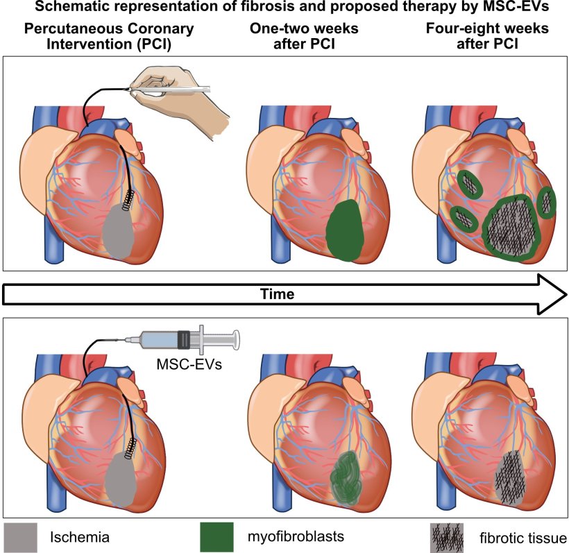
The figure shows how the heart changes after a major heart attack and how the new treatment using exosomes from mesenchymal stromal cells (MSC-EVs) can slow the progression of the disease.
ImageCell 2026 (CC BY 4.0)
In the study, they also demonstrate how a new PET (Positron Emission Tomography) method can be used to identify patients with ongoing scarring and, ultimately, enable personalised treatment.
Millions of people around the world suffer a heart attack each year. Although current treatments rapidly restore blood flow to the heart, many patients go on to develop heart failure. A key cause is the inflammation-driven scarring that occurs after the heart attack. There are currently no treatments that specifically target this process. In the current study, published in the journal Cell Stem Cell, the <a href="https://healthylife7.com/new-mexico-green-chile-researchers-explore-how-to-make-it-more-disease/” title=”New Mexico green chile researchers explore how to make it more disease”>researchers present a new therapy that reduces inflammation and subsequent scarring. The treatment has been developed for clinical use in conjunction with balloon angioplasty.
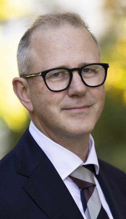 Karl-Henrik Grinnemo, Professor and Consultant in Cardio-Thoracic Surgery at Uppsala University and Uppsala University Hospital, and the study’s first author.
Image
“We’ve developed a completely new type of biological drug that can be described as an anti-inflammatory treatment. It can be administered during balloon angioplasty to dampen inflammation and the scarring process that can otherwise lead to heart failure. The study shows that the treatment is effective in both small and large animal models, and we now hope to proceed with a clinical trial this autumn. We believe the method has great potential to reduce the risk of heart failure after a heart attack and thus also reduce the risk of cardiac death,” says Karl-Henrik Grinnemo, Professor and Consultant in Cardio-Thoracic Surgery at Uppsala University and Uppsala University Hospital, and the study’s first author.
The biological drug is produced from donated human bone marrow. The researchers cultivate stromal cells from the bone marrow and then harvest the tiny particles known as exosomes that are produced by the cells. Exosomes are small membrane vesicles that transmit signalling molecules between cells. Although they occur naturally in the body, they are produced here in the laboratory so that they can be delivered to the damaged coronary artery in much higher concentrations. The study has shown that the exosomes both reduce the scarring process and preserve heart function over time in a small-animal model, and that they have an acute cardioprotective effect in pig trials when administered directly into the coronary arteries.
“The role of the exosomes is to ‘switch’ the activated inflammatory cells in the body away from further stimulating inflammation and towards doing the opposite – dampening inflammation and promoting healing. What we are doing is utilising the body’s own cells and amplifying a process that already occurs naturally.”
In previous studies on severe inflammatory conditions in the lungs, researchers used stromal cells with associated exosomes to achieve a similar effect. The method developed by the Uppsala researchers uses only the exosomes, which offers many advantages. They are cheaper, can be used for more patients and can easily be frozen, then thawed and used immediately
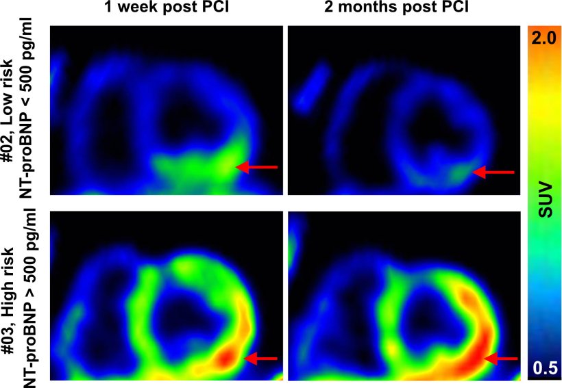
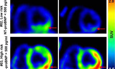
New PET imaging visualises how the heart is healing after a heart attack and is being evaluated to identify patients at increased risk of heart failure.
ImageCell 2026 (CC BY 4.0)
The researchers have developed a new PET method to monitor the effects of the treatment. Olof Eriksson, Professor of Drug Development at Uppsala University, has developed a tracer that makes it possible to visualise the active cells driving the scarring process using PET imaging. “For the first time, we can combine a treatment that targets the scarring process with an imaging technique that makes it possible to monitor how the patient’s heart is responding to the treatment. This paves the way for more personalised treatment after a heart attack,” says Karl-Henrik Grinnemo.
In an ongoing clinical trial, the researchers observed that some patients still had a high level of scarring two months after their heart attack, while others healed significantly faster. “Ultimately, we also hope to be able to use PET scans to identify patients in whom the scarring process remains active months after a heart attack. These patients could then receive this treatment at an early stage, which could slow the progression of heart failure. This would be of great benefit to patients.”
Survival rates among patients who suffer an ST-elevation myocardial infarction (i.e. a heart attack with complete blockage of a blood vessel) and are treated with balloon angioplasty have remained virtually unchanged since 2008, largely because current therapies cannot target the scarring process that is initiated after the balloon angioplasty and which leads to heart failure. By targeting this process, the researchers hope to reduce mortality in the long term. Due to its anti-inflammatory effect, the researchers see several potential applications for the treatment, even outside the field of cardiology.
“We see great potential for this treatment in conditions other than heart attacks as well. Stroke is an obvious example, but also acute inflammatory conditions such as acute respiratory distress syndrome. The same principle could also be applied to organ transplants. Transplants cause severe inflammation. If we can reduce this, we could protect the heart, lungs, kidneys and liver, thereby improving the function of these organs after a transplant.”The study is the result of an extensive collaboration between researchers at Uppsala University, Uppsala University Hospital, Karolinska Institutet, the University of Gothenburg, SLU and several international partners.
02.08.2026
More on the subject:
