


Abstract
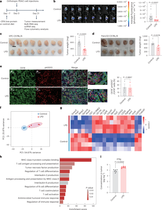
Dietary interventions can influence cancer progression, yet the role of low-protein diets (LPDs) in pancreatic ductal adenocarcinoma (PDAC) immunotherapy is unclear. Here we show that an LPD suppresses PDAC progression in male mice by remodeling the gut microbiota and activating antitumor immunity. LPD promoted immune activation and drove an immunostimulatory tumor-associated macrophage phenotype. Microbiota depletion abolished these effects and fecal microbiota transplantation from LPD-fed donors transferred the protective phenotype to recipients. Mechanistically, LPD enriched Blautiacoccoides, which produced uridine diphosphate (UDP)-galactose to activate the macrophage P2Y14R–STAT1 axis, inducing an immunostimulatory phenotype. Combining LPD, B. coccoides or UDP-galactose with anti-PD1 improved survival over anti-PD1 alone. In persons with advanced PDAC, reduced fecal B. coccoides and serum UDP-galactose correlated with poor outcomes. These findings establish that LPD reshapes the gut microbiota and metabolites to enhance antitumor immunity through the UDP-galactose–P2Y14R–STAT1 axis, offering a dietary strategy to improve PDAC immunotherapy.
This is a preview of subscription content, access
Access options
Access through your institution
- Purchase on SpringerLink
- Instant access to the full article PDF.
39,95 €
Prices may be subject to local taxes which are calculated during checkout
Subjects
- Pancreatic cancer
Data availability
Sequencing data were deposited to the Sequence Read Archive under BioProject PRJNA1196505. Targeted metabolomics data used in this publication were deposited to the EMBL-EBI MetaboLights database with identifier MTBLS14989. The remaining data that support the findings of this study are available within the article and its Supplementary Information and/or from the corresponding authors upon request
References
Huang, J. et al. Worldwide burden of, risk factors for, and trends in pancreatic cancer. Gastroenterology160, 744–754 (2021)
Article
PubMed
<a href="http://scholar.google.com/scholar_lookup?&title=Worldwide%20burden%20of%2C%20risk%20factors%20for%2C%20and%20trends%20in%20pancreatic%20cancer&journal=Gastroenterology&doi=10.1053%2Fj.gastro.2020.10.007&volume=160&pages=744-754&publication_year=2021&author=Huang%2CJ” rel=”nofollow noopener” target=”_blank”>Google ScholarBalachandran, V. P., Beatty, G. L. & Dougan, S. K. Broadening the impact of immunotherapy to pancreatic cancer: challenges and opportunities. Gastroenterology156, 2056–2072 (2019)
Klein, A. P. Pancreatic cancer epidemiology: understanding the role of lifestyle and inherited risk factors. Nat. Rev. Gastroenterol. Hepatol.18, 493–502 (2021)
Siegel, R. L., Miller, K. D., Fuchs, H. E. & Jemal, A. Cancer statistics, 2021. CA Cancer J. Clin.71, 7–33 (2021)
Farhangnia, P., Khorramdelazad, H., Nickho, H. & Delbandi, A.-A. Current and future immunotherapeutic approaches in pancreatic cancer treatment. J. Hematol. Oncol.17, 40 (2024)
Hezaveh, K. et al. Tryptophan-derived microbial metabolites activate the aryl hydrocarbon receptor in tumor-associated macrophages to suppress anti-tumor immunity. Immunity55, 324–340 (2022)
Rubio-Patiño, C. et al. Low-protein diet induces IRE1α-dependent anticancer immunosurveillance. Cell Metab.27, 828–842 (2018)
Zhang, X. et al. Reprogramming tumour-associated macrophages to outcompete cancer cells. Nature619, 616–623 (2023)
Nakatsu, G., Andreeva, N., MacDonald, M. H. & Garrett, W. S. Interactions between diet and gut microbiota in cancer. Nat. Microbiol.9, 1644–1654 (2024)
Nan, K. et al. Fasting-mimicking diet-enriched Bifidobacteriumpseudolongum suppresses colorectal cancer by inducing memory CD8+ T cells. Gut74, 775–786 (2025)
Orillion, A. et al. Dietary protein restriction reprograms tumor-associated macrophages and enhances immunotherapy. Clin. Cancer Res.24, 6383–6395 (2018)
Pushalkar, S. et al. The pancreatic cancer microbiome promotes oncogenesis by induction of innate and adaptive immune suppression. Cancer Discov.8, 403–416 (2018)
Mirji, G. et al. The microbiome-derived metabolite TMAO drives immune activation and boosts responses to immune checkpoint blockade in pancreatic cancer. Sci. Immunol.7, eabn0704 (2022)
Tintelnot, J. et al. Microbiota-derived 3-IAA influences chemotherapy efficacy in pancreatic cancer. Nature615, 168–174 (2023)
Saxton, R. A. & Sabatini, D. M. mTOR signaling in growth, metabolism, and disease. Cell169, 361–371 (2017)
Noy, R. & Pollard, J. W. Tumor-associated macrophages: from mechanisms to therapy. Immunity41, 49–61 (2014)
Yin, W. et al. CCRL2 promotes antitumor T-cell immunitytion. Proc. Natl Acad. Sci. USA118, e2024171118 (2021)
Mantovani, A., Allavena, P., Marchesi, F. & Garlanda, C. Macrophages as tools and targets in cancer therapy. Nat. Rev. Drug Discov.21, 799–820 (2022)
Pozzi, L.-A. M., Maciaszek, J. W. & Rock, K. L. Both dendritic cells and macrophages can stimulate naive CD8 T cells in vivo to proliferate, develop effector function, and differentiate into memory cells. J. Immunol.175, 2071–2081 (2005)
Pathria, P., Louis, T. L. & Varner, J. A. Targeting tumor-associated macrophages in cancer. Trends Immunol.40, 310–327 (2019)
Huang, Y. et al. Intratumor microbiome analysis identifies positive association between Megasphaera and survival of Chinese patients with pancreatic ductal adenocarcinomas. Front. Immunol.13, 785422 (2022)
Dickson, I. Microbiome promotes pancreatic cancer. Nat. Rev. Gastroenterol. Hepatol.15, 328 (2018)
Chen, Y. et al. Metagenomic microbial signatures for noninvasive detection of pancreatic cancer. Biomedicines13, 1000 (2025)
Yu, G., Xu, C., Zhang, D., Ju, F. & Ni, Y. MetOrigin: discriminating the origins of microbial metabolites for integrative analysis of the gut microbiome and metabolome. iMeta1, e10 (2022)
Gonzalez, J. T. & Betts, J. A. Dietary sugars, exercise and hepatic carbohydrate metabolism. Proc. Nutr. Soc.78, 246–256 (2019)
Ma, J. et al. Glycogen metabolism regulates macrophage-mediated acute inflammatory responses. Nat. Commun.11, 1769 (2020)
Lawrence, T. & Natoli, G. Transcriptional regulation of macrophage polarization: enabling diversity with identity. Nat. Rev. Immunol.11, 750–761 (2011)
Wang, W. et al. RIP1 kinase drives macrophage-mediated adaptive immune tolerance in pancreatic cancer. Cancer Cell34, 757–774 (2018)
Rafehi, M. & Müller, C. E. Tools and drugs for uracil nucleotide-activated P2Y receptors. Pharmacol. Ther.190, 24–80 (2018)
Schizas, D. et al. Immunotherapy for pancreatic cancer: a 2020 update. Cancer Treat. Rev.86, 102016 (2020)
Hanahan, D. & Coussens, L. M. Accessories to the crime: functions of cells recruited to the tumor microenvironment. Cancer Cell21, 309–322 (2012)
Ugel, S., Canè, S., De Sanctis, F. & Bronte, V. Monocytes in the tumor microenvironment. Annu. Rev. Pathol.16, 93–122 (2021)
Fidelle, M. et al. A microbiota-modulated checkpoint directs immunosuppressive intestinal T cells into cancers. Science380, eabo2296 (2023)
Caronni, N. et al. IL-1β+ macrophages fuel pathogenic inflammation in pancreatic cancer. Nature623, 415–422 (2023)
Niu, Y. et al. Blautiacoccoides is a newly identified bacterium increased by leucine deprivation and has a novel function in improving metabolic disorders. Adv. Sci.11, e2309255 (2024)
Ye, L. et al. Repressed Blautia–acetate immunological axis underlies breast cancer progression promoted by chronic stress. Nat. Commun.14, 6160 (2023)
Wang, B. et al. Blautiacoccoides and its metabolic products enhance the efficacy of bladder cancer immunotherapy by promoting CD8+ T cell infiltration. J. Transl. Med.22, 964 (2024)
Nomura, S. et al. Blautiacoccoides JCM1395T achieved intratumoral growth with minimal inflammation: evidence for live bacterial therapeutic potential by an optimized sample preparation and colony PCR method. Pharmaceutics15, 989 (2023)
Zhang, X. et al. Tissue-resident Lachnospiraceae family bacteria protect against colorectal carcinogenesis by promoting tumor immune surveillance. Cell Host Microbe31, 418–432 (2023)
Fatahi-Bafghi, M. Genome-based reclassification of Blautia coccoides (Kaneuchi et al. 1976) Liu et al. 2008 as a later heterotypic synonym of Blautia producta (Prévot 1941) Liu et al. 2008. Int. J. Syst. Evol. Microbiol.75, 006823 https://doi.org/10.1099/ijsem.0.006823 (2025)
Zhang, S.-L. et al. Lacticaseibacillusparacasei sh2020 induced antitumor immunity and synergized with anti-programmed cell death 1 to reduce tumor burden in mice. Gut Microbes14, 2046246 (2022)
Zhang, J.-Z. et al. UDP-glucose sensing P2Y14R: a novel target for inflammation. Neuropharmacology238, 109655 (2023)
Wang, X. et al. UDP-glucose accelerates SNAI1 mRNA decay and impairs lung cancer metastasis. Nature571, 127–131 (2019)
Du, X. et al. Diet-derived galactose reprograms hepatocytes to prevent T cell exhaustion and elicit antitumour immunity. Nat. Cell Biol.27, 1357–1366 (2025)
Xu, T. et al. P2RY14 downregulation in lung adenocarcinoma: a potential therapeutic target associated with immune infiltration. J. Thorac. Dis.14, 515–535 (2022)
Shin, A. et al. P2Y receptor signaling regulates phenotype and IFN-α secretion of human plasmacytoid dendritic cells. Blood111, 3062–3069 (2008)
Sesma, J. I. et al. UDP-glucose promotes neutrophil recruitment in the lung. Purinergic Signal.12, 627–635 (2016)
Amison, R. T. et al. Lipopolysaccharide (LPS) induced pulmonary neutrophil recruitment and platelet activation is mediated45, 62–68 (2017)
Demirci, S. et al. BCL11A +58/+55 enhancer-editing facilitates HSPC engraftment and HbF induction in rhesus macaques conditioned with a CD45 antibody–drug conjugate. Cell Stem Cell32, 209–226 (2025)
Feng, M. et al. PD-1/PD-L1 and immunotherapy for pancreatic cancer. Cancer Lett.407, 57–65 (2017)
Taylor, S. R., Falcone, J. N., Cantley, L. C. & Goncalves, M. D. Developing dietary interventions as therapy for cancer. Nat. Rev. Cancer22, 452–466 (2022)
Herbrich, S. et al. TET2-mutant clonal hematopoiesis enhances macrophage antigen presentation and improves immune checkpoint therapy in solid tumors. Cancer Cell44, 187–202 (2026)
Shinde, R. et al. Apoptotic cell-induced AhR activity is required for immunological tolerance and suppression of systemic lupus erythematosus in mice and humans. Nat. Immunol.19, 571–582 (2018)
Zhang, F. et al. Activation of NOD1 on tumor-associated macrophages augments CD8+ T cell-mediated antitumor immunity in hepatocellular carcinoma. Sci. Adv.10, eadp8266 (2024)
Chen, S., Zhou, Y., Chen, Y. & Gu, J. fastp: an ultra-fast all-in-one FASTQ preprocessor. Bioinformatics34, i884–i890 (2018)
Kim, D., Langmead, B. & Salzberg, S. L. HISAT: a fast spliced aligner with low memory requirements. Nat. Methods12, 357–360 (2015)
Roberts, A., Trapnell, C., Donaghey, J., Rinn, J. L. & Pachter, L. Improving RNA-seq expression estimates by correcting for fragment bias. Genome Biol.12, R22 (2011)
Anders, S., Pyl, P. T. & Huber, W. HTSeq—a Python framework to work with high-throughput sequencing data. Bioinformatics31, 166–169 (2015)
Love, M. I., Huber, W. & Anders, S. Moderated estimation of fold change and dispersion for RNA-seq data with DESeq2. Genome Biol.15, 550 (2014)
Gene Ontology Consortium. The Gene Ontology ReD338 (2019)
Kanehisa, M. et al. KEGG for linking genomes to life and the environment. Nucleic Acids Res.36, D480–D484 (2008)
Hao, Y. et al. Integrated analysis of multimodal single-cell data. Cell184, 3573–3587 (2021)
McGinnis, C. S., Murrow, L. M. & Gartner, Z. J. DoubletFinder: doublet detection in single-cell RNA sequencing data using artificial nearest neighbors. Cell Syst.8, 329–337 (2019)
Li, H. & Durbin, R. Fast and accurate short read alignment with Burrows–Wheeler transform. Bioinformatics25, 1754–1760 (2009)
Li, D., Liu, C.-M., Luo, R., Sadakane, K. & Lam, T.-W. MEGAHIT: an ultra-fast single-node solution for large and complex metagenomics assembly
Hyatt, D. et al. Prodigal: prokaryotic gene recognition and translation initiation site identification. BMC Bioinformatics11, 119 (2010)
Li, W. & Godzik, A. CD-HIT: a fast program for clustering and comparing large sets of protein or nucleotide sequences. Bioinformatics22, 1658–1659 (2006)
Langmead, B. & Salzberg, S. L. Fast gapped-read alignment with Bowtie 2. Nat. Methods9, 357–359 (2012)
Rinttilä, T., Kassinen, A., Malinen, E., Krogius, L. & Palva, A. Development of an extensive set of 16S rDNA-targeted primers for quantification of pathogenic and indigenous bacteria in faecal samples by real-time PCR. J. Appl. Microbiol.97, 1166–1177 (2004)
Acknowledgements
We are grateful to B. Du (East China Normal University) for generously supplying the P2ry14-knockout mice that were essential for this research. We gratefully thank OE Biotech for all scRNA-seq experiments performed at the Single-Cell Core facility. We thank Majorbio for metagenomics sequencing
Funding
This work was supported by grants from the National Natural Science Foundation of China (82173122, 81972234, 82273027 and 82170868) and Natural Science Foundation of Chongqing (2024NSCQ-MSX3956)
Author information
Author notes
These authors contributed equally: Yueying Chen, Fulin Nian, Shengdi Wu, Tianli Yuan, Yifan Ma
Authors and Affiliations
Department of Gastroenterology and Hepatology, Zhongshan Hospital, Fudan University, Shanghai, China
Yueying Chen, Fulin Nian, Shengdi Wu, Yifan Ma, Wenfeng Liu, Wenqing Tang, Danying Zhang, Xizhong Shen & Ling Dong
Shanghai Institute of Liver Diseases, Shanghai, China
Yueying Chen, Fulin Nian, Shengdi Wu, Yifan Ma, Wenfeng Liu, Wenqing Tang, Danying Zhang, Xizhong Shen & Ling Dong
Department of Oncology, Renji Hospital, Shanghai Jiao Tong University School of Medicine, Shanghai, China
Yueying Chen
Department of Gastrointestinal Surgery, Renji Hospital, Shanghai Jiao Tong University School of Medicine, Shanghai, China
Tianli Yuan
Health Science Center, East China Normal University, Shanghai, China
Junyang Cao, Fengwanni Wang, Xiulong Xia & Xiaoming Hu
Institutes of Biomedical Sciences and Liver Cancer Institute, Zhongshan Hospital, Fudan University, Shanghai, China
Yuxiao Zhang
Department of Gastroenterology, Second Affiliated Hospital of Zhejiang University School of Medicine, Hangzhou, China
Wenfeng Liu
Shanghai Institute of Nutrition and Health, University of Chinese Academy of Sciences, Chinese Academy of Sciences, Shanghai, China
Zi Li & Wei Lu
Department of Gastroenterology, The Shanghai Tenth People’s Hospital, Tongji University, Shanghai, China
Zhanju Liu
NHC Key Laboratory of Glycoconjugates Research, Department of Biochemistry and Molecular Biology, School of Basic Medical Sciences, Fudan University, Shanghai, China
Si Zhang
Authors
- Yueying ChenView author publications
Search author on:PubMed Google Scholar
- Fulin NianView author publications
Search author on:PubMed Google Scholar
- Shengdi WuView author publications
Search author on:PubMed Google Scholar
- Tianli YuanView author publications
Search author on:PubMed Google Scholar
- Yifan MaView author publications
Search author on:PubMed Google Scholar
- Junyang CaoView author publications
Search author on:PubMed Google Scholar
- Yuxiao ZhangView author publications
Search author on:PubMed Google Scholar
- Wenfeng LiuView author publications
Search author on:PubMed Google Scholar
- Wenqing TangView author publications
Search author on:PubMed Google Scholar
- Danying ZhangView author publications
Search author on:PubMed Google Scholar
- Zi LiView author publications
Search author on:PubMed Google Scholar
- Wei LuView author publications
Search author on:PubMed Google Scholar
- Fengwanni WangView author publications
Search author on:PubMed Google Scholar
- Xiulong XiaView author publications
Search author on:PubMed Google Scholar
- Zhanju LiuView author publications
Search author on:PubMed Google Scholar
- Si ZhangView author publications
Search author on:PubMed Google Scholar
- Xizhong ShenView author publications
Search author on:PubMed Google Scholar
- Xiaoming HuView author publications
Search author on:PubMed Google Scholar
- Ling DongView author publications
Search author on:PubMed Google Scholar
Contributions
Y.C., F.N., S.W., T.Y. and Y.M. contributed equally to this work. L.D., X.H., X.S., S.Z. and Y.C. planned and supervised the experimental work and data analysis. Y.C., F.N., J.C., T.Y., Y.Z., W. Liu and Z. Li performed all the experiments. S.W., W. Liu, W.T. and D.Z. recruited the human participants and performed the related data analysis. Y.C., F.N., S.W., W. Liu, J.C., W. Lu, Z. Liu and S.Z. analyzed the data. Y.C., F.W. and X.X. cultured the anaerobes. Y.C., F.N. and X.H. wrote the paper. L.D., X.H. and X.S. conceptualized and supervised the study.
Ethics declarations
Competing interests
The authors declare no competing interests
Peer review
Peer review information
Nature Cancer thanks Elisabeth Letellier, Alexander Visekruna and the other, anonymous, reviewer(s) for their contribution to the peer review of this work
Additional information
Publisher’s note Springer Nature remains neutral with regard to jurisdictional claims in published maps and institutional affiliations
Extended data
Extended Data Fig. 1 A −25% low-protein diet dose not directly influence the tumor growth or cell cycle progression
(a) Schematic overview of the experiments with treatments of control diet, -12.5%, -25% and -50% LPD in subcutaneous KPC tumor-bearing mice. Mice were pre-fed with the control diet or LPD for 7 days prior to tumor inoculation. Analyses were performed 21 days later, including tumor measurement. (b) Monitoring of food intake (shown on left, n = 7 per group) and body weight in subcutaneous tumor-bearing mice with supplementation of control, -12.5%, -25% and -50% LPD for 28 days (n = 6 per group). (c) Tumor volume was measured in mice fed the control diet, -12.5%, -25% and -50% LPD over a 21-day period following subcutaneous implantation (n = 6 per group). The black line in the tumor dissection images indicates 1 cm. (d) Representative image and the positive area of Ki67 in the tumor samples from control and -25% LPD (n = 3 per group).(e) qRT-PCR analysis of Ccna2, Ccnb1, Ccnd2, and Cdk4, in the orthotopic tumor samples from control and -25% LPD (n = 5 per group). (f) The levels of total cholesterol and triglycerides in the serum of mice receiving control diet or -25% LPD (n = 5 per group). (g) Western blot analysis showed the protein levels of mTOR, p-mTOR, AKT and p-AKT in the tumor samples from control and -25% LPD (n = 3 per group). (h-i) Concentrations of 20 amino acids in serum (h) and tumors (i) of mice treated with control and -25% LPD (n = 5 in -25% LPD, n = 6 in control). (j) Heatmap showing gene expression associated with cell cycle, carbohydrate metabolism, lipid metabolism and amino metabolism of indicated RNA transcripts in tumors of day 21 orthotopic KPC tumor-bearing mice treated with control or LPD (n = 4 per group). (k) Gene Set Enrichment Analysis (GSEA) reveals the enrichment of metabolic pathways in tumors from mice fed control or LPD (n = 4 per group). GSEA was performed using a two-tailed permutation test with the Benjamini-Hochberg (BH) correction. (l) Schematic overview of the experiments with treatments of control diet or -25% LPD in orthotopic KPC tumor-bearing mice. Mice were randomized to receive a control diet or an LPD one week after tumor implantation. Tumors were harvested and weighed at the endpoint, following 2 weeks of dietary intervention. (n = 5 per group). (m) Tumor weight in KPC tumor-bearing mice fed a control or LPD for 14 days (n = 5 per group). The black line in the tumor dissection images indicates 1 cm. Data were mean ± SEM (Two-tailed student’s t test in d, f, m; h, i; Two-tailed multiple t-tests with FDR (B–H) correction in e, g, h, i; two-way ANOVA in b, c). -12.5%pro, -12.5% low protein diet; -25%pro, -25% low protein diet; -50%pro, -50% low protein diet; LPD, -25% low protein diet.
Extended Data Fig. 2 A −25% low-protein diet activates tumor immune responses in PDAC
(a) Heatmap depicts the relative expression of marker genes for distinct macrophage clusters identified by single-cell RNA sequencing. (b) UMAP plot of scRNA-seq on T cells sorted from pooled orthotopic KPC tumors of day 21 treated with control or LPD. Tumors from n = 4 mice were pooled together per group. (c) Bar plot of scRNA-seq data in (a) showing the proportion of cell numbers in each T cell cluster from control and LPD-treated group. (d) Gating strategies used for flow analysis of CD45+ immune infiltrates in tumor tissues from orthotopic KPC tumor-bearing mice. (e) Histogram shows percent of CD11B+ cells, MFI for CD86 on CD11B+ cells in bone marrow; MFI for CD86 on F480+ CD11B+ cells, and IFNγ on CD8+ T cells in spleen; percent of F480+ CD11B+ cells, MFI for CD86 on F480+ CD11B+ cells and IFNγ on CD8+ T cells in colonic lamina propria (n = 5 per group). Data were mean ± SEM (Two-tailed student’s t test). LPD, -25% low protein diet.
Extended Data Fig. 3 The effect of anti-CSF plus clodronate liposomes and CD antibodies in depleting the macrophages and CD+ T cells in tumors
(a) Schematic overview of the experiments with treatments of anti-CSF plus CL in orthotopic KPC tumor-bearing mice. Mice were treated with intraperitoneal injections of PBS or anti-CSF plus CL every 3 days, starting from the day of tumor inoculation until the endpoint analysis at day 21. (b) Flow cytometry analysis of the percent of F480+ CD11B+ cells in orthotopic tumors from control and anti-CSF plus CL group (n = 5 per group). (c) Flow cytometry analysis of the percent of CD8+ T and CD4+ T cells in orthotopic tumors from control and anti-CSF plus CL group (n = 5 per group). (d) Histogram shows MFI for IFNγ on CD8+ T and CD4+ T cells in tumors from control and anti-CSF plus CL group (n = 5 per group). (e) Flow cytometry analysis of the percent of CD8+ T cells in tumors from control and anti-CD8 group (n = 5 per group). Data were mean ± SEM (Two-tailed welch’s t-test in a, e, c (%CD8+ T); Two-tailed student’s t test in b, c (CD4+ T), d). CL, clodronate liposomes.
Extended Data Fig. 4 A −25% low-protein diet reshapes the gut microbiota and metabolites in PDAC
(a) Protein levels of GRP78 and XBP1 of tumors in the control or LPD diet as determined by Western blot analysis (n = 3 per group). (b) Scheme for the experiment design. Microbiota-depleted orthotopic KPC tumor-bearing mice were administered feces from mice receiving control or LPD. (c) Tumor weight in tumor-bearing mice administered feces from mice receiving control or LPD for 28 days (n = 5 per group). The white line in the tumor dissection images indicates 1 cm. (d) Histogram shows the MFI of IFNγ in tumor-infiltrating CD8+ T cells, and the MFI of CD86 and CD206 in TAMs, from mice that received FMT from control die or LPD donors (n = 5 per group). (e) Abundance of B. coccoides in the feces of mice received microbiota transplantation (n = 5 per group). (f) Box plots showing alpha (left) and beta diversity (right) of the intratumoral microbiota in mice fed control or LPD (n = 3 per group). Data are presented as mean with error bars indicating the minimum to maximum range. (g) Relative abundance of the Blautia genus in tumors from control and LPD groups (n = 3 per group). (h) Lefse analysis on significantly differential intratumoral microbiota between control and LPD groups (n = 3 per group). (i) Heatmap showing the relative levels of 60 differentially regulated metabolites between the control group and LPD group (n = 6 per group). Data were mean ± SEM (Two-tailed Mann-Whitney test in f, and MFI of CD86 in d; Two-tailed student’s t test in a, c, d, e, g). FMT, fecal microbiota transplantation; LPD, -25% low protein diet.
Extended Data Fig. 5 B. coccoides-derived UDP-galactose promoted the immunostimulatory phenotype of macrophage and inhibited the tumor growth
(a) ELISA analysis of IL-6 and TNFα concentrations in the supernatant of BMDMs treated with CDP-Choline (100 μM), GPC (100 μM), PC (100 μM), stearoylcarnitine (50 μM), and UDP-Gal (100 μM) for 24 h prior to stimulation with IFN-γ (100 ng/ml) plus LPS (100 ng/ml) (n = 5 per group). (b) qRT-PCR analysis of IL6, TNFA, and IL10 in PMA-treated THP-1 cells that treated with PBS or UDP-Gal (100 μM) for 24 h prior to stimulation with IFN-γ (20 ng/ml) plus LPS (100 ng/ml) or IL-4 (20 ng/ml) plus IL-13 (20 ng/ml) (n = 5 per group). (c) Cell proliferation evaluation of KPC cells treated with CDP-Choline (100 μM), GPC (100 μM), PC (100 μM), stearoylcarnitine (50 μM), and UDP-Gal (100 μM), using the CCK8 assay (n = 5 per group). (d) qRT-PCR analysis of Il6, Nos2, and Arg1 in BMDMs treated with or without TCM and UDP-Gal (100 μM) for 24 h prior to stimulation with IL-4 (10 ng/ml) (n = 5 per group). (e-f) The protein levels of GALM, GALK, GALT and GALE in mouse livers and tumors treated with control or LPD analyzed by western blot (n = 3 per group). (g) The mRNA levels of Galm, Galk, Galt and Gale in mouse livers and tumors treated with control or LPD analyzed by qRT-PCR (n = 5 per group). (h) The concentration of UDP-galactose in mouse tumors administration control diet or LPD with or without ABX (n = 5 per group). (i) The concentration of UDP-galactose in the bacterial liquid of B. coccoides incubated with PBS or D-galactose (n = 5 per group). (j) Fecal abundance of B. coccoides and serum concentration of UDP-galactose were assessed in mice after 7 days of control or LPD diet (n = 5 per group). (k) The mRNA expression of galM, galK, galT and galE in B. coccoides incubated with PBS or D-galactose for 6 and 12 hours (n = 5 per group). (l) Assessment of tumor volumes in subcutaneous KPC tumor-bearing mice treated with 100 mg/kg, 150 mg/kg or 200 mg/kg UDP-galactose (n = 4 per group). The white line in the tumor dissection images indicates 1 cm. (m) The concentration of UDP-Gal in serum and tumors from mice in (k) (n = 4 per group). Data were mean ± SEM (Two-tailed student’s t test in b, f, g, i, j, k; one way ANOVA in a, d, h, m; two-way ANOVA in c, l) GPC, glycerophosphocholine; PC, phosphocholine; UDP-Gal, uridine 5’-diphospho-α-D-galactose; CDP-choline, cytidine 5’-diphosphocholine; GALM, aldose 1-epimerase; GALK, galactokinase; GALT, UDP-glucose-hexose-1-phosphate uridylyltransferase; GALE, UDP-glucose 4-epimerase; TCM, tumor conditioned media.
Extended Data Fig. 6 UDP-galactose enhances the PYR/STAT signaling pathway in macrophages
(a) P2ry14 expression at the transcriptomic level in various immune cell types in single RNA as shown by violin plots (n = 4 mice were pooled together per group). (b) P2ry14 expression at the transcriptomic level in macrophage clusters in single RNA as shown by violin plots (n = 4 mice were pooled together per group). (c) Schematic representation of UDP-galactose promoted STAT1 expression and phosphorylation via P2Y14R activation (n = 3 mice per group). (d) qRT-PCR analysis of Il6, Nos2 and Arg1 in BMDMs treated with UDP-galactose (100 μM) with or without PPTN for 24 h prior to stimulation with IFN-γ (100 ng/ml) plus LPS (100 ng/ml) or IL-4 (10 ng/ml) (n = 5 biologically independent cell cultures). (e) BMDMs were labeled with CTV and adoptively transferred into mice via intravenous injection. The histogram shows the proportion of donor-derived (CTV+) macrophages (F4/80+ CD11b+) infiltrating the tumor, as analyzed by flow cytometry (n = 3 mice per group). (f) The mRNA expression of STAT1, P2RY14 and RARβ in PMA-treated THP-1 cells treated with PBS or UDP-galactose (100 μM) for 24 h were determined by RT-qPCR (n = 5 biologically independent cell cultures). (g) Scheme for the experiment design. Mice were administered AAV9 via injection 14 days prior to tumor inoculation. A control diet or LPD intervention was initiated 7 days before inoculation. A booster dose of AAV9 was delivered on the day of inoculation. Tumor weight and immune cell profiles were assessed by flow cytometry 3 weeks post-inoculation. (h) Assessment of tumor size in orthotopic KPC tumor-bearing mice receiving treatments with PBS plus AAV9-F4/80-Vector, UDP-Gal plus AAV9-F4/80-Vector, and UDP-Gal plus AAV9-F4/80-P2ry14-shRNA (n = 5 mice per group). The white line in the tumor dissection images indicates 1 cm. (i) Flow cytometric analysis of peripheral blood CD45.2 chimerism in bone marrow chimeras generated from P2ry14‒/‒ (CD45.2) donors (n = 5 per group). (j) Assessment of orthotopic tumor size in WT or P2ry14‒/‒ bone marrow chimeras treated with PBS or UDP-Gal (n = 5 per group). The white line in the tumor dissection images indicates 1 cm. (k) Histogram shows MFI for CD86 on TAMs and IFNγ on CD8+ T cells from tumors in (j) (n = 5 per group). Data were mean ± SEM (Two-tailed Mann-Whitney test in a, b; Two-tailed student’s t test in e, f; Two-tailed welch’s t-test in i; one way ANOVA in d, h, j, k). BMDM, bone marrow derived macrophages; CTV, Cell Trace Violet; UDP-Gal, uridine 5’-diphospho-α-D-galactose.
Supplementary information
Reporting Summary (download PDF )
Supplementary Tables 1–6 (download XLSX )
Supplementary Tables 1–6
Source data
Source Data Figs. 1–8 (download PDF )
Unprocessed western blots
Source Data Figs. 1–8 (download XLSX )
Statistical
Rights and permissions
Springer Nature or its licensor (e.g. a society or other partner) holds exclusive rights to this article under a publishing agreement with the author(s) or other rightsholder(s); author self-archiving of the accepted manuscript version of this article is solely governed by the terms of such publishing agreement and applicable law
About this article
Cite this article
Chen, Y., Nian, F., Wu, S. et al. Low-protein diet enhances antitumor immunity in pancreatic cancer through microbiota-derived UDP-galactose.
Nat Cancer (2026). https://doi.org/10.1038/s43018-026-01222-2
Received:10 January 2025
Accepted:17 July 2026
Published:17 August 2026
Version of record:17 August 2026
DOI
:https://doi.org/10.1038/s43018-026-01222-2
