


Download PDF
Abstract
Viral persistence refers to the ability of a virus to remain in its host for an extended period. Although most non-retroviral RNA viruses are traditionally known for causing acute, self-limiting infections, accumulating evidence of the detection of viral products long after the infectious virus is cleared suggests that RNA viruses can establish persistent infections. Persistent viral products include replication-competent viral genomes, viral proteins, mutated viruses or non-standard viral genomes. The persistence of viral products entails a continuous interaction with the host and is often associated with prolonged tissue inflammation and chronic disease. Here, we discuss emerging evidence of the persistence of viral products from viruses commonly considered to cause acute infections. We explore the known requirements for virus persistence and the implications for the development of post-acute sequelae and predisposition to chronic inflammatory syndromes.
RNA viruses include some of the most common human viruses, such as seasonal respiratory syncytial virus (RSV), coronaviruses (CoV), human influenza viruses and human parainfluenza viruses (HPIVs); some of the most infectious viruses, such as measles virus (MeV); some of the most debilitating viruses, such as chikungunya (CHIKV); and some of the most lethal viruses, such as Ebola virus (EBOV) and the henipaviruses. Except for retroviruses, hepatitis C virus (HCV) and arenaviruses, RNA viruses are generally thought to cause acute infections that resolve quickly, leading to the development of protective immunity. However, recent technological advances reveal that several RNA viruses or their viral products can persist long term in the host, impacting health for months or years after initial infection1,2,3,4,5,6,7,8,9. Although evidence of transmission of persistent non-retroviral RNA viruses in humans remains limited, compelling examples do exist. For example, several reports have shown that EBOV can be transmitted from the male reproductive tract to sexual partners months after infection10,11,12,13. Evidence for the persistence of HPIVs in humans dates back to the early 1980s, when HPIVs were detected in infection outbreaks among individuals isolated for several months in Antarctica14. More recently, persistent HPIV shedding has been documented in individuals who are immunosuppressed, even when asymptomatic15.
Furthermore, increasing evidence indicates that persistent viral products can lead to sustained tissue inflammation and chronic diseases. This is likely due to the presence of pathogen-associated molecular patterns that trigger the host’s innate immune response2,16,17,18. Examples include MeV infections and the onset of subacute sclerosing panencephalitis (SSPE)18,19,20,21,22, SARS-CoV-2 infection and the development of long COVID5,6,17,23, CHIKV infection and the onset of persistent myalgia7,24,25,26 and EBOV infection and post-EBOV disease8,27,28,29. Demonstrating a causal relationship between the persistence of viral products and the development of chronic disease in humans is challenging owing to the need for isolated populations, the inaccessibility of persistently infected cells in live individuals and ethical issues with human experimentation. However, a more conclusive causal relationship was recently demonstrated for parainfluenza viruses (PIVs) and CHIKV. Using the murine PIV Sendai virus (SeV), our group identified lung macrophages, type 2 innate lymphoid cells (ILC2s) and dendritic cells (DCs) containing viral RNA and protein long after the acute infection actively produced proinflammatory molecules. Importantly, eliminating these cells drastically reduced chronic lung inflammation2. For CHIKV, recent evidence in mice showed that inflammatory macrophages accumulate in joint-associated tissues and harbor viral RNA during chronic disease, and suggests that CD4+ T cells infiltrating these tissues have a role in inflammation9.
Persistent infections require immune evasion or subversion, allowing continuous virus replication and/or long-term retention of viral material. Because all RNA viruses face robust immune surveillance, their persistence in the host may result from evolved strategies to evade or suppress the host’s innate immune defenses or disrupt antigen presentation, which is essential for the development of cytotoxic effector cells that clear the infection. Infection of cellular niches that offer favorable conditions, such as longevity, location or impaired antiviral signaling, can also contribute to virus persistence. Here, we review advances in our understanding of the mechanisms driving the persistence of viral products and viruses traditionally considered to cause acute infections, as well as emerging evidence linking persistent viral products to post-acute sequelae and predisposition to chronic inflammatory syndromes. The mechanisms driving persistence of the most classical and well-studied RNA viruses that cause chronic infections, such as HCV and arenaviruses, have been recently reviewed elsewhere30,31. Instead, this review focuses on emerging evidence of the persistence of viruses and viral products traditionally considered to cause acute infections.
Immune mechanisms of RNA virus persistence
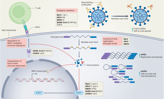
DNA viruses use genetic strategies to persist, including maintaining their genomes as episomes in the nucleus of dividing cells (for example, herpesviruses and papillomaviruses) or integrating their genome into the host genome. Retroviruses, such as HIV, integrate a cDNA version of their genome into the host genome and have a high viral mutation rate that facilitates evasion of neutralizing antibodies. Persistence of other RNA viruses relies mainly on evading or modulating the host immune response, and, in some cases (for example, mumps virus and PIVs), persistence has been associated with the formation of viral condensates that presumably protect the virus from immune recognition32,33. The critical role of the immune response in preventing the persistence of acute RNA viruses is evident in individuals who are immunocompromised in response to specific treatments, in individuals with congenital immunodeficiency and in individuals experiencing age-related immune decline, conditions where viral replication is prolonged with an increased risk of developing persistent infections34,35,36,37,38. Adaptive immune mechanisms that support the maintenance of persistent infections, such as T cell exhaustion and suppression of T cell responses, have been thoroughly covered in recent reviews31,39. Here, we discuss virus-driven mechanisms that restrict early immune responses, enabling high replication of acute RNA viruses and contributing to virus persistence (Fig. 1).
RNA viruses use diverse strategies to evade immune detection and establish persistence. These include (1) antigenic variation (acquisition of advantageous mutations that enable the virus to escape immune recognition), (2) production of nsVGs (generation of truncated viral genomes that reduce overall viral replication), (3) suppression of IFN production or signaling (disruption of IFN signaling pathways, thereby inhibiting the activation of downstream effectors essential for the antiviral response), and (4) inhibition of antigen presentation (interference with the expression of MHC to prevent effective antigen presentation to immune cells); F, fusion; G, glycoprotein; HN, hemagglutinin-neuraminidase; L, large protein (polymerase); M, matrix; NSP1, non-structural protein 1; stVGs, standard viral genomes.
Suppression of early host antiviral responses
The host immune response to RNA viruses is initiated when viral pathogen-associated molecular patterns are recognized by virus sensor molecules that drive the expression of type I and III interferons (IFNs), as well as cytokines and chemokines. Type I and III IFNs signal for the expression of hundreds of genes that block virus replication. In addition, type I IFNs amplify and activate cellular responses that contribute to controlling virus spread. For example, type I IFNs are critical for the effective function of natural killer (NK) cells that participate in early infection control40 and for the maturation of DCs, a process that is essential to generate type 1 adaptive responses that clear the virus and protect from reinfection via the action of cytotoxic T cells41. RNA viruses that are adapted to their host encode proteins that directly block early steps of the antiviral response, allowing the virus to replicate and spread while limiting immune intervention. Therefore, interfering with type I and III IFN production leads to higher rates of virus replication and spread, which can facilitate the establishment of a chronic infection.
The list of viral proteins from acute RNA viruses that interfere with the type I/III IFN pathway is extensive. Some examples are the V and C proteins of MeV and HPIVs42,43,44, non-structural protein 2 (nsP2) of CHIKV45,46 and viral protein 35 (VP35) of EBOV47,48,49. SARS-CoV-2 allocates over one-third of its genome to encode proteins that antagonize almost every level of the IFN response50. Viral-encoded antagonists can interfere at multiple levels within the IFN signaling cascade by inhibiting sensing by RIG-I-like receptors or other specialized cellular proteins, blocking signaling molecules of the IFN induction pathway or blocking type I/III IFN signaling itself42,43,44,47,48,49,51. RNA viruses can also directly or indirectly interfere with DC and NK cell function. Examples include the MeV hemagglutinin protein and EBOV VP35 that inhibit maturation and activity of DCs52,53,54.
Evidence suggests a causal link between early immune response inhibition by viruses, high viral load during the acute infection phase and the establishment of persistent infections. For example, CHIKV persistence in the spleen of aged rhesus monkeys is linked to reduced innate and adaptive immune responses45. MeV creates an immunosuppressed environment by decreasing the numbers of T cells and B cells in circulation, thereby facilitating the establishment of persistence in lymphoid tissues and immune-privileged sites like the brain55,56. SARS-CoV-2 inhibition of the type I IFN response and associated DC dysfunction observed in severe acute cases is linked to systemic viral spread. This dysfunction persists for several months, likely contributing to the long-term presence of SARS-CoV-2 antigens in various tissues, including the intestines and brain57,58,59,60,61. In addition, immune-escape mutations in the SARS-CoV-2 spike protein and deletions in ORF1ab and ORF8, accessory genes with immune-modulatory functions, are frequently detected in hosts with persistent infection3. Last, HPIV shedding is often observed in individuals who are immunosuppressed, particularly in recipients of hematopoietic cell transplants35,62. These examples demonstrate that failure to mount an effective antiviral response early in infection results in increased virus replication that directly or indirectly creates permissive conditions for the establishment of viral persistence.
Regulation of MHC expression and function
Another strategy for RNA viruses to suppress the establishment of an effective antiviral immune response is to disrupt the expression of major histocompatibility complex (MHC) molecules and/or the presentation of viral antigens on the surface of infected cells. These processes are essential for the killing of infected cells by cytotoxic T cells. Viruses can downregulate the expression of MHC molecules both at the transcriptional and post-translational levels. One example is during HCV infection, where the virus core protein induces the degradation of MHC class I (MHCI), impairing the presentation of viral antigens to cytotoxic T lymphocytes and allowing the virus to evade clearance by the immune system63. SARS-CoV-2 also disrupts MHCI expression in humans, cultured cells and mice by targeting IFN signaling64,65. In addition, the SARS-CoV-2 ORF3a accessory protein interferes with MHCI intracellular trafficking, and the ORF7a protein prevents proper MHCI assembly66. Similarly, CHIKV nsP2 disrupts MHCI antigen presentation, allowing the virus to escape immune recognition by T cells67. Although not all RNA viruses that exploit this pathway are proven to persist, hijacking MHC antigen presentation is a strategy that impairs downstream activation of effector immune cells. This facilitates the survival of infected cells that serve as reservoirs for virus persistence.
Viral mechanisms of RNA virus persistence
After establishing infection in potential viral reservoirs, many RNA viruses acquire adaptive mutations that reduce their replication rate, allowing the infected cell to evade immune detection and facilitating virus persistence. In some persistent infections, mutations that favor cell-to-cell spread over virus particle production, together with antigenic variation, allow the virus to escape antibody detection22. In addition, most, if not all, RNA viruses generate defective or truncated versions of the viral genome known as non-standard viral genomes (nsVGs) or defective viral genomes that interfere with virus replication and can also minimize the exposure of viral antigens to the immune system. nsVGs arise during viral replication and interfere with replication of the standard virus by several mechanisms, including competing for viral and host resources, inducing potent IFN production early after infection and regulating transcription of viral genes40,68,69,70,71. Evidence since the early days of virology has associated the production of nsVGs with the establishment of persistent RNA virus infections in vitro72,73,74,75, and emerging evidence supports their role in vivo. Here, we provide specific examples of these viral-driven strategies as contributors to the establishment of persistence by acute RNA viruses (Fig. 1).
Antigenic variation
Antigenic variation is a common strategy to escape antibody recognition by RNA viruses. Their high mutation rate allows them to change their external proteins enough to be undetectable by antibodies76,77. Antigenic variation can also change the way the virus spreads. For example, MeV brain tropism is associated with hyperfusogenic mutations in the viral fusion (F) protein that allow for increased virus propagation via cell-to-cell fusion while reducing viral particle production19,78,79,80,81. In addition, mutations in the viral matrix (M) protein typically detected in postmortem SSPE tissue are thought to impair the formation of viral particles18,82,83. Mutations in the RNA-dependent RNA polymerase (L protein) that result in reduced viral replication were described in an SSPE-causing virus22,84. By reducing viral protein expression and infectious particle release, MeV persists by evading immune detection and minimizing neuronal death. A similar strategy is observed in EBOV infection, as mutations in the virus glycoprotein alter viral entry dynamics and promote non-lytic infection85,86,87,88. Similarly, analysis of HPIV3 persisting in individuals who are immunocompromised showed accumulation of mutations in the virus hemagglutinin-neuraminidase attachment protein that led to altered particle fusion dynamics, increased avidity to cellular receptors and preference for cell-to-cell transmission of the virus35.
Generation of nsVGs
In the early 1970s, RNA virus persistence was described as the onset of an ‘asynchronous cycle’ that fluctuates between standard virus replication and the production of defective interfering particles in the infected cell89. In this model, defective interfering particles are produced gradually during virus replication until they predominate over the standard virus. The high concentration of defective interfering particles, in turn, interferes with standard virus particle production and reduces the amount of standard virus. Once the standard virus levels are low, defective interfering particles are produced at a lower rate, allowing the standard virus to resurface and initiate the cycle again89. A newer model for nsVG-driven persistence was generated based on the observation that during infection with SeV or RSV, a subset of cells is dominated by standard virus genomes, whereas others are dominated by nsVGs90,91,92. The heterogeneous composition of viral genomes in these cells was associated with distinct host responses to the virus, including the switch of the TNF receptor from proapoptotic TNFR1 to prosurvival TNFR2 in nsVG-enriched cells, allowing their survival over other infected cells and facilitating the long-term persistence of the virus in vitro90.
The demonstration that nsVGs are present in human samples infected with MeV, RSV and EBOV, together with their detection in persistent infections, support the hypothesis that nsVGs facilitate the establishment of RNA virus persistence during natural infections40,93,94,95. Analysis of brain tissue from an individual who succumbed to SSPE two decades after MeV infection identified nsVGs across multiple brain regions93. In studies of EBOV infection in rhesus macaques, nsVGs were detected in the serum and testes of infected animals at 5, 7 and 9 days after infection, and nsVGs have also been detected in semen from men with persistent EBOV infection4,95. Although direct evidence of an in vivo role for nsVGs in prolonging the duration of RNA virus infections is lacking, their ubiquitous presence across RNA viruses suggests that nsVG generation is a conserved proviral strategy. We propose that by stimulating the host IFN response, promoting cell survival mechanisms and reducing viral RNA and protein, nsVGs act as a switch between high and low viral replication, enabling the virus to evade immunity and extend its presence in the host96.
Establishment of cellular reservoirs of viral products
To persist in immunocompetent hosts, RNA viruses rely on finding a niche that is or can become resistant to immune clearance. Although cellular reservoirs have been well studied for retroviruses such as HIV-1, our understanding of such reservoirs for other RNA viruses remains limited97. Immune-privileged organs, such as the brain and testes, long-lived cells and cells with low expression of sensor molecules that trigger antiviral immunity in response to virus recognition (for example, RIG-I-like receptors) are good candidate host reservoirs for RNA viruses17,98,99. However, recent studies suggest that RNA viruses or their viral products can also persist in other cell types, including mucosal structural cells100,101, sensory cells102,103, muscle cells24,25, synovial joints104, macrophages2,9,105,106, DCs and ILC2s2, significantly expanding the cell types that could be long-term viral reservoirs.
RNA virus persistence in immune-privileged sites
The central nervous system is considered an immune-privileged system as it mounts limited immune responses to threats. Limited immune responses prevent swelling and damage of the tissues98,107. In some cases, neurotrophic viruses, such as MeV, harness such an immunosuppressed environment to establish persistence within neurons. Autopsy studies of individuals who succumbed to SSPE confirmed the presence of MeV RNA and protein in the brain22,93,108,109. Interestingly, MeV can infect and persist in neurons and astrocytes that lack the MeV receptors CD150 and nectin-4, indicating the use of alternative entry mechanisms or cell-to-cell transmission for the virus to establish itself in the brain110,111. Similarly, EBOV has been found in the brain ventricular system of non-human primates, even after standard antiviral treatment with monoclonal antibodies112,113. Moreover, onset of meningoencephalitis associated with viral persistence was reported in two survivors of EBOV infection, where the virus was detected in the blood and cerebrospinal fluid 342 days after initial infection for one individual and 137 days after initial infection for the other28. SARS-CoV-2 RNA, as well as spike protein and nucleoprotein, have been reported in the skull, meninges and brain tissue of individuals who succumbed to COVID-19 and in those that died from non-COVID-19-related causes, suggesting long-term persistence of SARS-CoV-2 in the brain23,100. Experiments in mice have also shown persistence of RNA viruses in the brain, including enteroviruses114,115 and Zika virus98,116, suggesting that long-term neurological sequelae of viral infections may be in part due to the persistence of these viruses in the brain.
In the testes, physical barriers together with an anti-inflammatory microenvironment that protects sperm from autoimmunity create an immune-privileged organ where acute RNA viruses can persist. EBOV RNA and infectious virus have been detected in the semen of male survivors up to 200 days after initial infection. Additionally, persistent virus was shown to be transmitted to female partners, leading to their subsequent infection10,11,29. Similarly, Lassa virus RNA and infectious virus were recovered from the seminal fluid of survivors up to 12 months into the study117, and Zika virus has been detected in seminal fluid for up to 83 days after acute disease and was shown to be transmitted to female partners118,119. The eye is another immune-privileged site where local immune responses are more suppressed to limit inflammation-induced impairment of vision. The eye has been identified as a site for persistent EBOV120. These examples highlight the role of immune-privileged organs as niches for acute RNA virus persistence and emphasize the importance of studying and identifying persistent viral reservoirs to reduce the transmission of pathogenic viruses and alleviate the public health burden of post-viral chronic conditions.
RNA virus persistence in immune cells
Infection of mobile lymphocytes and myeloid cells offers an opportunity for viruses to circulate throughout an organism and reach immune-privileged sites. In addition, non-circulating immune cells such as macrophages, which can take up infected cells and are relatively long lived, can serve as reservoirs for RNA viruses (Fig. 2). Although SARS-CoV-2 infection is primarily associated with respiratory pathology, it is now recognized to infect multiple systems, including cells of the immune system5,17. Studies in non-human primate models showed that replication-competent SARS-CoV-2 can persist in alveolar macrophages for over 200 days after infection121. SARS-CoV-2 proteins and replicative RNA intermediates have also been detected in circulating monocytes, B cells, T cells and pulmonary inflammatory macrophages from individuals with COVID-19 for up to 15 months after acute infection5,17,122.
The persistence of RNA viruses, whether as fully infectious particles or viral subproducts, can significantly impact the biology of various non-immune and immune cells. This persistence can disrupt cellular functions and lead to alterations in tissue homeostasis. IAV, influenza A virus; NoV, norovirus; TH2, type 2 helper T cell
Recently, our group demonstrated that the mouse PIV SeV establishes long-term infection in the lung, with persistent viral products detected in alveolar macrophages, ILC2s, DCs and other immune cells2. Similarly, in rhesus macaques, MeV RNA is retained in lymphoid tissues for up to 6 months, mostly in B cells55,56. Moreover, clinical studies have demonstrated that children with chronic adenotonsillar disease harbor viral RNA and antigens from influenza, RSV and other viruses in tonsillar lymphocytes long after primary infection123,124,125.
Apart from respiratory RNA viruses, arthritogenic viruses like CHIKV cause infections associated with persistent RNA in immune cells. Studies in non-human primates have identified macrophages as cellular reservoirs that contain significant amounts of CHIKV antigen and RNA for up to 3 months after infection. Inflammatory macrophages were also recently found in joint-associated tissues9. These studies confirm findings in humans, where synovial macrophages were found to harbor persistent CHIKV105,106. Infection of motile immune cells can be a means of increased viral dissemination, commonly referred to as ‘trojan horse’-mediated dissemination. One of the most well-studied examples, Zika virus, targets human CD14+ monocytes in circulating blood and can be found in monocyte-derived cells in the central nervous system of human fetuses99,126. Zika virus infection of monocytes is productive and alters the expression of adhesion factors that increase the likelihood of blood–brain barrier crossing by infected cells and delivery of infectious virus to the central nervous system126. These observations highlight the complexity of virus–immune cell interactions and reinforce the need for a thorough understanding of viral persistence within immune cells.
RNA virus persistence in other cell types
Structural cells lining mucosal surfaces, such as respiratory and intestinal epithelial cells, are attractive niches for viral persistence owing to their slow turnover and accessible location. SARS-CoV-2 RNA and spike protein are detected in intestinal epithelial cells months after acute infection100,101, and fecal shedding of viral RNA is detected up to 7 months after infection127,128. One study reported the detection of SARS-CoV-2 spike protein-encoding RNA in colorectal tissue 676 days after initial infection129. Owing to the potential role of persistent viral products in chronic intestinal symptoms, increasing efforts are focused on identifying the specific intestinal cell types harboring SARS-CoV-2 material with the purpose of developing targeted strategies to alleviate long COVID symptoms. Another example of an RNA virus persisting in non-immune cells in the gastrointestinal tract is norovirus, which establishes persistent infection in mouse intestinal tuft cells. These cells are chemosensory epithelial cells found in mucosal epithelia and can escape CD8+ T cell killing, thus they are considered an immune-privileged cell type103.
Persistence in musculoskeletal tissue is a hallmark of arthritogenic RNA virus pathogenesis. CHIKV has long been recognized to cause infections in which viral RNA persists in musculoskeletal tissue in animal models24,25,130. High levels of viral RNA are found chronically in myofibers and muscular fibroblasts of mice infected with CHIKV and Mayaro virus (MAYV), and this correlates with lingering expression of interleukin-10 and transforming growth factor-β and reduced muscle fiber mass25,131. Similarly, but not restricted to arthritogenic arboviruses, enteroviruses are known to persist in the myocardium with potential links to cardiomyopathies132. Cardiomyopathies are frequently idiopathic, and more studies are needed to determine if persistent viral infections and/or consequent immune dysregulation are responsible for these diseases.
Sensory tissues have also been demonstrated to be niches for the persistence of RNA viruses and viral products. SARS-CoV-2 can persist in the taste papillae of humans for months after infection, specifically in basal epithelial cells and lamina propria cells. This persistence is associated with structural damage and an aberrant immune response that contributes to long-term taste dysfunction102. Similarly, SeV products can be detected long after acute infection in long-lived olfactory neurons, providing a potential link between respiratory viral infections and post-viral anosmia133,134.
Whether RNA viruses can persist in basal cells of the epithelium, which are responsible for generating most epithelial cells, is unknown. There is evidence that tissue stem cells are resistant to T cell killing, which would make them an attractive long-term home for viruses135. Structural cells that are reservoirs for RNA viruses may experience damage or long-term changes that could impact how the tissue senses and responds to future infections. Understanding how viral persistence can alter the identity and function of non-immune cells may be key to explaining some of the lingering symptoms observed after acute viral infection and could help develop strategies to prevent or reverse chronic tissue dysfunction.
Long-term consequences of viral persistence
The prolonged detection of RNA viruses or viral products in diverse tissues suggests that many of these viruses can persist in the host for a long time after initial infection. This persistence has been associated with the development of chronic manifestations long after the resolution of the acute phase of infection. These manifestations, collectively referred to as post-acute or post-viral syndromes, challenge the long-standing understanding of acute RNA viral infections and raise questions not only about the host–viral interactions but also about the treatment and prognosis of acute infections136.
The occurrence and severity of post-viral syndromes are diverse and may depend on the virus involved (Table 1). The emergence of long COVID, also referred to as post-acute sequelae of SARS-CoV-2 infection, suggests that viral persistence may cause a wide range of symptoms, including fatigue, cognitive dysfunction, gastrointestinal symptoms, cardiovascular complications and taste and smell dysfunction that persist for weeks or months after infection102,127,137,138. The detection of SARS-CoV-2 RNA and viral protein in various tissues, such as the gut, brain and lymph nodes, months after infection demonstrates incomplete clearance of viral products by the host immune system100. For example, prolonged shedding of SARS-CoV-2 RNA in the stool is associated with gastrointestinal symptoms including abdominal pain, nausea and vomiting101,127,128. Another example is the reported persistence of SARS-CoV-2 spike protein in basal epithelial cells of taste papillae, which is linked to long-term taste dysfunction102, while persistent RNA in the brain is associated with neurological impairment100.
EBOV persistence can also drive systemic and delayed complications affecting multiple organs139,140,141. In some cases, post-EBOV disease results from reactivation of the virus that had been hiding from the immune system for months after the primary infection. For example, EBOV was recovered from the eye of a survivor who suffered from sudden uveitis, a chronic inflammation in the eye that can lead to vision loss27,120. Post-viral persistence has also been associated with fatal outcomes, such as the development of SSPE years after initial MeV infection. SSPE cases are often observed at higher rates among unvaccinated children, and individuals who are immunosuppressed are also at risk of suffering severe infection and SSPE, likely owing to the high levels of virus reached during acute infection20,21.
More generalized post-viral symptoms have also been reported, such as chronic fatigue syndrome/myalgic encephalomyelitis in a cohort who survived a pandemic H1N1/09 infection or persistent arthralgia in severe cases of CHIKV infection136,142. Although the persistence of respiratory RNA viruses in the respiratory tract has long been recognized in individuals who are immunocompromised, numerous studies during the COVID-19 pandemic demonstrated that immunocompetent and otherwise healthy individuals can experience the persistence of viral products100,101,143. It is also established that severe infection with respiratory viruses like RSV or PIVs can predispose individuals to develop chronic respiratory conditions of the lung, including asthma and chronic obstructive pulmonary disease144,145, and recent studies using the murine PIV SeV demonstrated a direct causal relationship between persistent PIV products and the development of asthma-like disease in mice2,144,145.
Inflammation is a key component of the antiviral response, involving the activation of immune and non-immune cells and the production of proinflammatory cytokines to help control viral infections. However, when acute inflammatory responses fail to resolve the infection, sustained immune cell infiltration and cytokine production can promote tissue damage, remodeling and chronic symptoms. One major challenge in understanding the molecular mechanisms linking acute viral infection to chronic disease is the lack of animal models that accurately recapitulate human pathology. In addition, sensitive virus detection methods that could be applied to finding the rare virus-containing cells in human organs are lacking. The development of these tools or the use of representative models is therefore essential to advance our understanding of viral persistence and its long-term consequences. Using SeV as a model PIV, together with a battery of tools that eliminate concerns of false positives, including the use of humanized antibodies and transgenic reporter mice, our group demonstrated that the persistence of SeV RNA and protein is a major driver of chronic type 2 inflammation in the lung. This inflammation is like asthma in humans, showing a direct link between respiratory viral persistence and chronic inflammatory responses2. Similarly, in vivo lineage tracing of CHIKV-infected cells identified myofibers, dermal and muscle fibroblasts and macrophages as harboring persistent CHIKV RNA at chronic time points, likely contributing to musculoskeletal inflammation associated with the chronic stage of CHIKV disease9,25,146. Unresolved viral persistence may contribute to the onset of post-viral syndromes by directly or indirectly disrupting tissue-specific immune responses. Developing more sensitive detection methods and better animal models that help elucidate the cellular and molecular basis of these syndromes is critical for the identification of therapeutic or diagnostic targets to mitigate the long-term burden of viral infections.
Conclusions
The traditional view of RNA viruses as strictly acute agents is being reshaped by emerging evidence of their capacity to persist as intact or mutated genomes, nsVGs or proteins found in diverse cellular reservoirs. These findings reveal that beyond the acute phase, common RNA viruses leave molecular footprints that have functional consequences in contributing to chronic inflammation, immune dysregulation and long-term organ dysfunction. The causal link between acute RNA virus persistence and post-viral sequalae should raise awareness of the long-term risks of what are usually assumed to be short-course viral infections. If these acute infections are severe, there is a significant chance that a chronic condition will manifest years later. Preventing severe acute infections in susceptible individuals through vaccination or isolation may diminish this risk147,148.
Several questions remain unanswered. Can all acute RNA viruses persist? What are the mechanisms of viral RNA retention in the diverse cell reservoirs? What is the fate of cells that survive infection, and how are these surviving cells different from noninfected or bystander cells? What are the host cell molecular determinants that characterize a productive RNA virus infection versus a persistent infection? What is the exact role of the immune system in the development or resolution of chronic disease? It is also essential to investigate whether different RNA viruses or their products induce common transcriptomic or epigenomic reprogramming in their host cells and whether such footprints can serve as biomarkers for chronic inflammatory syndromes.
References
Griffin, D. E. Why does viral RNA sometimes persist after recovery from acute infections? PLoS Biol. 20, e3001687 (2022)
Castro, Í. A. et al. Murine parainfluenza virus persists in lung innate immune cells sustaining chronic lung pathology. Nat. Microbiol.9, 2803–2816 (2024)
Ghafari, M. et al. Prevalence of persistent SARS-CoV-2 in a large community surveillance study. Nature626, 1094–1101 (2024)
Johnson, R. I. et al. Identification and characterization of defective viral genomes in Ebola virus-infected rhesus macaques. J. Virol.95, e0071421 (2021)
Zuo, W. et al. The persistence of SARS-CoV-2 in tissues and its association with long COVID symptoms: a cross-sectional cohort study in China. Lancet Infect. Dis.24, 845–855 (2024)
Liu, F. et al. Prolonged dysregulation and pathological changes in the upper respiratory tract of SARS-CoV-2 infected hamsters. NPJ Viruses4, 15 (2026)
Silveira-Freitas, J. E. P. et al. Long chikungunya? An overview to immunopathology of persistent arthralgia. World J. Virol.13, 89985 (2024)
Stawicki, S. P. et al. Reflections on the Ebola Public Health Emergency of International Concern, Part 1: post-Ebola syndrome: the silent outbreak. J. Glob. Infect. Dis.9, 41–44 (2017)
Zarrella, K. M. et al. Chikungunya virus persists in joint-associated macrophages and promotes chronic disease in mice. Nat. Microbiol.11, 1302–1317 (2026)
Mate, S. E. et al. Molecular evidence of sexual transmission of Ebola virus. N. Engl. J. Med.373, 2448–2454 (2015)
Uyeki, T. M. et al. Ebola virus persistence in semen of male survivors. Clin. Infect. Dis.62, 1552–1555 (2016)
Sissoko, D. et al. Persistence and clearance of Ebola virus RNA from seminal fluid of Ebola virus disease survivors: a longitudinal analysis and modelling study. Lancet Glob. Health5, e80–e88 (2017)
Thorson, A. E. et al. Persistence of Ebola virus in semen among Ebola virus disease survivors in Sierra Leone: a cohort study of frequency, duration, and risk factors. PLoS Med. 18, e1003273 (2021)
Muchmore, H. G. et al. Persistent parainfluenza virus shedding during isolation at the South Pole. Nature289, 187–189 (1981)
Maziarz, R. T. et al. Control of an outbreak of human parainfluenza virus 3 in hematopoietic stem cell transplant recipients. Biol. Blood Marrow Transplant.16, 192–198 (2010)
McCarthy, M. K. & Morrison, T. E. Persistent RNA virus infections: do PAMPS drive chronic disease? Curr. Opin. Virol.23, 8–15 (2017)
Patterson, B. K. et al. Persistence of SARS CoV-2 S1 protein in CD16+ monocytes in post-acute sequelae of COVID-19 (PASC) up to 15 months post-infection. Front. Immunol.12, 746021 (2021)
Patterson, J. B. et al. Evidence that the hypermutated M protein of a subacute sclerosing panencephalitis measles virus actively contributes to the chronic progressive CNS disease. Virology291, 215–225 (2001)
Schmid, A. et al. Subacute sclerosing panencephalitis is typically characterized by alterations in the fusion protein cytoplasmic domain of the persisting measles virus. Virology188, 910–915 (1992)
Bellini, W. J. et al. Subacute sclerosing panencephalitis: more cases of this fatal disease are prevented by measles immunization than was previously recognized. J. Infect. Dis.192, 1686–1693 (2005)
Wendorf, K. A. et al. Subacute sclerosing panencephalitis: the devastating measles complication that might be more common than previously estimated. Clin. Infect. Dis.65, 226–232 (2017)
Schmitz, K. S. et al. Functional properties of measles virus proteins derived from a subacute sclerosing panencephalitis patient who received repeated remdesivir treatments. J. Virol.98, e0187423 (2024)
Rong, Z. et al. Persistence of spike protein at the skull–meninges–brain axis may contribute to the neurological sequelae of COVID-19. Cell Host Microbe32, 2112–2130 (2024)
Ozden, S. et al. Human muscle satellite cells as targets of chikungunya virus infection. PLoS ONE2, e527 (2007)
Young, A. R. et al. Dermal and muscle fibroblasts and skeletal myofibers survive chikungunya virus infection and harbor persistent RNA. PLoS Pathog.15, e1007993 (2019)
Imad, H. A. et al. Post-chikungunya virus infection musculoskeletal disorders: syndromic sequelae after an outbreak. Trop. Med. Infect. Dis.6, 52 (2021)
Shantha, J. G., Crozier, I. & Yeh, S. An update on ocular complications of Ebola virus disease. Curr. Opin. Ophthalmol.28, 600–606 (2017)
Mukadi-Bamuleka, D. et al. Fatal meningoencephalitis associated with Ebola virus persistence in two survivors of Ebola virus disease in the Democratic Republic of the Congo: a case report study. Lancet Microbe5, 100905 (2024)
Muwonge, H. et al. Long-term clinical sequelae among Sudan ebolavirus disease survivors 2 years post-infection: a matched cohort study. BMC Med.23, 432 (2025)
Barathan, M. et al. Viral persistence and chronicity in hepatitis C virus infection: role of T-cell apoptosis, senescence and exhaustion. Cells7, 165 (2018)
Studstill, C. J. & Hahm, B. Chronic LCMV infection is fortified with versatile tactics to suppress host T cell immunity and establish viral persistence. Viruses13, 1951 (2021)
Zhang, X. et al. Molecular mechanisms of stress-induced reactivation in mumps virus condensates. Cell186, 1877–1894 (2023)
Kurebayashi, Y. et al. Human parainfluenza virus type 1 regulates cholesterol biosynthesis and establishes quiescent infection in human airway cells. PLoS Pathog.17, e1009908 (2021)
Tuxhorn, I. E. Familial subacute sclerosing panencephalitis in two siblings. Pediatr. Neurol.31, 291–294 (2004)
Greninger, A. L. et al. Human parainfluenza virus evolution during lung infection of immunocompromised individuals promotes viral persistence. J. Clin. Invest.131, e150506 (2021)
Zhang, Q. et al. Inborn errors of type I IFN immunity in patients with life-threatening COVID-19. Science370, eabd4570 (2020)
Abolhassani, H. et al. Inherited IFNAR1 deficiency in a child with both critical COVID-19 pneumonia and multisystem inflammatory syndrome. J. Clin. Immunol.42, 471–483 (2022)
Kee, A. C., Yang, S. & Tambyah, P. Atypical chikungunya virus infections in immunocompromised patients. Emerg. Infect. Dis.16, 1038–1040 (2010)
Kahan, S. M., Wherry, E. J. & Zajac, A. J. T cell exhaustion during persistent viral infections. Virology479–480, 180–193 (2015)
Madera, S. et al. Type I IFN promotes NK cell expansion during viral infection by protecting NK cells against fratricide. J. Exp. Med.213, 225–233 (2016)
Longhi, M. P. et al. Dendritic cells require a systemic type I interferon response to mature and induce CD4+ TH1 immunity with poly(IC) as adjuvant. J. Exp. Med.206, 1589–1602 (2009)
Gotoh, B., Komatsu, T., Takeuchi, K. & Yokoo, J. Paramyxovirus accessory proteins as interferon antagonists. Microbiol. Immunol.45, 787–800 (2001)
Nakatsu, Y. et al. Measles virus circumvents the host interferon response by different actions of the C and V proteins. J. Virol.82, 8296–8306 (2008)
Mesman, A. W. et al. Measles virus suppresses RIG-I-like receptor activation in dendritic cellsicrobe16, 31–42 (2014)
Messaoudi, I. et al. Chikungunya virus infection results in higher and persistent viral replication in aged rhesus macaques due to defects in anti-viral immunity. PLoS Negl. Trop. Dis.7, e2343 (2013)
Raj, S., Irfan, M. & Ranjith-Kumar, C. T. Chikungunya virus non-structural protein 2 (nsP2) inhibits RIG-I and TLR-mediated immune response. Preprint at bioRxivhttps://doi.org/10.1101/2025.04.30.651505 (2025)
Basler, C. F. et al. The Ebola virus VP35 protein functions as a type I IFN antagonist. Proc. Natl Acad. Sci. USA97, 12289–12294 (2000)
Cardenas, W. B. et al. Ebola virus VP35 protein binds double-stranded RNA and inhibits α/β interferon production induced by RIG-I signaling. J. Virol.80, 5168–5178 (2006)
Zhu, L. et al. Ebola virus sequesters IRF3 in viral inclusion bodies to evade host antiviral immunity. eLife12, RP88122 (2024)
Minkoff, J. M. & tenOever, B. Innate immune evasion strategies of SARS-CoV-2. Nat. Rev. Microbiol.21, 178–194 (2023)
Kotliar, D. et al. Single-cell profiling of Ebola virus disease in vivo reveals viral and host dynamics. Cell183, 1383–1401 (2020)
Hahm, B., Trifilo, M. J., Zuniga, E. I. & Oldstone, M. B. Viruses evade the immune system through type I interferon-mediated STAT2-dependent, but STAT1-independent, signaling. Immunity22, 247–257 (2005)
Romanets-Korbut, O., Kovalevska, L. M., Seya, T., Sidorenko, S. P. & Horvat, B. Measles virus hemagglutinin triggers intracellular signaling in CD150-expressing dendritic cells and inhibits immune response. Cell. Mol. Immunol.13, 828–838 (2016)
Yen, B., Mulder, L. C., Martinez, O. & Basler, C. F. Molecular basis for ebolavirus VP35 suppression of human dendritic cell maturation. J. Virol.88, 12500–12510 (2014)
Nelson, A. N. et al. Association of persistent wild-type measles virus RNA with long-term humoral immunity in rhesus macaques. JCI Insight5, e134992 (2020)
Lin, W. H., Kouyos, R. D., Adams, R. J., Grenfell, B. T. & Griffin, D. E. Prolonged persistence of measles virus RNA is characteristic of primary infection dynamics. Proc. Natl Acad. Sci. USA109, 14989–14994 (2012)
Zhou, R. et al. Acute SARS-CoV-2 infection impairs dendritic cell and T cell responses. Immunity53, 864–877 (2020)
Hadjadj, J. et al. Impaired type I interferon activity and inflammatory responses in severe COVID-19 patients. Science369, 718–724 (2020)
Blanco-Melo, D. et al. Imbalanced host response to SARS-CoV-2 drives development of COVID-19. Cell181, 1036–1045 (2020)
Liu, J. et al. Longitudinal characteristics of lymphocyte responses and cytokine profiles in the peripheral blood of SARS-CoV-2 infected patients. EBioMedicine55, 102763 (2020)
Perez-Gomez, A. et al. Dendritic cell deficiencies persist seven months after SARS-CoV-2 infection. Cell. Mol. Immunol.18, 2128–2139 (2021)
Peck, A. J. et al. Respiratory virus infection among hematopoietic cell transplant recipients: evidence for asymptomatic parainfluenza virus infection. Blood110, 1681–1688 (2007)
Hirano, J. et al. Hepatitis C virus modulates signal peptide peptidase to alter host protein processing. Proc. Natl Acad. Sci. USA118, e2026184118 (2021)
Yoo, J. S. et al. SARS-CoV-2 inhibits induction of the MHC class I pathway by targeting the STAT1–IRF1–NLRC5 axis. Nat. Commun.12, 6602 (2021)
Moriyama, M., Lucas, C., Monteiro, V. S., Yale, S.-C.-G. S. I. & Iwasaki, A. Enhanced inhibition of MHC-I expression by SARS-CoV-2 Omicron subvariants. Proc. Natl Acad. Sci. USA120, e2221652120 (2023)
Arshad, N. et al. SARS-CoV-2 accessory proteins ORF7a and ORF3a use distinct mechanisms to down-regulate MHC-I surface expression. Proc. Natl Acad. Sci. USA120, e2208525120 (2023)
Ware, B. C., Parks, M. G., da Silva, M. O. L. & Morrison, T. E. Chikungunya virus infection disrupts MHC-I antigen presentation
Vignuzzi, M. & Lopez, C. B. Defective viral genomes are key drivers of the virus–host interaction. Nat. Microbiol.4, 1075–1087 (2019)
Genoyer, E. & Lopez, C. B. The impact of defective viruses on infection and immunity. Annu. Rev. Virol.6, 547–566 (2019)
Gonzalez Aparicio, L. J., Yang, Y., Hackbart, M. & Lopez, C. B. Copy-back viral genomes induce a cellular stress response that interferes with viral protein expression without affecting antiviral immunity. PLoS Biol.21, e3002381 (2023)
Xiao, Y. et al. A defective viral genome strategy elicits broad protective immunity against respiratory viruses. Cell184, 6037–6051 (2021)
Moscona, A. & Peluso, R. W. Persistent infection with human parainfluenza virus 3 in CV-1 cells: analysis of the role of defective interfering particles. Virology194, 399–402 (1993)
Calain, P., Monroe, M. C. & Nichol, S. T. Ebola virus defective interfering particles and persistent infection. Virology262, 114–128 (1999)
Valdovinos, M. R. & Gomez, B. Establishment of respiratory syncytial virus persistence in cell lines: association with defective interfering particles. Intervirology46, 190–198 (2003)
Roux, L. & Holland, J. J. Role of defective interfering particles of Sendai virus in persistent infections. Virology93, 91–103 (1979)
Wang, Z. et al. Interplay of host and viral genetic variations in modulating antibody responses to genotype 3a hepatitis C virus: implications for vaccine design. Cell Rep.44, 116418 (2025)
Keck, M. L., Wrensch, F., Pierce, B. G., Baumert, T. F. & Foung, S. K. H. Mapping determinants of virus neutralization and viral escape for rational design of a hepatitis C virus vaccine. Front. Immunol.9, 1194 (2018)
Watanabe, S. et al. Mutant fusion proteins with enhanced fusion activity promote measles virus spread in human neuronal cells and brains of suckling hamsters. J. Virol.87, 2648–2659 (2013)
Sato, Y. et al. Cell-to-cell measles virus spread between human neurons is dependent on hemagglutinin and hyperfusogenic fusion protein. J. Virol.92, e02166-17 (2018)
Mathieu, C. et al. Molecular features of the measles virus viral fusion complex that favor infection and spread in the brain. mBio12, e0079921 (2021)
Yousaf, I. et al. Brain tropism acquisition: the spatial dynamics and evolution of a measles virus collective infectious unit that drove lethal subacute sclerosing panencephalitis. PLOS Pathog.19, e1011817 (2023)
Cattaneo, R. et al. Accumulated measles virus mutations in a case of subacute sclerosing panencephalitis: interrupted matrix protein reading frame and transcription alteration. Virology154, 97–107 (1986)
Ayata, M., Hirano, A. & Wong, T. C. Altered translation of the matrix genes in Niigata and Yamagata neurovirulent measles virus strains. Virology180, 166–174 (1991)
Sakamoto, K. et al. Suppression of viral RNA polymerase activity is necessary for persistent infection during the transformation of measles virus into SSPE virus. PLoS Pathog.19, e1011528 (2023)
Martinez, O. et al. A mutation in the Ebola virus envelope glycoprotein restricts viral entry in a host species- and cell-type-specific manner. J. Virol.87, 3324–3334 (2013)
Wang, M. K., Lim, S. Y., Lee, S. M. & Cunningham, J. M. Biochemical basis for increased activity of Ebola glycoprotein in the 2013–16 epidemic. Cell Host Microbe21, 367–375 (2017)
Fels, J. M. et al. A glycoprotein mutation that emerged during the 2013–2016 Ebola virus epidemic alters proteolysis and accelerates membrane fusion. mBio12, e03616-20 (2021)
Banadyga, L. et al. Atypical Ebola virus disease in a nonhuman primate following monoclonal antibody treatment is associated with glycoprotein mutations within the fusion loop. mBio12, e01438-20 (2021)
Huang, A. S. & Baltimore, D. Defective viral particles and viral disease processes. Nature226, 325–327 (1970)
Xu, J. et al. Replication defective viral genomes exploit a cellular pro-survival mechanism to establish paramyxovirus persistence. Nat. Commun.8, 799 (2017)
Genoyer, E. & Lopez, C. B. Defective viral genomes alter how Sendai virus interacts with cellular trafficking machinery, leading to heterogeneity in the production of viral particles among infected cells. J. Virol.93, e01579-18 (2019)
Aparicio, L. J. G., López, C. B. & Felt, S. A. A virus is a community: diversity within negative-sense RNA virus populations. Microbiol. Mol. Biol. Rev.86, e0008621 (2022)
Taye, B. et al. A measles virus collective infectious unit that caused lethal human brain disease includes many locally restricted and few widespread copy-back defective genomes. J. Virol.98, e0123224 (2024)
Felt, S. A., Achouri, E., Faber, S. R. & Lopez, C. B. Accumulation of copy-back viral genomes during respiratory syncytial virus infection is preceded by diversification of the copy-back viral genome population followed by selection. Virus Evol.8, veac091 (2022)
Whitmer, S. L. M. et al. Active Ebola virus replication and heterogeneous evolutionary rates in EVD survivors. Cell Rep.22, 1159–1168 (2018)
Gonzalez Aparicio, L. J. & Lopez, C. B. Selection of nonstandard viral genomes during the evolution of RNA viruses: a virus survival strategy or a pesky inconvenience? Adv. Virus Res.119, 39–61 (2024)
Lorenzo-Redondo, R. et al. Persistent HIV-1 replication maintains the tissue reservoir during therapy. Nature530, 51–56 (2016)
Kristensson, K. & Norrby, E. Persistence of RNA viruses in the central nervous system. Annu. Rev. Microbiol.40, 159–184 (1986)
Michlmayr, D., Andrade, P., Gonzalez, K., Balmaseda, A. & Harris, E. CD14+CD16+ monocytes are the main target of Zika virus infection in peripheral blood mononuclear cells in a paediatric study in Nicaragua. Nat. Microbiol.2, 1462–1470 (2017)
Stein, S. R. et al. SARS-CoV-2 infection and persistence in the human body and brain at autopsy. Nature612, 758–763 (2022)
Gaebler, C. et al. Evolution of antibody immunity to SARS-CoV-2. Nature591, 639–644 (2021)
Yao, Q. et al. Long-term dysfunction of taste papillae in SARS-CoV-2. NEJM Evid.2, 10.1056/evidoa2300046 (2023)
Strine, M. S. et al. Intestinal tuft cell immune privilege enables norovirus persistence. Sci. Immunol.9, eadi7038 (2024)
Soden, M. et al. Detection of viral ribonucleic acid and histologic analysis of inflamed synovium in Ross River virus infection. Arthritis Rheum.43, 365–369 (2000)
Labadie, K. et al. Chikungunya disease in nonhuman primates involves long-term viral persistence in macrophages. J. Clin. Invest.120, 894–906 (2010)
Hoarau, J. J. et al. Persistent chronic inflammation and infection by chikungunya arthritogenic alphavirus in spite of a robust host immune response. J. Immunol.184, 5914–5927 (2010)
Miller, K. D., Schnell, M. J. & Rall, G. F. Keeping it in check: chronic viral infection and antiviral immunity in the brain. Nat. Rev. Neurosci.17, 766–776 (2016)
Baczko, K. et al. Expression of defective measles virus genes in brain tissues of patients with subacute sclerosing panencephalitis. J. Virol.59, 472–478 (1986)
Baczko, K. et al. Clonal expansion of hypermutated measles virus in a SSPE brain. Virology197, 188–195 (1993)
Generous, A. R. et al. Trans-endocytosis elicited by nectins transfers cytoplasmic cargo, including infectious material, between cells. J. Cell Sci.132, jcs235507 (2019)
Poelaert, K. C. K., Williams, R. M., Matullo, C. M. & Rall, G. F. Noncanonical transmission of a measles virus vaccine strain from neurons to astrocytes. mBio12, e00288-21 (2021)
Larsen, T. et al. Pathologic findings associated with delayed death in nonhuman primates experimentally infected with Zaire Ebola virus. J. Infect. Dis.196, S323–S328 (2007)
Liu, J. et al. Ebola virus persistence and disease recrudescence in the brains of antibody-treated nonhuman primate survivors. Sci. Transl. Med.14, eabi5229 (2022)
Feuer, R. et al. Viral persistence and chronic immunopathology in the adult central nervous system following coxsackievirus infection during the neonatal period. J. Virol.83, 9356–9369 (2009)
Rhoades, R. E., Tabor-Godwin, J. M., Tsueng, G. & Feuer, R. Enterovirus infections of the central nervous system. Virology411, 288–305 (2011)
Ireland, D. D. C. et al. Long-term persistence of infectious Zika virus: inflammation and behavioral sequela in mice. PLoS Pathog.16, e1008689 (2020)
Thielebein, A. et al. Virus persistence after recovery from acute Lassa fever in Nigeria: a 2-year interim analysis of a prospective longitudinal cohort study. Lancet Microbe3, e32–e40 (2022)
Russell, K. et al. Male-to-female sexual transmission of Zika virus—United States, January–April 2016. Clin. Infect. Dis.64, 211–213 (2017)
Paz-Bailey, G. et al. Persistence of Zika virus in body fluids—final report. N. Engl. J. Med.379, 1234–1243 (2018)
Varkey, J. B. et al. Persistence of Ebola virus in ocular fluid during convalescence. N. Engl. J. Med.372, 2423–2427 (2015)
Huot, N. et al. SARS-CoV-2 viral persistence in lung alveolar macrophages is controlled by IFN-γ and NK cells. Nat. Immunol.24, 2068–2079 (2023)
Pontelli, M. C. et al. SARS-CoV-2 productively infects primary human immune system cells in vitro and in COVID-19 patients. J. Mol. Cell Biol.14, mjac021 (2022)
Proenca-Modena, J. L. et al. High rates of detection of respiratory viruses in tonsillar tissues from children with chronic adenotonsillar disease. PLoS ONE7, e42136 (2012)
Castro, I. A. et al. Silent infection of B and CD8+ T lymphocytes by influenza A virus in children with tonsillar hypertrophy. J. Virol. 94, e01969-19 (2020)
Lima, T. M. et al. Tonsils are major sites of persistence of SARS-CoV-2 in children. Microbiol. Spectr.11, e0134723 (2023)
Ayala-Nunez, N. V. et al. Zika virus enhances monocyte adhesion and transmigration favoring viral dissemination to neural cells. Nat. Commun.10, 4430 (2019)
Natarajan, A. et al. Gastrointestinal symptoms and fecal shedding of SARS-CoV-2 RNA suggest prolonged gastrointestinal infection. Med3, 371–387 (2022)
Zollner, A. et al. Postacute COVID-19 is characterized by gut viral antigen persistence in inflammatory bowel diseases. Gastroenterology163, 495–506 (2022)
Peluso, M. J. et al. Tissue-based T cell activation and viral RNA persist for up to 2 years after SARS-CoV-2 infection. Sci. Transl. Med.16, eadk3295 (2024)
Morrison, T. E. et al. A mouse model of chikungunya virus-induced musculoskeletal inflammatory disease: evidence of arthritis, tenosynovitis, myositis, and persistence. Am. J. Pathol.178, 32–40 (2011)
da Silva, M. O. L. et al. Chikungunya and Mayaro viruses induce chronic skeletal muscle atrophy triggered by pro-inflammatory and oxidative response. Int. J. Mol. Sci.25, 8909 (2024)
Andreoletti, L. et al. Enteroviruses can persist with or without active viral replication in cardiac tissue of patients with end-stage ischemic or dilated cardiomyopathy. J. Infect. Dis.182, 1222–1227 (2000)
Mori, I. et al. Parainfluenza virus type 1 infects olfactory neurons and establishes long-term persistence in the nerve tissue. J. Gen. Virol.76, 1251–1254 (1995)
Lin, L., Zhang, M., Stoilov, P., Chen, L. & Zheng, S. Developmental attenuation of neuronal apoptosis by neural-specific splicing of Bak1 microexon. Neuron107, 1180–1196 (2020)
Agudo, J. et al. Quiescent tissue stem cells evade immune surveillance. Immunity48, 271–285 (2018)
Choutka, J., Jansari, V., Hornig, M. & Iwasaki, A. Author Correction: Unexplained post-acute infection syndromes. Nat. Med.28, 1723 (2022)
Zhang, H. et al. Data-driven identification of post-acute SARS-CoV-2 infection subphenotypes. Nat. Med.29, 226–235 (2023)
Prakash, S. et al. Insights into persistent SARS-CoV-2 reservoirs in chronic long COVID. Viruses17, 1310 (2025)
Carod-Artal, F. J. Post-Ebolavirus disease syndrome: what do we know? Expert Rev. Anti. Infect. Ther.13, 1185–1187 (2015)
Rojas, M. et al. Ebola virus disease: an emerging and re-emerging viral threat. J. Autoimmun.106, 102375 (2020)
Durant, O. & Marzi, A. Ebola virus disease sequelae and viral persistence in animal models: implications for the future. PLoS Pathog.20, e1012065 (2024)
Magnus, P. et al. Chronic fatigue syndrome/myalgic encephalomyelitis (CFS/ME) is associated with pandemic influenza infection, but not with an adjuvanted pandemic influenza vaccine. Vaccine33, 6173–6177 (2015)
Santiago-Olivares, C., Martinez-Alvarado, E. & Rivera-Toledo, E. Persistence of RNA viruses in the respiratory tract: an overview. Viral Immunol.36, 3–12 (2023)
Kurai, D., Saraya, T., Ishii, H. & Takizawa, H. Virus-induced exacerbations in asthma and COPD. Front. Microbiol.4, 293 (2013)
Rosas-Salazar, C. et al. Respiratory syncytial virus infection during infancy and asthma during childhood in the USA (INSPIRE): a population-based, prospective birth cohort study. Lancet401, 1669–1680 (2023)
Hawman, D. W. et al. Chronic joint disease caused by persistent chikungunya virus infection is controlled by the adaptive immune response. J. Virol.87, 13878–13888 (2013)
Xie, Y., Choi, T. & Al-Aly, Z. Postacute sequelae of SARS-CoV-2 infection in the pre-Delta, Delta, and Omicron eras. N. Engl. J. Med.391, 515–525 (2024)
Trinh, N. T. et al. Effectiveness of COVID-19 vaccines to prevent long COVID: data from Norway. Lancet Respir. Med.12, e33–e34 (2024)
Davis, H. E. et al. Long COVID: major findings, mechanisms and recommendations. Nat. Rev. Microbiol.21, 133–146 (2023)
Rocke, Z. & Belyayeva, M. Subacute sclerosing panencephalitis. StatPearlshttps://www.ncbi.nlm.nih.gov/books/NBK560673/ (2025)
Acknowledgements
C.B.L. acknowledges the National Institutes of Health (AI R01AI188900) and the BJC Investigators Program
Author information
Authors and Affiliations
Department of Molecular Microbiology, Center for Women’s Infectious Diseases Research, Washington University School of Medicine, Saint Louis, MO, USA
Daniela Vidal, Ítalo de Araújo Castro & Carolina B. López
Authors
- Daniela VidalView author publications
Search author on:PubMed Google Scholar
- Ítalo de Araújo CastroView author publications
Search author on:PubMed Google Scholar
- Carolina B. LópezView author publications
Search author on:PubMed Google Scholar
Contributions
The authors contributed equally to all aspects of the review article
Ethics declarations
Competing interests
The authors declare no competing interests
Peer review
Peer review information
Nature Immunology thanks the anonymous reviewer(s) for their contribution to the peer review of this work. Primary Handling Editor: Paula Jauregui, in collaboration with the Nature Immunology team
Additional information
Publisher’s note Springer Nature remains neutral with regard to jurisdictional claims in published maps and institutional affiliations
Rights and permissions
Springer Nature or its licensor (e.g. a society or other partner) holds exclusive rights to this article under a publishing agreement with the author(s) or other rightsholder(s); author self-archiving of the accepted manuscript version of this article is solely governed by the terms of such publishing agreement and applicable law
About this article
Cite this article
Vidal, D., Castro, Í.d.A. & López, C.B. Implications of RNA virus persistence for post-acute sequelae and chronic inflammatory syndromes.
Nat Immunol (2026). https://doi.org/10.1038/s41590-026-02577-5
Received:11 July 2025
Accepted:01 June 2026
Published:07 July 2026
Version of record:07 July 2026
DOI
:https://doi.org/10.1038/s41590-026-02577-5
