


Abstract
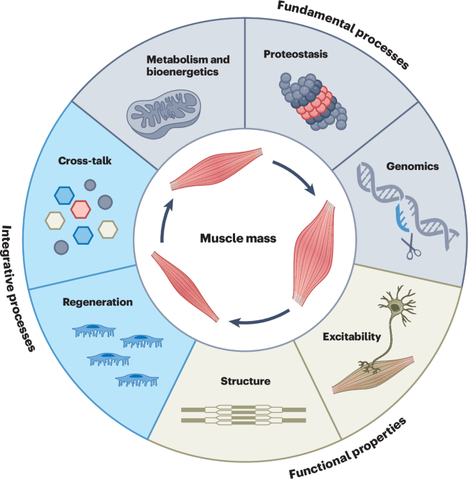
Skeletal muscle is a central determinant of organismal health. Preserving muscle quality is therefore critical for preventing disease and sustaining quality of life across the lifespan. Despite its central role, the field lacks a unifying framework that defines the core properties of skeletal muscle health. Here, we propose a conceptual framework for muscle homeostasis built around seven interconnected hallmarks—metabolism and bioenergetics, proteostasis, genomics, excitability, structure, regeneration and cross-talk—that collectively govern muscle integrity, adaptability and resilience. Each hallmark is mechanistically grounded, quantifiable and potentially modifiable. This framework provides a unifying blueprint for the next generation of precision diagnostics and targeted therapies for preserving skeletal muscle health.
Subjects
- Ageing
- Metabolism
- Musculoskeletal system
Enjoying our latest content?
Log in or create an account to continue
- Access the most recent journalism from Nature’s award-winning team
- Explore the latest features & opinion covering groundbreaking research
Access through your institution
Sign in or create an account
Continue with Google
Continue with ORCiD
References
Argilés, J. M., Campos, N., Lopez-Pedrosa, J. M., Rueda, R. & Rodriguez-Mañas, L. Skeletal muscle regulates metabolismd. Dir. Assoc.17, 789–796 (2016)
Zurlo, F., Larson, K., Bogardus, C. & Ravussin, E. Skeletal muscle metabolism is a major determinant of resting energy expenditure. J. Clin. Invest.86, 1423–1427 (1990)
Meyer, C., Dostou, J. M., Welle, S. L. & Gerich, J. E. Role of human liver, kidney, and skeletal muscle in postprandial glucose homeostasis. Am. J. Physiol. Endocrinol. Metab.282, 419–427 (2002)
Wolfe, R. R. The underappreciated role of muscle in health and disease. Am. J. Clin. Nutr.84, 475–482 (2006)
Pedersen, B. K. Muscles and their myokines. J. Exp. Biol.214, 337–346 (2011)
Cruz-Jentoft, A. J. et al. Sarcopenia: revised European consensus on definition and diagnosis. Age Ageing48, 16–31 (2019)
Argilés, J. M., López-Soriano, F. J., Stemmler, B. & Busquets, S. Cancer-associated cachexia—understanding the tumour macroenvironment and microenvironment to improve management. Nat. Rev. Clin. Oncol.20, 250–264 (2023)
von Haehling, S. & Anker, S. D. Prevalence, incidence and clinical impact of cachexia: facts and numbers-update 2014. J. Cachexia. Sarcopenia Muscle5, 261–263 (2014)
Rausch, V., Sala, V., Penna, F., Porporato, P. E. & Ghigo, A. Understanding the common mechanisms of heart and skeletal muscle wasting in cancer cachexia. Oncogenesis10, 1–13 (2021)
Damluji, A. A. et al. Sarcopenia and cardiovascular diseases. Circulation147, 1534–1553 (2023)
Gao, K. et al. Association between sarcopenia and cardiovascular disease among middle-aged and older adults: findings from the China health and retirement longitudinal study. EClinicalMedicine44, 101264 (2022)
von Haehling, S. et al. Muscle wasting as an independent predictor of survival in patients with chronic heart failure. J. Cachexia Sarcopenia Muscle11, 1242–1249 (2020)
Perry, B. D. et al. Muscle atrophy in patients with type 2 diabetes mellitus: roles of inflammatory pathways, physical activity and exercise. Exerc. Immunol. Rev.22, 94–109 (2016)
Haines, M. S., Leong, A., Porneala, B. C., Meigs, J. B. & Miller, K. K. Association between muscle mass and diabetes prevalence independent of body fat distribution in adults under 50 years old. Nutr. Diabetes12, 29 (2022)
Wang, X. H., Mitch, W. E. & Price, S. R. Pathophysiological mechanisms leading to muscle loss in chronic kidney disease. Nat. Rev. Nephrol.18, 138–152 (2022)
Barreiro, E. & Jaitovich, A. Muscle atrophy in chronic obstructive pulmonary disease: molecular basis and potential therapeutic targets. J. Thorac. Dis.10, S1415 (2018)
Wüst, R. C. I. & Degens, H. Factors contributing to muscle wasting and dysfunction in COPD patients. Int. J. Chron. Obstruct. Pulmon. Dis.2, 289–300 (2007)
Hanahan, D. & Weinberg, R. A. Hallmarks of cancer: the next generation. Cell144, 646–674 (2011)
López-Otín, C., Blasco, M. A., Partridge, L., Serrano, M. & Kroemer, G. The hallmarks of aging. Cell153, 1194–1217 (2013)
Rooks, D. et al. Bimagrumab vs optimized standard of care for treatment of sarcopenia in community-dwelling older adults: a randomized clinical trial. JAMA Netw. Open3, e2020836 (2020)
Hofbauer, L. C. et al. Bimagrumab to improve recovery after hip fracture in older adults: a multicentre, double-blind, randomised, parallel-group, placebo-controlled, phase 2a/b trial. Lancet Healthy Longev.2, e263–e274 (2021)
Sivakumar, K. et al. Long-term safety and tolerability of bimagrumab (BYM338) in sporadic inclusion body myositis. Neurology95, E1971–E1978 (2020)
Wagner, K. R. et al. A phase I/II trial of MYO-029 in adult subjects with muscular dystrophy. Ann. Neurol.63, 561–571 (2008)
Wagner, K. R. et al. Randomized phase 2 trial and open-label extension of domagrozumab in Duchenne muscular dystrophy. Neuromuscul. Disord.30, 492–502 (2020)
Campbell, C. et al. Myostatin inhibitor ACE-031 treatment of ambulatory boys with Duchenne muscular dystrophy: Results of a randomized, placebo-controlled clinical trial. Muscle Nerve55, 458–464 (2017)
Polkey, M. I. et al. Activin type II receptor blockade for treatment of muscle depletion in chronic obstructive pulmonary disease. A randomized trial. Am. J. Respir. Crit. Care Med.199, 313–320 (2019)
Sargeant, J. A. et al. A review of the effects of glucagon-like peptide-1 receptor agonists and sodium-glucose cotransporter 2 inhibitors on lean body mass in humans. Endocrinol. Metab.34, 247–262 (2019)
Neeland, I. J., Linge, J. & Birkenfeld, A. L. Changes in lean body mass with glucagon-like peptide-1-based therapies and mitigation strategies. Diabetes. Obes. Metab.26, 16–27 (2024)
Langer, H. T. et al. Weight loss with GLP-1 medicines does not result in a disproportionate loss of muscle mass or function in obese mice and humans. Cell Rep. Med.7, 102665 (2026)
Prokopidis, K., Daly, R. M. & Suetta, C. Weighing the risk of GLP-1 treatment in older adults: should we be concerned about sarcopenic obesity? J. Nutr. Heal. Aging29, 100652 (2025)
Liu, G. et al. Biomarkers for sarcopenia, muscle mass, muscle strength, and physical performance: an umbrella review. J. Transl. Med.23, 650 (2025)
Bolte, J., Smelter, A. A. & Norton, L. Are we giving too much weight to lean mass loss? Mol. Metab.101, 102253 (2025)
McGregor, R. A., Cameron-Smith, D. & Poppitt, S. D. It is not just muscle mass: a review of muscle quality, composition and metabolism during ageing as determinants of muscle function and mobility in later life. Longev. Healthspan3, 9 (2014)
Daugaard, J. R. & Richter, E. A. Muscle- and fibre type-specific expression of glucose transporter 4, glycogen synthase and glycogen phosphorylase proteins in human skeletal muscle. Pflugers Arch.447, 452–456 (2004)
Deshmukh, A. S. et al. Deep muscle-proteomic analysis of freeze-dried human muscle biopsies reveals fiber type-specific adaptations to exercise training. Nat. Commun.12, 304 (2021)
Schiaffino, S. & Reggiani, C. Fiber types in mammalian skeletal muscles. Physiol. Rev.91, 1447–1531 (2011)
San-Millán, I. & Brooks, G. A. Assessment of metabolic flexibility by means of measuring blood lactate, fat, and carbohydrate oxidation responses to exercise in professional endurance athletes and less-fit individuals. Sports Med.48, 467–479 (2018)
Kiens, B. Skeletal muscle lipid metabolism in exercise and insulin resistance. Physiol. Rev.86, 205–243 (2006)
Smith, J. A. B., Murach, K. A., Dyar, K. A. & Zierath, J. R. Exercise metabolism and adaptation in skeletal muscle. Nat. Rev. Mol. Cell Biol.24, 607–632 (2023)
Lin, J. et al. Transcriptional co-activator PGC-1 alpha drives the formation of slow-twitch muscle fibres. Nature418, 797–801 (2002)
Monzel, A. S., Enríquez, J. A. & Picard, M. Multifaceted mitochondria: moving mitochondrial science beyond function and dysfunction. Nat. Metab.5, 546–562 (2023)
Schytz, C. T. et al. Skeletal muscle mitochondria demonstrate similar respiration per cristae surface area independent of training status and sex in healthy humans. J. Physiol.602, 129–151 (2024)
Larsen, S. et al. Biomarkers of mitochondrial content in skeletal muscle of healthy young human subjects. J. Physiol.590, 3349–3360 (2012)
Romanello, V. & Sandri, M. The connection between the dynamic remodeling of the mitochondrial network and the regulation of muscle mass. Cell. Mol. Life Sci.78, 1305–1328 (2021)
Leduc-Gaudet, J. P., Hussain, S. N. A., Barreiro, E. & Gouspillou, G. Mitochondrial dynamics and mitophagy in skeletal muscle health and aging. Int. J. Mol. Sci.22, 8179 (2021)
Ritenis, E. J. et al. The acute and chronic influence of exercise on mitochondrial dynamics in skeletal muscle. Am. J. Physiol. Endocrinol. Metab.328, E198–E209 (2025)
Botella, J. et al. Increased mitochondrial surface area and cristae density in the skeletal muscle of strength athletes. J. Physiol.601, 2899–2915 (2023)
Handschin, C. & Spiegelman, B. M. Peroxisome proliferator-activated receptor gamma coactivator 1 coactivators, energy homeostasis, and metabolism. Endocr. Rev.27, 728–735 (2006)
Vainshtein, A., Tryon, L. D., Pauly, M. & Hood, D. A. Role of PGC-1α during acute exercise-induced autophagy and mitophagy in skeletal muscle. Am. J. Physiol. Cell Physiol.308, C710–C719 (2015)
Henríquez-Olguin, C. et al. Cytosolic ROS production by NADPH oxidase 2 regulates muscle glucose uptake during exercise. Nat. Commun.10, 4623 (2019)
Ristow, M. et al. Antioxidants prevent health-promoting effects of physical exercise in humans. Proc. Natl Acad. Sci. USA106, 8665–8670 (2009)
Ross, M., Kargl, C. K., Ferguson, R., Gavin, T. P. & Hellsten, Y. Exercise-induced skeletal muscle angiogenesis: impact of age, sex, angiocrines and cellular mediators. Eur. J. Appl. Physiol.123, 1415–1432 (2023)
Lillioja, S. et al. Skeletal muscle capillary density and fiber type are possible determinants of in vivo insulin resistance in man. J. Clin. Invest.80, 415–424 (1987)
Mølmen, K. S., Almquist, N. W. & Skattebo, Ø Effects of exercise training on mitochondrial and capillary growth in human skeletal muscle: a systematic review and meta-regression. Sports Med.55, 115–144 (2025)
McIntosh, M. C., Anglin, D. A., Robinson, A. T., Beck, D. T. & Roberts, M. D. Making the case for resistance training in improving vascular function and skeletal muscle capillarization. Front. Physiol.15, 1338507 (2024)
Landers-Ramos, R. Q. & Prior, S. J. The microvasculature and skeletal muscle health in aging. Exerc. Sport Sci. Rev.46, 172–179 (2018)
DeFronzo, R. A. et al. The effect of insulin on the disposal of intravenous glucose. Results from indirect calorimetry and hepatic and femoral venous catheterization. Diabetes30, 1000–1007 (1981)
Richter, E. A., Bilan, P. J. & Klip, A. A comprehensive view of muscle glucose uptake: regulation by insulin, contractile activity, and exercise. Physiol. Rev.105, 1867–1945 (2025)
Wahren, J., Felig, P., Ahlborg, G. & Jorfeldt, L. Glucose metabolism during leg exercise in man. J. Clin. Invest.50, 2715–2725 (1971)
Galbo, H., Holst, J. J. & Christensen, N. J. Glucagon and plasma catecholamine responses to graded and prolonged exercise in man. J. Appl. Physiol.38, 70–76 (1975)
Richter, E. A., Mikines, K. J., Galbo, H. & Kiens, B. Effect of exercise on insulin action in human skeletal muscle. J. Appl. Physiol.66, 876–885 (1989)
Dela, F., Mikines, K. J., von Linstow, M., Secher, N. H. & Galbo, H. Effect of training on insulin-mediated glucose uptake in human muscle. Am. J. Physiol.263, E1134–E1143 (1992)
CAS
PubMed
Google ScholarFrøsig, C. et al. Effects of endurance exercise training on insulin signaling in human skeletal muscle: interactions at the level of phosphatidylinositol 3-kinase, Akt, and AS160. Diabetes56, 2093–2102 (2007)
Holloszy, J. O. Regulation by exercise of skeletal muscle content of mitochondria and GLUT4. J. Physiol. Pharmacol.59, 5–18 (2008)
Gorski, T. & De Bock, K. Metabolic regulation of exercise-induced angiogenesis. Vasc. Biol.1, H1–H8 (2019)
Andersen, P. & Henriksson, J. Capillary supply of the quadriceps femoris muscle of man: adaptive response to exercise. J. Physiol.270, 677–690 (1977)
Mootha, V. K. et al. PGC-1α-responsive genes involved in oxidative phosphorylation are coordinately downregulated in human diabetes. Nat. Genet.34, 267–273 (2003)
Migliavacca, E. et al. Mitochondrial oxidative capacity and NAD+ biosynthesis are reduced in human sarcopenia across ethnicities. Nat. Commun.10, 5808 (2019)
DeFronzo, R. A. & Ferrannini, E. Insulin resistance. A multifaceted syndrome responsible for NIDDM, obesity, hypertension, dyslipidemia, and atherosclerotic cardiovascular disease. Diabetes Care14, 173–194 (1991)
James, D. E., Stöckli, J. & Birnbaum, M. J. The aetiology and molecular landscape of insulin resistance. Nat. Rev. Mol. Cell Biol.22, 751–771 (2021)
Lanza, I. R., Bhagra, S., Nair, K. S. & Port, J. D. Measurement of human skeletal muscle oxidative capacity by 31P-MRS: a cross-validation with in vitro measurements. J. Magn. Reson. Imaging34, 1143–1150 (2011)
Furrer, R. & Handschin, C. Biomarkers of aging: from molecules and surrogates to physiology and function. Physiol. Rev.105, 1609–1694 (2025)
DeFronzo, R. A., Tobin, J. D. & Andres, R. Glucose clamp technique: a method for quantifying insulin secretion and resistance. Am. J. Physiol.237, E214–E223 (1979)
CAS
PubMed
Google ScholarAntuna-Puente, B. et al. How can we measure insulin sensitivity/resistance? Diabetes Metab.37, 179–188 (2011)
Li, Y. et al. Circulating FGF21 and GDF15 as biomarkers for screening, diagnosis, and severity assessment of primary mitochondrial disorders in children. Front. Pediatr.10, 851534 (2022)
Hoppeler, H. & Flock, M. Plasticity of skeletal muscle mitochondria: structure and function. Med. Sci. Sports Exerc.35, 95–104 (2003)
Mather, K. J. et al. Greater improvement in insulin sensitivity per unit weight loss associated with tirzepatide versus semaglutide: an exploratory analysis. Diabetes. Obes. Metab.27, 1507–1514 (2025)
Nunn, E. et al. Antibody blockade of activin type II receptors preserves skeletal muscle mass and enhances fat loss during GLP-1 receptor agonism. Mol. Metab.80, 101880 (2024)
Heymsfield, S. B. et al. Bimagrumab plus semaglutide alone or in combination for the treatment of obesity: a randomized phase 2 trial. Nat. Med.32, 869–882 (2026)
Chubanava, S. et al. NAD depletion in skeletal muscle does not compromise muscle function or accelerate aging. Cell Metab.37, 1460–1481 (2025)
Pallafacchina, G., Calabria, E., Serrano, A. L., Kalhovde, J. M. & Schiaffino, S. A protein kinase B-dependent and rapamycin-sensitive pathway controls skeletal muscle growth but not fiber type specification. Proc. Natl Acad. Sci. USA99, 9213–9218 (2002)
Bodine, S. C. et al. Akt/mTOR pathway is a crucial regulator of skeletal muscle hypertrophy and can prevent muscle atrophy in vivo. Nat. Cell Biol.3, 1014–1019 (2001)
Castets, P. et al. Sustained activation of mTORC1 in skeletal muscle inhibits constitutive and starvation-induced autophagy and causes a severe, late-onset myopathy. Cell Metab.17, 731–744 (2013)
Sandri, M. et al. Signalling pathways regulating muscle mass in ageing skeletal muscle: the role of the IGF1-Akt-mTOR-FoxO pathway. Biogerontology14, 303–323 (2013)
Langer, H. T., Rohm, M., Goncalves, M. D. S. & Sylow, L. AMPK as a mediator of tissue preservation: time for a shift in dogma? Nat. Rev. Endocrinol.20, 526–540 (2024)
Figueiredo, V. C. & McCarthy, J. J. Regulation of ribosome biogenesis in skeletal muscle hypertrophy. Physiology34, 30–42 (2019)
Von Walden, F. Ribosome biogenesis in skeletal muscle: coordination of transcription and translation. J. Appl. Physiol.127, 591–598 (2019)
Roberts, M. D. et al. Mechanisms of mechanical overload-induced skeletal muscle hypertrophy: current understanding and future directions. Physiol. Rev.103, 2679–2757 (2023)
Martinez-Miguel, V. E. et al. Increased fidelity of protein synthesis extends lifespan. Cell Metab.33, 2288–2300 (2021)
Anisimova, A. S., Alexandrov, A. I., Makarova, N. E., Gladyshev, V. N. & Dmitriev, S. E. Protein synthesis and quality control in aging. Aging10, 4269–4288 (2018)
Marabita, M. et al. S6K1 is required for increasing skeletal muscle force during hypertrophy. Cell Rep.17, 501–513 (2016)
Teske, B. F. et al. The eIF2 kinase PERK and the integrated stress response facilitate activation of ATF6 during endoplasmic reticulum stress. Mol. Biol. Cell22, 4390–4405 (2011)
Deldicque, L. Endoplasmic reticulum stress in human skeletal muscle: any contribution to sarcopenia? Front. Physiol.4, 236 (2013)
Koh, H. J. et al. Tribbles 3 mediates endoplasmic reticulum stress-induced insulin resistance in skeletal muscle. Nat. Commun.4, 1871 (2013)
Carnio, S. et al. Autophagy impairment in muscle induces neuromuscular junction degeneration and precocious aging. Cell Rep.8, 1509–1521 (2014)
Santiago-Fernández, O. et al. Age-related decline of chaperone-mediated autophagy in skeletal muscle leads to progressive myopathy. Nat. Metab.7, 2589–2611 (2025)
Mammucari, C. et al. FoxO3 controls autophagy in skeletal muscle in vivo. Cell Metab.6, 458–471 (2007)
Milan, G. et al. Regulation of autophagy and the ubiquitin-proteasome system by the FoxO transcriptional network during muscle atrophy. Nat. Commun.6, 6670 (2015)
Fielitz, J. et al. Myosin accumulation and striated muscle myopathy result from the loss of muscle RING finger 1 and 3. J. Clin. Invest.117, 2486–2495 (2007)
Lodka, D. et al. Muscle RING-finger 2 and 3 maintain striated-muscle structure and function. J. Cachexia. Sarcopenia Muscle7, 165–180 (2016)
Zaglia, T. et al. Atrogin-1 deficiency promotes cardiomyopathy and premature death
Kitajima, Y. et al. Inducible Rpt3, a proteasome component, knockout in adult skeletal muscle results in muscle atrophy. Front. Cell Dev. Biol.8, 859 (2020)
Bodine, S. C. et al. Identification of ubiquitin ligases required for skeletal muscle atrophy. Science294, 1704–1708 (2001)
Gomes, M. D., Lecker, S. H., Jagoe, R. T., Navon, A. & Goldberg, A. L. Atrogin-1, a muscle-specific F-box protein highly expressed during muscle atrophy. Proc. Natl Acad. Sci. USA98, 14440–14445 (2001)
Acquarone, D., Bertero, A., Brancaccio, M. & Sorge, M. Chaperone proteins: the rising players in muscle atrophy. J. Cachexia Sarcopenia Muscle16, e13659 (2025)
Ham, D. J. et al. Distinct and additive effects of calorie restriction and rapamycin in aging skeletal muscle. Nat. Commun.13, 2025 (2022)
Masiero, E. et al. Autophagy is required to maintain muscle mass. Cell Metab.10, 507–515 (2009)
Strucksberg, K. H., Tangavelou, K., Schröder, R. & Clemen, C. S. Proteasomal activity in skeletal muscle: a matter of assay design, muscle type, and age. Anal. Biochem.399, 225–229 (2010)
Ham, D. J. et al. The neuromuscular junction is a focal point of mTORC1 signaling in sarcopenia. Nat. Commun.11, 4510 (2020)
Baraldo, M. et al. Skeletal muscle mTORC1 regulates neuromuscular junction stability. J. Cachexia. Sarcopenia Muscle11, 208–225 (2020)
Dumitras, A. G. et al. Neural stimulation suppresses mTORC1-mediated protein synthesis in skeletal muscle. Sci. Adv.11, eadt4955 (2025)
Schmidt, E. K., Clavarino, G., Ceppi, M. & Pierre, P. SUnSET, a nonradioactive method to monitor protein synthesis. Nat. Methods6, 275–277 (2009)
Coen, P. M. et al. Autophagy gene expression in skeletal muscle of older individuals is associated with physical performance, muscle volume and mitochondrial function in the study of muscle, mobility and aging (SOMMA). Aging Cell23, e14118 (2024)
Nascimbeni, A. C., Fanin, M., Masiero, E., Angelini, C. & Sandri, M. The role of autophagy in the pathogenesis of glycogen storage disease type II (GSDII). Cell Death Differ.19, 1698–1708 (2012)
Sartori, R., Romanello, V. & Sandri, M. Mechanisms of muscle atrophy and hypertrophy: implications in health and disease. Nat. Commun.12, 330 (2021)
Kelu, J. J. & Hughes, S. M. Muscle peripheral circadian clock drives nocturnal protein degradation. Proc. Natl Acad. Sci. USA122, e2422446122 (2025)
He, C. et al. Exercise-induced BCL2-regulated autophagy is required for muscle glucose homeostasis. Nature481, 511–515 (2012)
Lo Verso, F., Carnio, S., Vainshtein, A. & Sandri, M. Autophagy is not required to sustain exercise and PRKAA1/AMPK activity but is important to prevent mitochondrial damage during physical activity. Autophagy10, 1883–1894 (2014)
Laker, R. C. et al. Ampk phosphorylation of Ulk1 is required for targeting of mitochondria to lysosomes in exercise-induced mitophagy. Nat. Commun.8, 548 (2017)
Grumati, P. et al. Autophagy is defective in collagen VI muscular dystrophies, and its reactivation rescues myofiber degeneration. Nat. Med.16, 1313–1320 (2010)
Wilkinson, S. B. et al. Differential effects of resistance and endurance exercise in the fed state on signalling molecule phosphorylation and protein synthesis in human muscle. J. Physiol.586, 3701–3717 (2008)
Singh, A. et al. Urolithin A improves muscle strength, exercise performance, and biomarkers of mitochondrial health in a randomized trial in middle-aged adults. Cell Rep. Med.3, 100633 (2022)
Gambarotto, L. et al. Sustained oral spermidine supplementation rescues functional and structural defects in COL6-deficient myopathic mice. Autophagy19, 3221–3229 (2023)
Civiletto, G. et al. Herbal terpenoids activate autophagy and mitophagy through modulation of bioenergetics and protect from metabolic stress, sarcopenia and epigenetic aging. Nat. Aging5, 2003–2021 (2025)
Zhang, N. et al. Muscle progenitor specification and myogenic differentiation are associated with changes in chromatin topology. Nat. Commun.11, 6222 (2020)
Jarvis, J. C. Muscle plasticity, adaptation and epigenetics. Adv. Exp. Med. Biol.1478, 475–489 (2025)
Lindholm, M. E. et al. An integrative analysis reveals coordinated reprogramming of the epigenome and the transcriptome in human skeletal muscle after training. Epigenetics9, 1557–1569 (2014)
Quiat, D. et al. Concerted regulation of myofiber-specific gene expression and muscle performance by the transcriptional repressor Sox6. Proc. Natl Acad. Sci. USA108, 10196–10201 (2011)
Potthoff, M. J. et al. Histone deacetylase degradation and MEF2 activation promote the formation of slow-twitch myofibers. J. Clin. Invest.117, 2459–2467 (2007)
Chin, E. R. et al. A calcineurin-dependent transcriptional pathway controls skeletal muscle fiber type. Genes Dev.12, 2499–2509 (1998)
Dos Santos, M. et al. Opposing gene regulatory programs governing myofiber development and maturation revealed at single nucleus resolution. Nat. Commun.14, 4333 (2023)
Wu, H. et al. MEF2 responds to multiple calcium-regulated signals in the control of skeletal muscle fiber type. EMBO J.19, 1963–1973 (2000)
Li, J., Zhang, Z., Bo, H. & Zhang, Y. Exercise couples mitochondrial function with skeletal muscle fiber type213, 409–425 (2024)
Schiaffino, S., Chemello, F. & Reggiani, C. The diversity of skeletal muscle fiber types. Cold Spring Harb. Perspect. Biol.17, a041477 (2025)
Dos Santos, M. et al. Single-nucleus RNA-seq and FISH identify coordinated transcriptional activity in mammalian myofibers. Nat. Commun.11, 5102 (2020)
Ham, A. S. et al. Single-nuclei sequencing of skeletal muscle reveals subsynaptic-specific transcripts involved in neuromuscular junction maintenance. Nat. Commun.16, 2220 (2025)
Petrany, M. J. et al. Single-nucleus RNA-seq identifies transcriptional heterogeneity in multinucleated skeletal myofibers. Nat. Commun.11, 6374 (2020)
Sun, C. et al. Lineage tracing of nuclei in skeletal myofibers uncovers distinct transcripts and interplay between myonuclear populations. Nat. Commun.15, 9372 (2024)
Chemello, F. et al. Degenerative and regenerative pathways underlying Duchenne muscular dystrophy revealed by single-nucleus RNA sequencing. Proc. Natl Acad. Sci. USA117, 29691–29701 (2020)
Rudnicki, M. A., Le Grand, F., McKinnell, I. & Kuang, S. The molecular regulation of muscle stem cell function. Cold Spring Harb. Symp. Quant. Biol.73, 323–331 (2008)
Campanario, S. et al. A skeletal muscle atlas shows neuromuscular junction adaptations to growth and atrophy. Dev. Cell61, 1028–1043 (2026)
Zempo, H. et al. Heritability estimates of muscle strength-related phenotypes: a systematic review and meta-analysis. Scand. J. Med. Sci. Sports27, 1537–1546 (2017)
Jones, G. et al. Genome-wide meta-analysis of muscle weakness identifies 15 susceptibility loci in older men and women. Nat. Commun.12, 654 (2021)
Zillikens, M. C. et al. Large meta-analysis of genome-wide association studies identifies five loci for lean body mass. Nat. Commun.8, 80 (2017)
El Ouali, E. M. et al. A systematic review and meta-analysis of the association between ACTN3 R577X genotypes and performance in endurance versus power athletes and non-athletes. Sports Med. Open10, 37 (2024)
McPherron, A. C., Lawler, A. M. & Lee, S. J. Regulation of skeletal muscle mass in mice by a new TGF-beta superfamily member. Nature387, 83–90 (1997)
Schuelke, M. et al. Myostatin mutation associated with gross muscle hypertrophy in a child. N. Engl. J. Med.350, 2682–2688 (2004)
Amthor, H. et al. Lack of myostatin results in excessive muscle growth but impaired force generation. Proc. Natl Acad. Sci. USA104, 1835–1840 (2007)
Suárez-Calvet, X. et al. Decoding the transcriptome of Duchenne muscular dystrophy to the single nuclei level reveals clinical-genetic correlations. Cell Death Dis.14, 596 (2023)
Petrof, B. J., Shrager, J. B., Stedman, H. H., Kelly, A. M. & Sweeney, H. L. Dystrophin protects the sarcolemma from stresses developed during muscle contraction. Proc. Natl Acad. Sci. USA90, 3710–3714 (1993)
Arends, T., Hamm, D. C., van der Maarel, S. & Tapscott, S. J. Facioscapulohumeral dystrophy: molecular basis and therapeutic opportunities. Cold Spring Harb. Perspect. Biol.17, a041492 (2025)
Ravel-Chapuis, A., Atieh, S. A., Fahmi, C. & Jasmin, B. J. Disruptions of cell signaling pathways in myotonic dystrophy type 1 (DM1) skeletal muscle, their pathogenic impact and potential for combinatorial therapeutics. J. Biol. Chem.302, 111219 (2026)
Kirby, T. J., Zahr, H. C., Fong, E. H. H. & Lammerding, J. Eliminating elevated p53 signaling fails to rescue skeletal muscle defects or extend survival in lamin A/C-deficient mice. Cell Death Discov.10, 245 (2024)
Sousa-Victor, P., García-Prat, L., Serrano, A. L., Perdiguero, E. & Muñoz-Cánoves, P. Muscle stem cell aging: regulation and rejuvenation. Trends Endocrinol. Metab.26, 287–296 (2015)
Quarta, M. et al. An artificial niche preserves the quiescence of muscle stem cells and enhances their therapeutic efficacy. Nat. Biotechnol.34, 752–759 (2016)
Chakkalakal, J. V., Jones, K. M., Basson, M. A. & Brack, A. S. The aged niche disrupts muscle stem cell quiescence. Nature490, 355–360 (2012)
Kedlian, V. R. et al. Human skeletal muscle aging atlas. Nat. Aging4, 727–744 (2024)
Lai, Y. et al. Multimodal cell atlas of the ageing human skeletal muscle. Nature629, 154–164 (2024)
Li, Y. et al. Multiomics and cellular senescence profiling of aging human skeletal muscle uncovers Maraviroc as a senotherapeutic approach for sarcopenia. Nat. Commun.16, 6207 (2025)
Brorson, J. et al. Complementing muscle regeneration-fibro-adipogenic progenitor and macrophage-mediated repair of elderly human skeletal muscle. Nat. Commun.16, 5233 (2025)
Turner, D. C. et al. DNA methylation across the genome in aged human skeletal muscle tissue and muscle-derived cells: the role of HOX genes and physical activity. Sci. Rep.10, 15360 (2020)
Murach, K. A. et al. Late-life exercise mitigates skeletal muscle epigenetic aging. Aging Cell21, e13527 (2022)
Yang, S. et al. Epigenetic aging signatures and age prediction in human skeletal muscle. Aging17, 2809–2843 (2025)
Voisin, S. et al. An epigenetic clock for human skeletal muscle. J. Cachexia Sarcopenia Muscle11, 887–898 (2020)
Ubaida-Mohien, C. et al. Discovery proteomics in aging human skeletal muscle finds change in spliceosome, immunity, proteostasis and mitochondria. Elife8, e49874 (2019)
Donega, S., Gorospe, M., Harries, L. W. & Ferrucci, L. Loss of splicing homeostasis as a hallmark of aging. Mol. Cell. Biol.46, 673–691 (2026)
Roberts, M. D. et al. A novel deep proteomic approach in human skeletal muscle unveils distinct molecular signatures affected by aging and resistance training. Aging16, 6631–6651 (2024)
Seaborne, R. A. et al. Human skeletal muscle possesses an epigenetic memory of hypertrophy. Sci. Rep.8, 1898 (2018)
Yanai, K. et al. MicroRNAs in sarcopenia: a systematic review. Front. Med.7, 526883 (2020)
Srivastava, S., Rathor, R., Singh, S. N. & Suryakumar, G. Emerging role of MyomiRs as biomarkers and therapeutic targets in skeletal muscle diseases. Am. J. Physiol. Cell Physiol.321, C859–C875 (2021)
Laker, R. C. et al. Exercise prevents maternal high-fat diet–induced hypermethylation of the PGC-1α gene and age-dependent metabolic dysfunction in the offspring. Diabetes63, 1605–1611 (2014)
Mozzetta, C., Sartorelli, V., Steinkuhler, C. & Puri, P. L. HDAC inhibitors as pharmacological treatment for Duchenne muscular dystrophy: a discovery journey from bench to patients. Trends Mol. Med.30, 278–294 (2024)
Chemello, F., Bassel-Duby, R. & Olson, E. N. Correction of muscular dystrophies by CRISPR gene editing. J. Clin. Invest.130, 2766–2776 (2020)
de Lázaro, I. et al. Non-viral, tumor-free induction of transient cell reprogramming in mouse skeletal muscle to enhance tissue regeneration. Mol. Ther.27, 59–75 (2018)
Kim, N. et al. Lrp4 is a receptor for agrin and forms a complex with MuSK. Cell135, 334–342 (2008)
Glass, D. J. et al. Agrin acts
Belotti, E. & Schaeffer, L. Regulation of gene expression at the neuromuscular junction. Neurosci. Lett.735, 135163 (2020)
Rudolf, R., Kettelhut, I. C. & Navegantes, L. C. C. Sympathetic innervation in skeletal muscle and its role at the neuromuscular junction. J. Muscle Res. Cell Motil.45, 79–86 (2024)
Mantegazza, M., Cestèele, S. & Catterall, W. A. Sodium channelopathies of skeletal muscle and brain. Physiol. Rev.101, 1633–1689 (2021)
Cannon, S. C. Channelopathies of skeletal muscle excitability. Compr. Physiol.5, 761–790 (2015)
Clausen, T. Na+-K+ pump regulation and skeletal muscle contractility. Physiol. Rev.83, 1269–1324 (2003)
Pirkmajer, S. & Chibalin, A. V. Na,K-ATPase regulation in skeletal muscle. Am. J. Physiol. Endocrinol. Metab.311, E1–E31 (2016)
Handschin, C. et al. PGC-1α regulates the neuromuscular junction program and ameliorates Duchenne muscular dystrophy. Genes Dev21, 770–783 (2007)
Angus, L. M. et al. Calcineurin-NFAT signaling, together with GABP and peroxisome PGC-1{alpha}, drives utrophin gene expression at the neuromuscular junction. Am. J. Physiol. Cell Physiol.289, C908–C917 (2005)
Correia, J. C. et al. Muscle-secreted neurturin couples myofiber oxidative metabolism and slow motor neuron identity. Cell Metab.33, 2215–2230 (2021)
Lessard, L. E. R. et al. Mitochondrial disorders are associated with morphological neuromuscular junction defects. Neuromuscul. Disord.45, 105235 (2024)
Braz, L. P. et al. Neuromuscular junction abnormalities in mitochondrial disease: an observational cohort study. Neurol. Clin. Pract.11, 97–104 (2021)
Garcia, S. et al. Overexpression of PGC-1α in aging muscle enhances a subset of young-like molecular patterns. Aging Cell17, e12707 (2018)
Dupuis, L. et al. Muscle mitochondrial uncoupling dismantles neuromuscular junction and triggers distal degeneration of motor neurons. PLoS ONE4, e5390 (2009)
Chevessier, F. et al. MUSK, a new target for mutations causing congenital myasthenic syndrome. Hum. Mol. Genet.13, 3229–3240 (2004)
Huzé, C. et al. Identification of an agrin mutation that causes congenital myasthenia and affects synapse function. Am. J. Hum. Genet.85, 155–167 (2009)
Abicht, A. et al. A newly identified chromosomal microdeletion and an N-box mutation of the AChRϵ gene cause a congenital myasthenic syndrome. Brain125, 1005–1013 (2002)
Selcen, D. et al. Impaired synaptic development, maintenance, and neuromuscular transmission in LRP4-related myasthenia. JAMA Neurol72, 889–896 (2015)
Piasecki, M. et al. Failure to expand the motor unit size to compensate for declining motor unit numbers distinguishes sarcopenic from non-sarcopenic older men. J. Physiol.596, 1627–1637 (2018)
Hepple, R. T. & Rice, C. L. Innervation and neuromuscular control in ageing skeletal muscle. J. Physiol.594, 1965–1978 (2016)
Andersson, D. C. et al. Ryanodine receptor oxidation causes intracellular calcium leak and muscle weakness in aging. Cell Metab.14, 196–207 (2011)
Lynch, G. S. & Ryall, J. G. Role of beta-adrenoceptor signaling in skeletal muscle: implications for muscle wasting and disease. Physiol. Rev.88, 729–767 (2008)
Delbono, O., Rodrigues, A. C. Z., Bonilla, H. J. & Messi, M. L. The emerging role of the sympathetic nervous system in skeletal muscle motor innervation and sarcopenia. Ageing Res. Rev.67, 101305 (2021)
Piasecki, M. et al. Age-related neuromuscular changes affecting human vastus lateralis. J. Physiol.594, 4525–4536 (2016)
Conte, M. et al. The combination of GDF15, FGF21, sRAGE and NfL plasma levels can identify frailty in community-dwelling people across old age. Mech. Ageing Dev.226, 112077 (2025)
Monti, E. et al. C-terminal agrin fragment as a biomarker of muscle wasting and weakness: a narrative review. J. Cachexia Sarcopenia Muscle14, 730–744 (2023)
Monti, E. et al. Effects of a 2-year exercise training on neuromuscular system health in older individuals with low muscle function. J. Cachexia Sarcopenia Muscle14, 794–804 (2023)
Jones, E. J., Chiou, S. Y., Atherton, P. J., Phillips, B. E. & Piasecki, M. Ageing and exercise-induced motor unit remodelling. J. Physiol.600, 1839–1849 (2022)
Ascenzi, F. et al. Effects of IGF-1 isoforms on muscle growth and sarcopenia. Aging Cell18, e12954 (2019)
Lim, J. L. et al. Patient-specific therapeutic benefit of MuSK agonist antibody ARGX-119 in MuSK myasthenia gravis passive transfer models. iScience28, 111684 (2025)
Boido, M. et al. Increasing agrin function antagonizes muscle atrophy and motor impairment in spinal muscular atrophy. Front. Cell. Neurosci.12, 332237 (2018)
Osseni, A. et al. Pharmacological inhibition of HDAC6 improves muscle phenotypes in dystrophin-deficient mice by downregulating TGF-β
Winter, J. M. D. et al. Mutation-specific effects on thin filament length in thin filament myopathy. Ann. Neurol.79, 959–969 (2016)
Capetanaki, Y., Bloch, R. J., Kouloumenta, A., Mavroidis, M. & Psarras, S. Muscle intermediate filaments and their links to membranes and membranous organelles. Exp. Cell. Res.313, 2063–2076 (2007)
Allen, D. L., Roy, R. R. & Edgerton, V. R. Myonuclear domains in muscle adaptation and disease. Muscle Nerve22, 1350–1360 (1999)
Cramer, A. A. W. et al. Nuclear numbers in syncytial muscle fibers promote size but limit the development of larger myonuclear domains. Nat. Commun.11, 6287 (2020)
Peter, A. K., Cheng, H., Ross, R. S., Knowlton, K. U. & Chen, J. The costamere bridges sarcomeres to the sarcolemma in striated muscle. Prog. Pediatr. Cardiol.31, 83–88 (2011)
Ervasti, J. M. Costameres: the Achilles’ heel of Herculean muscle. J. Biol. Chem.278, 13591–13594 (2003)
Wohlgemuth, R. P., Brashear, S. E. & Smith, L. R. Alignment, cross linking, and beyond: a collagen architect’s guide to the skeletal muscle extracellular matrix. Am. J. Physiol. Cell Physiol.325, C1017–C1030 (2023)
Gillies, A. R. & Lieber, R. L. Structure and function of the skeletal muscle extracellular matrix. Muscle Nerve44, 318–331 (2011)
Nag, S. & Trivedi, D. V. To lie or not to lie: super-relaxing with myosins. Elife10, e63703 (2021)
Stewart, M. A., Franks-Skiba, K., Chen, S. & Cooke, R. Myosin ATP turnover rate is a mechanism involved in thermogenesis in resting skeletal muscle fibers. Proc. Natl Acad. Sci. USA107, 430–435 (2010)
De Napoli, C. et al. Reduced ATP turnover during hibernation in relaxed skeletal muscle. Nat. Commun.16, 80 (2025)
van den Berg, M. et al. Super-relaxed myosins contribute to respiratory muscle hibernation in mechanically ventilated patients. Sci. Transl. Med.16, eadg3894 (2024)
Bodine, S. C. & Baehr, L. M. Skeletal muscle atrophy and the E3 ubiquitin ligases MuRF1 and MAFbx/atrogin-1. Am. J. Physiol. Endocrinol. Metab.307, E469–E484 (2014)
Powers, S. K., Ji, L. L., Kavazis, A. N. & Jackson, M. J. Reactive oxygen species: impact on skeletal muscle. Compr. Physiol.1, 941–969 (2011)
Mogk, A., Bukau, B. & Kampinga, H. H. Cellular handling of protein aggregates by disaggregation machines. Mol. Cell69, 214–226 (2018)
Blaauw, B. et al. Inducible activation of Akt increases skeletal muscle mass and force without satellite cell activation. FASEB J.23, 3896–3905 (2009)
Sousa-Victor, P., García-Prat, L. & Muñoz-Cánoves, P. Control of satellite cell function in muscle regeneration and its disruption in ageing. Nat. Rev. Mol. Cell Biol.23, 204–226 (2022)
Rando, T. A., Brunet, A. & Goodell, M. A. Hallmarks of stem cell aging. Cell Stem Cell32, 1038–1054 (2025)
Fuchs, E. & Blau, H. M. Tissue stem cells: architects of their niches. Cell Stem Cell27, 532–556 (2020)
Kuang, S., Kuroda, K., Le Grand, F. & Rudnicki, M. A. Asymmetric self-renewal and commitment of satellite stem cells in muscle. Cell129, 999–1010 (2007)
Troy, A. et al. Coordination of satellite cell activation and self-renewal by par-complex-dependent asymmetric activation of p38α/β MAPK. Cell Stem Cell11, 541–553 (2012)
Feige, P., Brun, C. E., Ritso, M. & Rudnicki, M. A. Orienting muscle stem cells for regeneration in homeostasis, aging, and disease. Cell Stem Cell23, 653–664 (2018)
Weintraub, H. et al. The myoD gene family: nodal point during specification of the muscle cell lineage. Science.251, 761–766 (1991)
Liu, N. et al. Requirement of MEF2A, C, and D for skeletal muscle regeneration. Proc. Natl Acad. Sci. USA111, 4109–4114 (2014)
Aziz, A., Liu, Q. C. & Dilworth, F. J. Regulating a master regulator: establishing tissue-specific gene expression in skeletal muscle. Epigenetics5, 691–695 (2010)
Yamaguchi, M. et al. Calcitonin receptor signaling inhibits muscle stem cells from escaping the quiescent state and the niche. Cell Rep.13, 302–314 (2015)
Joe, A. W. B. et al. Muscle injury activates resident fibro/adipogenic progenitors that facilitate myogenesis. Nat. Cell Biol.12, 153–163 (2010)
Babaeijandaghi, F. et al. TNFα and IFNγ cooperate for efficient pro- to anti-inflammatory transition of macrophages during muscle regeneration. Proc. Natl Acad. Sci. USA119, e2209976119 (2022)
Lemos, D. R. et al. Nilotinib reduces muscle fibrosis in chronic muscle injury by promoting TNF-mediated apoptosis of fibro/adipogenic progenitors. Nat. Med.21, 786–794 (2015)
Panci, G. & Chazaud, B. Inflammation during post-injury skeletal muscle regeneration. Semin. Cell Dev. Biol.119, 32–38 (2021)
Arnold, L. et al. Inflammatory monocytes recruited after skeletal muscle injury switch into antiinflammatory macrophages to support myogenesis. J. Exp. Med.204, 1057–1069 (2007)
Panduro, M., Benoist, C. & Mathis, D. Treg cells limit IFN-γ production to control macrophage accrual and phenotype during skeletal muscle regeneration. Proc. Natl Acad. Sci. USA115, E2585–E2593 (2018)
Verdijk, L. B. et al. Skeletal muscle hypertrophy following resistance training is accompanied by a fiber type–specific increase in satellite cell content in elderly men. J. Gerontol. A Biol. Sci. Med. Sci.64A, 332–339 (2009)
Shefer, G., Van de Mark, D. P., Richardson, J. B. & Yablonka-Reuveni, Z. Satellite-cell pool size does matter: defining the myogenic potency of aging skeletal muscle. Dev. Biol.294, 50–66 (2006)
Sousa-Victor, P. et al. Geriatric muscle stem cells switch reversible quiescence into senescence. Nature506, 316–321 (2014)
García-Prat, L. et al. Autophagy maintains stemness by preventing senescence. Nature529, 37–42 (2016)
Ramírez-Pardo, I. et al. Chaperone-mediated autophagy sustains muscle stem cell regenerative functions but declines with age. Nat. Metab.7, 2571–2588 (2025)
García-Prat, L. et al. FoxO maintains a genuine muscle stem-cell quiescent state until geriatric age. Nat. Cell Biol.22, 1307–1318 (2020)
Palacios, D. et al. TNF/p38α/polycomb signaling to Pax7 locus in satellite cells links inflammation to the epigenetic control of muscle regeneration. Cell Stem Cell7, 455–469 (2010)
Baghdadi, M. B. et al. Reciprocal signalling by Notch-collagen V-CALCR retains muscle stem cells in their niche. Nature557, 714–718 (2018)
Ma, N. et al. Piezo1 regulates the regenerative capacity of skeletal muscles eabn0485 (2022)
Kann, A. P. et al. An injury-responsive Rac-to-Rho GTPase switch drives activation of muscle stem cells through rapid cytoskeletal remodeling. Cell Stem Cell29, 933–947 (2022)
Massenet, J., Gardner, E., Chazaud, B. & Dilworth, F. J. Epigenetic regulation of satellite cell fate during skeletal muscle regeneration. Skelet. Muscle11, 4 (2021)
Sartorelli, V. & Puri, P. L. Shaping gene expression by landscaping chromatin architecture: lessons from a master. Mol. Cell71, 375–388 (2018)
Nicoletti, C. et al. MYOD represses gene expression from non-E-box motifs. Genes Dev.39, 1299–1317 (2025)
Sartorelli, V. & Ciuffoli, V. Metabolic regulation in adult and aging skeletal muscle stem cells. Genes Dev.39, 186–208 (2025)
Ho, A. T. V. et al. Prostaglandin E2 is essential for efficacious skeletal muscle stem-cell function, augmenting regeneration and strength. Proc. Natl Acad. Sci. USA114, 6675–6684 (2017)
Blau, H. M. & Porpiglia, E. From cell reprogramming to tissue rejuvenation: countering aging by targeting a gerozyme. Annu. Rev. Pharmacol. Toxicol.66, 7–26 (2025)
Schüler, S. C. et al. Extensive remodeling of the extracellular matrix during aging contributes to age-dependent impairments of muscle stem cell functionality. Cell Rep.35, 109223 (2021)
Baker, N. et al. The mitochondrial protein OPA1 regulates the quiescent state of adult muscle stem cells. Cell Stem Cell29, 1315–1332 (2022)
Hong, X. et al. Mitochondrial dynamics maintain muscle stem cell regenerative competence throughout adult life by regulating metabolism and mitophagy. Cell Stem Cell29, 1298–1314 (2022)
Conboy, I. M. et al. Rejuvenation of aged progenitor cells by exposure to a young systemic environment. Nature433, 760–764 (2005)
Dumont, N. A. et al. Dystrophin expression in muscle stem cells regulates their polarity and asymmetric division. Nat. Med.21, 1455–1463 (2015)
Brett, J. O. et al. Exercise rejuvenates quiescent skeletal muscle stem cells in old mice through restoration of cyclin D1. Nat. Metab.2, 307–317 (2020)
Lee, U. et al. A Tead1-Apelin axis directs paracrine communication from myogenic to endothelial cells in skeletal muscle. iScience25, 104589 (2022)
Cerletti, M., Jang, Y. C., Finley, L. W. S., Haigis, M. C. & Wagers, A. J. Short-term calorie restriction enhances skeletal muscle stem cell function. Cell Stem Cell10, 515–519 (2012)
Sica, V. et al. The central clock drives metabolic rhythms in muscle stem cells. Cell Rep.44, 116354 (2025)
Theret, M. et al. AMPKα1-LDH pathway regulates muscle stem cell self-renewal by controlling metabolic homeostasis. EMBO J.36, 1946–1962 (2017)
Bentzinger, C. F. et al. Wnt7a stimulates myogenic stem cell motility and engraftment resulting in improved muscle strength. J. Cell Biol.205, 97–111 (2014)
Riparini, G. et al. Epigenetic dysregulation in aged muscle stem cells drives mesenchymal progenitor expansion
Porpiglia, E. et al. High-resolution myogenic lineage mapping by single-cell mass cytometry. Nat. Cell Biol.19, 558–567 (2017)
Tierney, M. T. et al. STAT3 signaling controls satellite cell expansion and skeletal muscle repair. Nat. Med.20, 1182–1186 (2014)
Bentzinger, C. F. et al. Fibronectin regulates Wnt7a signaling and satellite cell expansion. Cell Stem Cell12, 75–87 (2013)
Ocampo, A. et al. In vivo amelioration of age-associated hallmarks by partial reprogramming. Cell167, 1719–1733 (2016)
Leuchtmann, A. B., Adak, V., Dilbaz, S. & Handschin, C. The role of the skeletal muscle secretome in mediating endurance and resistance training adaptations. Front. Physiol.12, 709807 (2021)
Henningsen, J., Rigbolt, K. T. G., Blagoev, B., Pedersen, B. K. & Kratchmarova, I. Dynamics of the skeletal muscle secretome during myoblast differentiation. Mol. Cell. Proteomics9, 2482–2496 (2010)
Pedersen, B. K., Steensberg, A. & Schjerling, P. Muscle-derived interleukin-6: possible biological effects. J. Physiol.536, 329–337 (2001)
FEBBRAIO, M. A. & PEDERSEN, B. K. Muscle-derived interleukin-6: mechanisms for activation and possible biological roles. FASEB J.16, 1335–1347 (2002)
Pedersen, B. K. & Febbraio, M. A. Muscles, exercise and obesity: skeletal muscle as a secretory organ. Nat. Rev. Endocrinol.8, 457–465 (2012)
Saghizadeh, M., Ong, J. M., Garvey, W. T., Henry, R. R. & Kern, P. A. The expression of TNF alpha by human muscle. Relationship to insulin resistance. J. Clin. Invest.97, 1111–1116 (1996)
Elkasrawy, M. N. & Hamrick, M. W. Myostatin (GDF-8) as a key factor linking muscle mass and bone structure. J. Musculoskelet. Neuronal Interact.10, 56–63 (2010)
White, T. A. & Lebrasseur, N. K. Myostatin and sarcopenia: opportunities and challenges – a mini-review. Gerontology60, 289–293 (2014)
Agudelo, L. Z. et al. Skeletal muscle PGC-1α1 modulates kynurenine metabolism and mediates resilience to stress-induced depression. Cell159, 33–45 (2014)
Agudelo, L. Z. et al. Kynurenic acid and Gpr35 regulate adipose tissue energy homeostasis and inflammation. Cell Metab.27, 378–392 (2018)
Li, V. L. et al. An exercise-inducible metabolite that suppresses feeding and obesity. Nature606, 785–790 (2022)
Whitham, M. et al. Extracellular vesicles provide a means for tissue crosstalk during exercise. Cell Metab.27, 237–251 (2018)
Nederveen, J. P., Warnier, G., Di Carlo, A., Nilsson, M. I. & Tarnopolsky, M. A. Extracellular vesicles and exosomes: insights from exercise science. Front. Physiol.11, 604274 (2021)
Lin, J. et al. Exercise alleeletal muscle-derived extracellular vesicles that enhance plaque clearance by microglia. Nat. Aging6, 579–596 (2026)
Boström, P. et al. A PGC1-α-dependent myokine that drives brown-fat-like development of white fat and thermogenesis. Nature481, 463–468 (2012)
Panati, K., Suneetha, Y. & Narala, V. R. Irisin/FNDC5—an updated review. Eur. Rev. Med. Pharmacol. Sci.20, 689–697 (2016)
CAS
PubMed
Google ScholarWhitham, M. & Febbraio, M. A. The ever-expanding myokinome: discovery challenges and therapeutic implications. Nat. Rev. Drug Discov.15, 719–729 (2016)
Wrann, C. D. et al. Exercise induces hippocampal BDNF through a PGC-1α/FNDC5 pathway. Cell Metab.18, 649–659 (2013)
Severinsen, M. C. K. & Pedersen, B. K. Muscle-organ crosstalk: the emerging roles of myokines. Endocr. Rev.41, 594–609 (2020)
Roberts, L. D. et al. β-Aminoisobutyric acid induces browning of white fat and hepatic β-oxidation and is inversely correlated with cardiometabolic risk factors. Cell Metab.19, 96–108 (2014)
Meex, R. C. R. & Watt, M. J. Hepatokines: linking nonalcoholic fatty liver disease and insulin resistance. Nat. Rev. Endocrinol.13, 509–520 (2017)
Kirk, B., Feehan, J., Lombardi, G. & Duque, G. Muscle, bone, and fat crosstalk: the biological role of myokines, osteokines, and adipokines. Curr. Osteoporos. Rep.18, 388–400 (2020)
Sheng, R. et al. Muscle-bone crosstalkrelated fracture. J. Orthop. Transl.43, 36–46 (2023)
Lahiri, S. et al. The gut microbiota influences skeletal muscle mass and function in mice. Sci. Transl. Med.11, eaan5662 (2019)
Mancin, L., Wu, G. D. & Paoli, A. Gut microbiota–bile acid–skeletal muscle axis. Trends Microbiol.31, 254–269 (2023)
DeFronzo, R. A. & Tripathy, D. Skeletal muscle insulin resistance is the primary defect in type 2 diabetes. Diabetes Care32, S157–S163 (2009)
Defronzo, R. A. Banting Lecture. From the triumvirate to the ominous octet: a new paradigm for the treatment of type 2 diabetes mellitus. Diabetes58, 773–795 (2009)
Coats, A. J. S., Clark, A. L., Piepoli, M., Volterrani, M. & Poole-Wilson, P. A. Symptoms and quality of life in heart failure: the muscle hypothesis. Br. Heart J.72, S36–S39 (1994)
Drexler, H. et al. Alterations of skeletal muscle in chronic heart failure. Circulation85, 1751–1759 (1992)
Fearon, K. et al. Definition and classification of cancer cachexia: an international consensus. Lancet Oncol.12, 489–495 (2011)
Baracos, V. E., Martin, L., Korc, M., Guttridge, D. C. & Fearon, K. C. H. Cancer-associated cachexia. Nat. Rev. Dis. Primers4, 17105 (2018)
Tezze, C., Sandri, M. & Tessari, P. Anabolic resistance in the pathogenesis of sarcopenia in the elderly: role of nutrition and exercise in young and old people. Nutrients15, 4073 (2023)
Barbieri, M. et al. Chronic inflammation and the effect of IGF-I on muscle strength and power in older persons. Am. J. Physiol. Endocrinol. Metab.284, E481–E487 (2003)
Franceschi, C. & Campisi, J. Chronic inflammation (inflammaging) and its potential contribution to age-associated diseases. J. Gerontol. A Biol. Sci. Med. Sci.69, S4–S9 (2014)
Rodríguez, C., Timóteo-Ferreira, F., Minchiotti, G., Brunelli, S. & Guardiola, O. Cellular interactions and microenvironment dynamics in skeletal muscle regeneration and disease. Front. Cell Dev. Biol.12, 1385399 (2024)
Amar, D. et al. Temporal dynamics of the multi-omic response to endurance exercise training. Nature629, 174–183 (2024)
Ticinesi, A. et al. Gut microbiota, muscle mass and function in aging: a focus on physical frailty and sarcopenia. Nutrients11, 1633 (2019)
Furrer, R., Hawley, J. A. & Handschin, C. The molecular athlete: exercise physiology from mechanisms to medals. Physiol. Rev.103, 1693 (2023)
Pilegaard, H., Saltin, B. & Neufer, D. P. Exercise induces transient transcriptional activation of the PGC-1α gene in human skeletal muscle. J. Physiol.546, 851–858 (2003)
Arany, Z. et al. HIF-independent regulation of VEGF and angiogenesis by the transcriptional coactivator PGC-1α. Nature451, 1008–1012 (2008)
Baar, K. et al. Adaptations of skeletal muscle to exercise: rapid increase in the transcriptional coactivator PGC-1. FASEB J.16, 1879–1886 (2002)
Olfert, I. M., Howlett, R. A., Wagner, P. D. & Breen, E. C. Myocyte vascular endothelial growth factor is required for exercise-induced skeletal muscle angiogenesis. Am. J. Physiol. Regul. Integr. Comp. Physiol.299, R1059–R1067 (2010)
Delavar, H. et al. Skeletal myofiber VEGF is essential for the exercise training response in adult mice. Am. J. Physiol. Regul. Integr. Comp. Physiol.306, R586–R595 (2014)
Imai, S. I. & Guarente, L. NAD+ and sirtuins in aging and disease. Trends Cell Biol.24, 464–471 (2014)
Dyar, K. A. et al. Muscle insulin sensitivity and glucose metabolism are controlled by the intrinsic muscle clock. Mol. Metab.3, 29–41 (2014)
Andrews, J. L. et al. CLOCK and BMAL1 regulate MyoD and are necessary for maintenance of skeletal muscle phenotype and function. Proc. Natl Acad. Sci. USA107, 19090–19095 (2010)
Doherty, T. J., Vandervoort, A. A., Taylor, A. W. & Brown, W. F. Effects of motor unit losses on strength in older men and women. J. Appl. Physiol.74, 868–874 (1993)
Gopinath, S. D., Webb, A. E., Brunet, A. & Rando, T. A. FOXO3 promotes quiescence in adult muscle stem cells during the process of self-renewal. Stem Cell Rep.2, 414–426 (2014)
Blau, H. M., Cosgrove, B. D. & Ho, A. T. V. The central role of muscle stem cells in regenerative failure with aging. Nat. Med.21, 854–862 (2015)
Demontis, F., Piccirillo, R., Goldberg, A. L. & Perrimon, N. The influence of skeletal muscle on systemic aging and lifespan. Aging Cell12, 943–949 (2013)
Larsson, L. et al. Sarcopenia: aging-related loss of muscle mass and function. Physiol. Rev.99, 427–511 (2019)
Valdez, G. et al. Attenuation of age-related changes in mouse neuromuscular synapses by caloric restriction and exercise. Proc. Natl Acad. Sci. USA107, 14863–14868 (2010)
Rowan, S. L. et al. Denervation causes fiber atrophy and myosin heavy chain co-expression in senescent skeletal muscle. PLoS ONE7, e29082 (2012)
Morley, J. E. et al. Nutritional recommendations for the management of sarcopenia. J. Am. Med. Dir. Assoc.11, 391–396 (2010)
Wang, X. H. & Mitch, W. E. Mechanisms of muscle wasting in chronic kidney disease. Nat. Rev. Nephrol.10, 504–516 (2014)
Piepoli, M. F. et al. Reduced peripheral skeletal muscle mass and abnormal reflex physiology in chronic heart failure. Circulation114, 126–134 (2006)
Haizlip, K. M., Harrison, B. C. & Leinwand, L. A. Sex-based differences in skeletal muscle kinetics and fiber-type composition. Physiology30, 30–39 (2015)
Mauvais-Jarvis, F., Clegg, D. J. & Hevener, A. L. The role of estrogens in control of energy balance and glucose homeostasis. Endocr. Rev.34, 309–338 (2013)
Enns, D. L. & Tiidus, P. M. The influence of estrogen on skeletal muscle: sex matters. Sports Med.40, 41–58 (2010)
Dubois, V., Laurent, M., Boonen, S., Vanderschueren, D. & Claessens, F. Androgens and skeletal muscle: cellular and molecular action mechanisms underlying the anabolic actions. Cell. Mol. Life Sci.69, 1651–1667 (2012)
Phillips, S. M. Menopause and muscle: closer to answers, but significant questions remain. J. Cachexia. Sarcopenia Muscle17, e70248 (2026)
Kemp, G. J., Ahmad, R. E., Nicolay, K. & Prompers, J. J. Quantification of skeletal muscle mitochondrial function by 31P magnetic resonance spectroscopy techniques: a quantitative review. Acta Physiol.213, 107–144 (2015)
Martinez-Valdes, E. et al. Tracking motor units longitudinally across experimental sessions with high-density surface electromyography. J. Physiol.595, 1479–1496 (2017)
Prado, C. M., Purcell, S. A. & Laancer. J. Cachexia. Sarcopenia Muscle11, 366–380 (2020)
Coffey, V. G. & Hawley, J. A. The molecular bases of training adaptation. Sports Med.37, 737–763 (2007)
Egan, B. & Zierath, J. R. Exercise metabolism and the molecular regulation of skeletal muscle adaptation. Cell Metab.17, 162–184 (2013)
Atherton, P. J. & Smith, K. Muscle protein synthesis in response to nutrition and exercise. J. Physiol.590, 1049–1057 (2012)
Granata, C., Oliveira, R. S. F., Little, J. P., Renner, K. & Bishop, D. J. Training intensity modulates changes in PGC-1α and p53 protein content and mitochondrial respiration, but not markers of mitochondrial content in human skeletal muscle. FASEB J.30, 959–970 (2016)
Goh, J., Wong, E., Soh, J., Maier, A. B. & Kennedy, B. K. Targeting the molecular & cellular pillars of human aging with exercise. FEBS J.290, 649–668 (2023)
Burd, N. A., Beals, J. W., Martinez, I. G., Salvador, A. F. & Skinner, S. K. Food-first approach to enhance the regulation of post-exercise skeletal muscle protein synthesis and remodeling. Sports Med.49, 59–68 (2019)
Cosgrove, B. D. et al. Rejuvenation of the muscle stem cell population restores strength to injured aged muscles. Nat. Med.20, 255–264 (2014)
Lukjanenko, L. et al. Aging disrupts muscle stem cell function by impairing matricellular wisp1 secretion from fibro-adipogenic progenitors. Cell Stem Cell24, 433–446 (2019)
Welle, S., Tawil, R. & Thornton, C. A. Sex-related differences in gene expression in human skeletal muscle. PLoS ONE3, e1385 (2008)
James, J. J. et al. Sex differences in human skeletal muscle fiber types and the influence of age, physical activity, and muscle group: a systematic review and meta-analysis. Physiol. Rep.13, e70616 (2025)
Christianto, A. et al. Sex differences in metabolic pathways are regulated by Pfkfb3 and Pdk4 expression in rodent muscle. Commun. Biol.4, 1264 (2021)
Miotto, P. M., McGlory, C., Holloway, T. M., Phillips, S. M. & Holloway, G. P. Sex differences in mitochondrial respiratory function in human skeletal muscle. Am. J. Physiol. Regul. Integr. Comp. Physiol.314, R909–R915 (2018)
Cardinale, D. A. et al. Superior intrinsic mitochondrial respiration in women than in men. Front. Physiol.9, 1133 (2018)
Lundsgaard, A. M. & Kiens, B. Gender differences in skeletal muscle substrate metabolism – molecular mechanisms and insulin sensitivity. Front. Endocrinol.5, 195 (2014)
Hunter, S. K., Critchlow, A., Shin, I. S. & Enoka, R. M. Men are more fatigable than strength-matched women when performing intermittent submaximal contractions. J. Appl. Physiol.96, 2125–2132 (2004)
Stupka, N. et al. Gender differences in muscle inflammation after eccentric exercise. J. Appl. Physiol.89, 2325–2332 (2000)
Hansen, M. & Kjaer, M. Influence of sex and estrogen on musculotendinous protein turnover at rest and after exercise. Exerc. Sport Sci. Rev42, 183–192 (2014)
Lowe, D. A., Baltgalvis, K. A. & Greising, S. M. Mechanisms behind estrogens’ beneficial effect on muscle strength in females. Exerc. Sport Sci. Rev.38, 61–67 (2010)
Greising, S. M., Baltgalvis, K. A., Lowe, D. A. & Warren, G. L. Hormone therapy and skeletal muscle strength: a meta-analysis. J. Gerontol. A. Biol. Sci. Med. Sci.64, 1071–1081 (2009)
Bhasin, S. et al. The effects of supraphysiologic doses of testosterone on muscle size and strength in normal men. N. Engl. J. Med.335, 1–7 (1996)
Bhasin, S. et al. Testosterone dose-response relationships in healthy young men. Am. J. Physiol. Endocrinol. Metab.281, E1172–E1181 (2001)
Storer, T. W. et al. Effects of testosterone supplementation for 3 years on muscle performance and physical function in older men. J. Clin. Endocrinol. Metab.102, 583–593 (2017)
Brown, M. Skeletal muscle and bone: effect of sex steroids and aging. Adv. Physiol. Educ.32, 120–126 (2008)
Fortino, S. A. et al. Sex-based differences in the myogenic response and inflammatory gene expression following eccentric contractions in humans. Front. Physiol.13, 880625 (2022)
Forbes, G. B. & Reina, J. C. Adult lean body mass declines with age: some longitudinal observations. Metabolism19, 653–663 (1970)
Asano, Y. et al. Sex- and age-related declines in muscle mass, strength, physical performance, and muscle quality among community-dwelling older adults: a cross-sectional study. Exp. Gerontol.210, 112862 (2025)
Park, Y. M. et al. Appendicular lean mass is lower in late compared with early perimenopausal women: potential role of FSH. J. Appl. Physiol.128, 1373–1380 (2020)
Petermann-Rocha, F. et al. Global prevalence of sarcopenia and severe sarcopenia: a systematic review and meta-analysis. J. Cachexia Sarcopenia Muscle13, 86–99 (2021)
Fanin, M., Nascimbeni, A. C. & Angelini, C. Gender difference in limb-girdle muscular dystrophy: a muscle fiber morphometric study in 101 patients. Clin. Neuropathol.33, 179–185 (2014)
Dogan, C. et al. Gender as a modifying factor influencing myotonic dystrophy type 1 phenotype severity and mortality: a nationwide multiple databases cross-sectional observational study. PLoS ONE11, e0148264 (2016)
Tourette, C. et al. Biophytis BIO101 in sarcopenia: update on the sara program: from SARA-INT towards the phase 3 study. Innov. Aging6, 815 (2022)
Mayhew, A. et al. Moving towards meaningful measurement: Rasch analysis of the North Star Ambulatory Assessment in Duchenne muscular dystrophy. Dev. Med. Child Neurol.53, 535–542 (2011)
Kanbay, M. et al. Effect of bimagrumab on body composition: a systematic review and meta-analysis. Aging Clin. Exp. Res.36, 185 (2024)
Sabatino, A. et al. Making the invisible visible: imaging techniques for assessing muscle mass and muscle quality in chronic kidney disease. Clin. Kidney J.17, sfae028 (2024)
Clark, R. V. et al. Creatine (methyl-D3) dilution in urine for estimation of total body skeletal muscle mass: accuracy and variability vs. MRI and DXA. J. Appl. Physiol.124, 1–9 (2018)
Pagano, A. P. et al. Estimating muscle mass using D3-creatine dilution: a narrative review of clinical implications and comparison with other methods. J. Gerontol. Ser. A Biol. Sci. Med. Sci.79, glad280 (2023)
Aryal, S., Bachman, S. L., Lyden, K. & Clay, I. Measuring what is meaningful in cancer cachexia clinical trials: a path forward with digital measures of real-world physical behavior. JCO Clin. Cancer Informaticshttps://doi.org/10.1200/cci.23.00055 (2023)
Reginster, J. Y. et al. Update on the ESCEO recommendation for the conduct of clinical trials for drugs aiming at the treatment of sarcopenia in older adults. Aging Clin. Exp. Res.33, 3–17 (2020)
Beaudart, C. et al. Quality of life in sarcopenia measured with the SarQoL questionnaire: a meta-analysis of individual patient data. Maturitas180, 107902 (2024)
Zhang, X. et al. A comprehensive analysis of the association between the EORTC QLQ-C30 questionnaire and cachexia in patients with gastric cancer. J. Cachexia. Sarcopenia Muscle16, e13859 (2025)
Barbat-Artigas, S., Rolland, Y., Zamboni, M. & Aubertin-Leheudre, M. How to assess functional status: a new muscle quality index. J. Nutr. Health Aging16, 67–77 (2012)
Yokoyama, I., Inoue, Y., Moritan, T., Ohtomo, K. & Nagai, R. Simple quantification of skeletal muscle glucose utilization by static 18F-FDG PET. J. Nucl. Med.44, 1592–1598 (2003)
CAS
PubMed
Google ScholarTam, C. S. et al. Defining insulin resistance from hyperinsulinemic-euglycemic clamps. Diabetes Care35, 1605–1610 (2012)
von Renesse, J. et al. Indirect calorimetry identifies hypermetabolism associated with muscle wasting and increased risk of energy deficit in ICU patients. Crit. Care29, 464 (2025)
Groarke, J. D. et al. Ponsegromab for the treatment of cancer cachexia. N. Engl. J. Med.391, 2291–2303 (2024)
Currow, D. et al. ROMANA 3: a phase 3 safety extension study of anamorelin in advanced non-small-cell lung cancer (NSCLC) patients with cachexia. Ann. Oncol.28, 1949–1956 (2017)
Yamamoto, K. et al. Efficacy and safety of anamorelin for cancer cachexia in patients with unresectable or recurrent gastric cancer: a multicentre, open-label, randomised controlled trial. EClinicalMedicine88, 103500 (2025)
Park, S. H. & Roh, Y. Which intervention is more effective in improving sarcopenia in older adults? A systematic review with meta-analysis of randomized controlled trials. Mech. Ageing Dev.210, 111773 (2023)
Hanada, K. et al. Combination therapy with anamorelin and a myostatin inhibitor is advantageous for cancer cachexia in a mouse model. Cancer Sci.113, 3547–3557 (2022)
Mantovani, G. et al. Randomized phase III clinical trial of five different arms of treatment in 332 patients with cancer cachexia. Oncologist15, 200–211 (2010)
Matsuo, M. Antisense oligonucleotide-mediated exon-skipping therapies: precision medicine spreading from Duchenne muscular dystrophy. JMA J.4, 232–240 (2021)
El Andari, J. et al. Semirational bioengineering of AAV vectors with increased potency and specificity for systemic gene therapy of muscle disorders. Sci. Adv.8, eabn4704 (2022)
Echevarría, L., Aupy, P. & Goyenvalle, A. Exon-skipping advances for Duchenne muscular dystrophy. Hum. Mol. Genet.27, R163–R172 (2018)
Engelbeen, S. et al. Challenges of assessing exon 53 skipping of the human DMD transcript with locked nucleic acid-modified antisense oligonucleotides in a mouse model for duchenne muscular dystrophy. Nucleic Acid Ther.33, 348–360 (2023)
Mendell, J. R. et al. Eteplirsen for the treatment of Duchenne muscular dystrophy. Ann. Neurol.74, 637–647 (2013)
Escobar, H. et al. Gene-editing in patient and humanized-mice primary muscle stem cells rescues dysferlin expression in dysferlin-deficient muscular dystrophy. Nat. Commun.16, 120 (2025)
Acknowledgements
This work was supported by the Next Generation EU initiative in the context of the National Recovery and Resilience Plan, Project CN00000041 (CUP C93C22002780006, Spoke 4 ‘Metabolic and Cardiovascular diseases’), the Next Generation EU initiative in the context of the National Recovery and Resilience Plan, Investment PE8 – Project Age-It: ‘Ageing Well in an Ageing Society’ (PE0000015 Decree of the Ministry of University and Research 1557, 11 October 2022, CUP C13C22000660001); Associazione Italiana per la Ricerca sul Cancro (AIRC; grants 23257 and 30366); the Italian Ministry of Health (PRIN 2022, grant 2022LZARA3); CARIPARO; ERC-2024-ADG Decode-And-Treat (grant 101201488) and AFM-Téléthon (grant 22982) to M.S. B.B. is supported by AFM-Téléthon (grant 24357) and AIRC (grant 27007). K.D.B. is endowed by the Schulthess Foundation. E.N.O. is supported by the National Institutes of Health (NIH; grants R01HL157281, P01HL160488 and P50HD087351), the Leducq Foundation Transatlantic Network of Excellence, the Robert A. Welch Foundation (grant 1-0025) and the British Heart Foundation Big Beat Challenge CureHeart award (BBC/F/21/220106). C.A.C.O. is supported by the NIH (grant R01HL121500). E.A.R. is supported by the Novo Nordisk Foundation (grants NNF20OC0063709 and NNF23OC0084246), and the Independent Research Fund Denmark (grant 2034-00102B). L.S. is supported by AFM telethon (MyoNeurALP) and ANR (RHU SMART, ANR-21-RHUS-0007).
Author information
Authors and Affiliations
Craft Science Inc., Toronto, Ontario, Canada
Anna Vainshtein
Department of Biomedical Sciences, University of Padova, Padova, Italy
Bert Blaauw & Marco Sandri
Veneto Institute of Molecular Medicine, Padova, Italy
Bert Blaauw & Marco Sandri
Department of Health Sciences and Technology, Swiss Federal Institute of Technology (ETH Zurich), Zurich, Switzerland
Katrien De Bock
Altos Labs Inc, Discovery Science, San Diego, CA, USA
Pura Munoz-Canoves
Department of Medicine and Life Sciences, Pompeu Fabra University (UPF), Barcelona, Spain
Pura Munoz-Canoves
Department of Molecular Biology, The University of Texas Southwestern Medical Center, Dallas, TX, USA
Eric N. Olson
Department of Physiology, Amsterdam UMC, location VUmc, Amsterdam, the Netherlands
Coen A. C. Ottenheijm
Department of Cellular and Molecular Medicine, University of Arizona, Tucson, AZ, USA
Coen A. C. Ottenheijm
Department of Nutrition, Exercise and Sports, Faculty of Science, University of Copenhagen, Copenhagen, Denmark
Erik A. Richter
Department of Pharmacology and Frankel Institute for Heart & Brain Health, University of Michigan Medical School, Ann Arbor, MI, USA
Jorge L. Ruas
Université Lyon1, Laboratory of Pathophysiology and Genetics of Neuron and Muscle, UMR5261 CNRS, Inserm, Lyon, France
Laurent Schaffer
Center of Cell Biology, Hospices Civils de Lyon, Lyon, France
Laurent Schaffer
Department of Cancer Biology, Dana-Farber Cancer Institute, Boston, MA, USA
Bruce Spiegelman
Department of Cell Biology, Harvard Medical School, Boston, MA, USA
Bruce Spiegelman
Department of Medicine, McGill University, Montreal, Quebec, Canada
Marco Sandri
Authors
- Anna VainshteinView author publications
Search author on:PubMed Google Scholar
- Bert BlaauwView author publications
Search author on:PubMed Google Scholar
- Katrien De BockView author publications
Search author on:PubMed Google Scholar
- Pura Munoz-CanovesView author publications
Search author on:PubMed Google Scholar
- Eric N. OlsonView author publications
Search author on:PubMed Google Scholar
- Coen A. C. OttenheijmView author publications
Search author on:PubMed Google Scholar
- Erik A. RichterView author publications
Search author on:PubMed Google Scholar
- Jorge L. RuasView author publications
Search author on:PubMed Google Scholar
- Laurent SchafferView author publications
Search author on:PubMed Google Scholar
- Bruce SpiegelmanView author publications
Search author on:PubMed Google Scholar
- Marco SandriView author publications
Search author on:PubMed Google Scholar
Contributions
A.V. and M.S. conceived and developed the hallmarks framework and led manuscript development. A.V. coordinated and integrated contributions across all sections and led overall manuscript development and revision. M.S., B.B., K.D.B., P.M.-C., E.N.O., C.A.C.O., E.A.R., J.L.R., L.S. and B.S. each wrote their respective hallmark sections, participated in consensus evaluation of the proposed hallmarks, and critically reviewed and revised the full manuscript. All authors approved the final version for submission.
Ethics declarations
Competing interests
A.V. is the founder and director of Craft Science Inc., a medical communications and scientific strategy consultancy. P.M.-C. is an employee of Altos Labs. B.S. is a cofounder and consultant of Aevum Therapeutics, which is developing exercise-induced molecules including irisin for therapeutic purposes. E.N.O. is a consultant for Vertex Pharmaceuticals, Cardurion and Tenaya Therapeutics. B.B., K.D.B., C.A.C.O., E.A.R., J.L.R., L.S. and M.S. declare no competing interests
Peer review
Peer review information
Nature Metabolism thanks the anonymous reviewers for their contribution to the peer review of this work. Primary Handling Editor: Jean Nakhle, in collaboration with the Nature Metabolism team
Additional information
Publisher’s note Springer Nature remains neutral with regard to jurisdictional claims in published maps and institutional affiliations
Supplementary information
Supplementary Information (download PDF )
Supplementary Tables 1–7 with references, Supplementary Table 8 with references and Supplementary Fig. 1
Rights and permissions
Springer Nature or its licensor (e.g. a society or other partner) holds exclusive rights to this article under a publishing agreement with the author(s) or other rightsholder(s); author self-archiving of the accepted manuscript version of this article is solely governed by the terms of such publishing agreement and applicable law
About this article
Cite this article
Vainshtein, A., Blaauw, B., De Bock, K. et al. The hallmarks of skeletal muscle health.
Nat Metab (2026). https://doi.org/10.1038/s42255-026-01595-9
Received:01 April 2026
Accepted:27 July 2026
Published:20 August 2026
Version of record:20 August 2026
DOI
:https://doi.org/10.1038/s42255-026-01595-9
