


Research and Innovation
Why Some Brain Cells May Be More Vulnerable to Parkinson’s Disease
By: Chad Hanson | July 14, 2026 | 9 min. read |
A University of Miami Miller School of Medicine study found that damaged mitochondrial DNA can continue accumulating in dopamine-producing neurons long after the initial injury occurs, offering new insight into why these cells are especially vulnerable in Parkinson’s disease
Scientists at the University of Miami Miller School of Medicine have uncovered new clues about why the brain cells most affected by Parkinson’s disease appear especially vulnerable to damage as people age
Their findings suggest that certain neurons may be more likely to accumulate harmful changes in their mitochondrial DNA. The discovery may explain why these cells are often the first to deteriorate in Parkinson’s disease
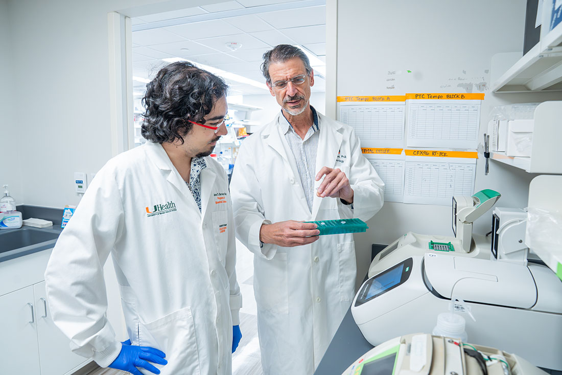
The study, led by Carlos Moraes, Ph.D., professor in the Department of Neurology at the Miller School, and Tania Arguello, Ph.D., a Department of Neurology researcher, focused on mitochondria, often described as the cell’s power plants because they produce the energy cells need to function and carry their own DNA. Over time, that DNA can become damaged, particularly in long-lived cells such as neurons
Why Dopamine-Producing Neurons Are Different
Previous research has shown that dopamine-producing neurons in the substantia nigra region of the brain accumulate unusually high levels of mitochondrial DNA abnormalities during both normal aging and Parkinson’s disease. What has remained unclear is why these defects build up so readily in those particular cells
“Twenty years ago it was shown that dopaminergic neurons have disproportionally high levels of mitochondrial DNA with large deletions,” Dr. Moraes said. “However, the mechanisms involved were not understood.”
Mitochondrial dysfunction is considered one of the hallmarks of Parkinson’s disease. Understanding how mitochondrial DNA damage forms and accumulates could help explain why certain neurons die as the disease progresses

To investigate, researchers used a specialized enzyme called Mito-PstI to create controlled breaks in mitochondrial DNA within dopamine-producing neurons. By introducing a known injury and then following what happened over time, the team hoped to better understand how mitochondrial DNA damage forms, spreads and persists
Using DNA sequencing, the researchers discovered that the initial DNA breaks triggered a surprisingly complex chain of events. Some mitochondrial DNA molecules lost large stretches of genetic material, creating deletions. Others developed duplications, in which segments of DNA were copied and inserted elsewhere in the mitochondrial genome
Importantly, these changes were not random. The team found that many of the rearrangements clustered around specific regions of mitochondrial DNA that help regulate how the genome is copied and used by the cell. These areas appear to function as “hotspots” where structural changes are more likely to occur after damage. The findings suggest that a break in one part of the mitochondrial genome can destabilize other regions, creating opportunities for additional DNA errors to emerge
Researchers also found that the two types of changes almost never appeared in the same DNA molecule. Instead, they seem to arise through different mechanisms, suggesting that mitochondria have multiple pathways for responding to genetic damage
Damage Doesn’t Stop When the Injury Ends
The study’s most significant finding emerged when researchers looked at what happened after the initial DNA damage stopped. The enzyme responsible for creating the mitochondrial DNA breaks was switched off after two months. At that point, no new damage should have occurred. Yet months later, the scientists found that mitochondrial DNA deletions had continued to increase in dopamine-producing neurons. In some brain regions, deletion levels increased more than tenfold, despite the absence of any new injuries.
“Were these deletions being formed all the time or could they accumulate once formed?” said Dr. Moraes. “The ability to manipulate the mitochondrial DNA in living organisms allowed us to test this longstanding question and the results were clear. They do accumulate after they are formed.”
In other words, the problem may be not only the initial damage, but also the ability of damaged mitochondrial genomes to expand over time
The study provides a compelling explanation for how age-related mitochondrial damage could gradually push vulnerable neurons toward dysfunction
The same pattern was not seen in other types of neurons. The researchers examined glutamatergic neurons, a major class of brain cells involved in learning, memory and communication throughout the brain. Although these neurons initially developed mitochondrial DNA deletions after the injury, they did not show the same ongoing buildup
That difference points to an intrinsic characteristic of dopamine-producing neurons that makes them particularly susceptible to mitochondrial DNA accumulation. These neurons have exceptionally high energy demands and depend heavily on healthy mitochondria to maintain their function. Dr. Moraes and team believe those demands may create conditions that allow damaged mitochondrial DNA molecules to gain a foothold and expand over time
The Impact of MGME1
The team found additional evidence supporting this theory when they studied MGME1, a protein involved in mitochondrial DNA replication. When MGME1 was absent, mitochondrial DNA deletions still formed after damage occurred. However, those deletions no longer accumulated over time. The finding suggests that the creation of defects and the expansion of those defects are distinct processes. While damage may initiate the problem, ongoing mitochondrial DNA replication appears necessary for defective genomes to build up inside neurons.
Researchers also observed important differences between two populations of dopamine-producing neurons. Although both accumulated mitochondrial DNA deletions, neurons in the substantia nigra also experienced a reduction in overall mitochondrial DNA content. This additional loss of healthy mitochondrial genomes may make these neurons even more vulnerable to dysfunction and eventual cell death
The study does not prove that mitochondrial DNA deletions directly cause Parkinson’s disease. However, it provides a compelling explanation for how age-related mitochondrial damage could gradually push vulnerable neurons toward dysfunction
While more research is needed to determine whether the same mechanisms operate in humans, the study offers important new insight into why some brain cells appear far less equipped than others to cope with mitochondrial damage and why they may be especially vulnerable in Parkinson’s disease
“If this accumulation has a role in the development of Parkinson’s disease,” Dr. Moraes said, “we could direct efforts to control it.”
Focused Ultrasound for Tremor Now Available at UHealth
UHealth offers focused ultrasound for tremor, a noninvasive treatment for essential tremor and Parkinson’s disease with no surgery
Read more

Shaping the Future of Neurodegenerative Research at the Miller School of Medicine
Dr. Leonard Petrucelli leads two major grants advancing collaborative Parkinson’s disease research at the Miller School
Read more

New Deep Brain Stimulation Target Shows Promise for Restoring Walking
A new deep brain stimulation strategy developed at The Miami Project improves walking in Parkinson’s disease and perhaps spinal cord injury
Read more
Neuronal Inflammasomes: How the Brain’s Immune Sensors Influence Health, Injury and Disease
Researchers at the Miller School are studying the ways neuronal inflammasomes help the brain respond to stress, infection or injury
Read more
