


Download PDF
Abstract
Background
Obesity is a risk factor for pancreatic cancer, but mechanisms remain unclear. We investigated how anthropometric traits, individually and combined, relate to pancreatic cancer risk and whether associations are mediated by metabolic biomarkers
Methods
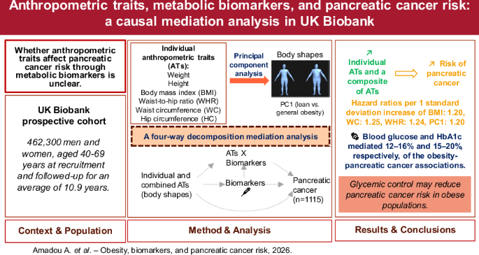
We analysed 462,300 adults (40–69 years) in the UK Biobank. Principal component analysis derived three body shape phenotypes combining body mass index (BMI), height, weight, waist and hip circumference, and waist-to-hip ratio (WHR). Mediation was assessed using four-way decomposition
Results
Over a median follow-up of 10.9 years, 1115 pancreatic cancer cases occurred. Each one-standard-deviation (SD) increase in BMI or WHR was associated with a higher incidence of pancreatic cancer, with hazard ratios (HRs) of 1.20 (confidence interval, CI: 1.12–1.28) and 1.24 (CI: 1.14–1.36), respectively. Body shape characterizing overall obesity showed a similar association (HR = 1.20; CI: 1.12–1.28 per 1-SD), with glucose and HbA1c accounting for mediated proportions (mediated interaction + pure indirect effect) of 12.2% (CI: 3.4–21.0%) and 15.0% (CI: 5.7–24.2%), respectively. For BMI, glucose accounted for 15.9% (CI: 2.8–28.9%) and HbA1c for 20.0% (CI: 6.3–33.7%) of the association.
Conclusions
Glucose and HbA1c mediate a large proportion of the obesity-pancreatic cancer association, highlighting the important role of glycemic control in obesity-related pancreatic carcinogenesis and targeted interventions in at-risk populations
Introduction
With rising incidence and poor prognosis, pancreatic cancer represents a growing challenge for public health and oncology. Globally, it is the 11th most common cancer in women and men. Worldwide, its incidence has more than doubled in recent decades, particularly in high-income countries. This upward trend is expected to persist across most regions, with projections estimating nearly one million new cases annually by 2050 [1]. The 5-year net survival rate remains around 7%, emphasizing the need for effective primary prevention strategies [2].
Obesity, including both general and abdominal adiposity, has emerged as a major modifiable risk factor for pancreatic cancer. In addition to established risk factors such as diabetes, smoking, chronic pancreatitis, and dietary patterns [3], observational and Mendelian randomization studies support a potential causal role of excess adiposity in pancreatic carcinogenesis [4,5,6]. Recent studies have reinforced this link, suggesting that abdominal obesity may be a stronger driver of pancreatic cancer risk than overall obesity, likely due to its closer association with metabolic dysfunction [7, 8]. However, the biological mechanisms through which obesity influences pancreatic cancer development remain incompletely understood.
Several plausible pathways have been proposed to explain the link between obesity and pancreatic cancer [9,10,11]. Obesity often causes insulin resistance, making the body’s cells less responsive to insulin and leading to elevated blood insulin levels (hyperinsulinemia). Both insulin and insulin-like growth factors (IGFs) promote cell proliferation and inhibit apoptosis, potentially contributing to pancreatic cancer initiation and progression [12]. Additionally, obesity is associated with chronic low-grade inflammation, as adipose tissue secretes pro-inflammatory cytokines. This inflammatory environment promotes DNA damage, enhances cellular proliferation, and supports tumour development [13, 14]. Beyond inflammation and insulin dysregulation, obesity also affects hormone levels, such as oestrogen and leptin, which have been linked to increased pancreatic cancer risk [15]. Moreover, obesity disrupts metabolic pathways related to glucose and lipid metabolism, leading to cellular stress and genomic instability [6, 14]. These metabolic disturbances are particularly pronounced in abdominal obesity, which is more strongly linked to insulin resistance and systemic inflammation [16]. Although these mechanisms are biologically plausible and supported by experimental studies [10], formal mediation analyses evaluating whether these pathways quantitatively explain the association between obesity and pancreatic cancer risk are lacking.
Additionally, it is important to consider which anthropometric measures best capture pancreatic cancer risk. Previous studies have primarily focused on individual anthropometric traits (ATs), such as body mass index (BMI), waist circumference (WC), and waist-to-hip ratio (WHR) [17]. However, these approaches may overlook the complex relationships between various body shape characteristics and pancreatic cancer risk. Recognizing the importance of fat distribution and overall body shape in disease development, recent studies have applied principal component analysis (PCA) to identify distinct body shape phenotypes (defined by distinct combinations of multiple ATs) associated with metabolic health indicators and cancer risks [18,19,20]. Using data from the EPIC cohort, our previous study identified two distinct body shape phenotypes associated with an increased risk of pancreatic cancer: one characterized by general obesity and another by tall stature and central obesity. Individuals with the general obesity phenotype had a hazard ratio (HR) and 95% confidence intervals (CI) of 1.12 (CI: 1.06–1.19) for each one standard deviation (SD) increase in overall adiposity, while those with the tall stature and central obesity phenotype had an HR of 1.10 (CI: 1.03–1.17) for each one SD increase in central obesity [21]. Nevertheless, it remains unclear whether these anthropometric patterns influence pancreatic cancer risk through specific metabolic pathways, such as glucose metabolism, insulin resistance, or chronic inflammation. Addressing this gap could help clarify the biological underpinnings of the obesity-pancreatic cancer link and identify potential targets for prevention.
Therefore, this study aimed to assess how associations of individual ATs (such as BMI, WC, and WHR) and body shape phenotypes (combined ATs) relate to pancreatic cancer risk and whether these associations are mediated by specific metabolic health biomarkers
Material and Methods
Study population and design
UK Biobank (Research Resource Identifier (RRID):SCR_012815; http://www.ukbiobank.ac.uk/) is a large national prospective cohort study involving approximately 500,000 participants, aged 40-69 years at recruitment across 22 centres in England, Wales, and Scotland, between 2006 and 2010 [22], with ongoing follow-up. At recruitment, participants provided extensive data through self-administered touchscreen questionnaires, covering health, demographics, lifestyle, and medical history. Biological samples (blood, saliva, and urine) were also collected at recruitment. The study received ethical approval from multiple committees, including the North West Multi-Center Research Ethics Committee, the National Information Governance Board for Health and Social Care in England and Wales, and the Community Health Index Advisory Group in Scotland (http://www.ukbiobank.ac.uk/ethics/). Informed consent was obtained from all participants. Of the 502,384 participants, we excluded those without information on sex (n = 1), those with prevalent cancer (n = 36,964), and those with missing or aberrant anthropometric data (n = 3119). After these exclusions, 462,300 individuals were included in the initial study population. For the main analysis, we further excluded participants with missing values in covariates (n = 94,685), resulting in a complete-case analytic sample of 367,615 participants (Supplementary Fig. 1).
Ascertainment of pancreatic cancer cases
Cancer cases were identified through cancer registries and national health databases, including National Health Service (NHS) Digital and Public Health England for participants in England and Wales, and the NHS Central Register (NHSCR) for those in Scotland. Follow-up data were complete up to February 29, 2020, for England and Wales, and January 31, 2021, for Scotland
First primary pancreatic cancer cases were identified using the 10th Revision of the International Classification of Diseases (ICD-10) (codes C25.0-C25.9)
Anthropometric assessments
At the baseline visit, trained personnel measured height, weight, WC, and hip circumference (HC) [23]. Weight was measured using a Tanita BC418MA body composition analyser, height with a Seca 240 cm stadiometer, and WC and HC with a Seca 200 cm tape measure. BMI and WHR were calculated from these measurements
Biomarker Assays
A broad range of biochemical markers, selected for their known associations with major diseases, were measured from baseline biological samples collected without fasting requirements, in accordance with the UK Biobank protocol [24]. The present study focused on biomarkers representing key metabolic pathways that may mediate the relationship between obesity and pancreatic cancer risk. These included markers of glucose metabolism (glucose, glycated haemoglobin (HbA1c)), insulin signalling (IGF-1, inflammation (C-reactive protein (CRP)), sex hormones (oestrogen, testosterone, sex hormone-binding globulin (SHBG)), lipid metabolism (triglycerides, high-density lipoprotein (HDL) cholesterol, low-density lipoprotein (LDL) cholesterol, total cholesterol), and liver or protein metabolism (total protein, albumin, alanine aminotransferase, gamma-glutamyltransferase, apolipoproteins A and B, cystatin C, total bilirubin, and urate). Biomarkers were quantified using validated biochemical assay techniques [25, 26]. For example, glucose was measured using the hexokinase method; triglycerides and cholesterol by enzymatic colorimetric assays (GPO-POD and CHOD-POD, respectively); CRP using a high-sensitivity immunoturbidimetric assay; total protein via the Biuret method; and HbA1c by high-performance liquid chromatography (Bio-Rad VARIANT II Turbo). SHBG and testosterone were measured using competitive analysis methods (Beckman Coulter, Unicel DxI 800).
Statistical analysis
To generate profiles of body shapes, PCA was performed on standardized residuals of height, weight, BMI, WC, HC, and WHR. The residuals were predicted from a separate regression of the six ATs with age, sex, and study center. Principal components (PCs) were retained based on the proportion of explained variance and their interpretability [27]. Each PC was a weighted combination of the six transformed ATs, remaining independent of the others, and represented a distinct body shape phenotype. Correlations between ATs and PCs were assessed using Pearson coefficients and the variance inflation factor (VIF).
Cox proportional hazards regression was used to estimate HR and 95% CI for the association between each individual ATs, PC, and pancreatic cancer risk. The proportional hazards assumptions were tested using scaled Schoenfeld residuals. Age at entry was defined as age at recruitment. Exit time was defined as the earliest of the following events: age at diagnosis of primary incident pancreatic cancer, age at diagnosis of another cancer (excluding non-melanoma skin cancer), age at the end of follow-up, age at loss to follow-up, or age at death. Non-linearity was assessed by comparing linear and cubic spline models using a likelihood ratio test. Each variable was analysed as a continuous measure per one-SD increase. In addition, we conducted complementary analyses using quartiles of BMI, WHR, HC, and WC, and categorical comparisons of PC scores (+1 and −1 vs 0), to facilitate interpretation, assess the robustness of associations, and compare the risk associated with more extreme body shapes. All multivariable models were adjusted for potential confounders identified using a directed acyclic graph (DAG) (Supplementary Fig. 2). These variables included age (in 5-year categories), centre, diet score, alcohol consumption frequency, smoking status, physical activity, qualifications, Townsend deprivation index, and sedentary behaviour. Physical activity was derived from self-reported walking, moderate, and vigorous activity (MET-min/week), using IPAQ-based scoring [28]. The healthy diet score was estimated from the consumption of fruits, vegetables, fish, processed meats, unprocessed red meats, whole grains, and refined grains ranging from 0 (least healthy) to 6 (healthy) [29] and modelled as a categorical variable with seven categories. Sedentary behaviour was defined as the sum of time spent watching TV, using a computer, and driving. Complete case analyses were conducted on all participants and separately for men and women.
As diabetes is a risk factor, but a mediator rather than confounder, a sensitivity analysis was conducted to adjust for this factor. To address potential reverse causation, a lag-time sensitivity analysis was conducted by excluding the first five years of follow-up for all participants (i.e., follow-up began at baseline age + 5 years). This was applied to both anthropometry–cancer and biomarker–cancer associations. To account for uncertainty due to missing data in adjustment variables, we performed multivariate imputation using chained equations (MICE) [30]. We ran 20 iterations of multiple imputation using fully conditional specification to impute missing values in alcohol consumption, physical activity, smoking status, and educational attainment.
Four-way decomposition mediation analysis was used to examine whether metabolic biomarkers mediated the relationship between individual ATs, body shapes, and pancreatic cancer risk. This method decomposes the total effect of the exposures into direct and indirect pathways, allowing for the simultaneous investigation of both mediation and interaction effects [31]. The total effect (TE) of each variable (single anthropometry, and generated PC) on pancreatic cancer was decomposed into four components: the controlled direct effect (CDE), which captures the effect of exposure independent of mediation and interaction; the reference interaction effect (INTref), due to interaction alone; the mediated interaction effect (INTmed), reflecting combined mediation and interaction; and the pure indirect effect (PIE), representing mediation alone. We assessed the causal effects of changes in ATs and body shapes, respectively, from the 25th to the 75th percentile, holding each mediator fixed at its median level. Each effect’s proportion is calculated relative to the TE, so their sum equals 100%. However, due to variability in statistical estimation and the complexity of effect decomposition, some proportions may appear to exceed 100%. In addition to the adjusted variables identified by the DAG, mutual adjustment for each biomarker was performed by adjusting each mediator model for all other biomarkers, except for highly correlated pairs (r > 0.5) (Fig. 1). Additionally, sensitivity analyses without mutual biomarker adjustment were conducted for BMI and body shape PC1.
Low-density lipoprotein cholesterol, SHBG Sex hormone-binding globulin
All statistical tests were two-sided, and a P-value < 0.05 was considered significant. We judged multiple testing correction as too restrictive because we focused on biomarkers that were associated with both pancreatic cancer and anthropometric measures and we regarded each anthropometric measure as an alternative indicator of adiposity rather than as a separate exposures. All analyses were conducted in R (RRID:SCR_001905), apart from the mediation analysis, which was done in Stata 14 (RRID:SCR_012763).
Results
Characteristics of the study population
Baseline characteristics of the study participants by BMI categories are presented in Table 1. Over a median follow-up of 10.9 years (interquartile range: 10.1–11.6 years), 1115 cases of pancreatic cancer were ascertained among 462,300 participants in the overall cohort. The average age at recruitment was 56.8 ± 8.1 years (mean ± SD). Participants with obesity (BMI ≥ 30 kg/m²) exhibited higher WC, HC, and greater sedentary behaviour compared to those in the underweight/normal-weight category (BMI < 25 kg/m²). Conversely, individuals in the underweight/normal-weight group were more likely to be women, have higher educational attainment, healthier diets, greater physical activity, and were more often never-smokers. Supplementary Table 1 shows that participants with pancreatic cancer had higher levels of liver enzymes, inflammatory markers (CRP), glucose metabolism markers, cystatin C, triglycerides, and urate compared to non-cases, who had higher HDL cholesterol and oestradiol.
Figure 1 presents a correlation matrix illustrating the relationships between biomarkers measured in the study. Most biomarkers exhibited weak to moderate positive correlations. As expected, the strongest correlation was observed between apolipoprotein B and LDL-cholesterol (r = 1), as well as between cholesterol and apolipoprotein B (r = 1). Similarly, cholesterol and LDL-cholesterol were also highly correlated (r = 0.9). Glucose and HbA1c showed a strong correlation (r = 0.6), while a very strong correlation was observed between alanine aminotransferase and aspartate aminotransferase (r = 0.8). Despite these high correlations, variance inflation factor (VIF) analysis indicated acceptable levels of multicollinearity (VIF < 5) (Supplementary Fig. 3).
Body shape phenotypes
Supplementary Table 2 provides the loadings and explained variance for the six PCs in all participants. The first three PCs were retained and together explained 98% of the total variance in anthropometric traits. PC1, accounting for 66.20% of the total variance, distinguished individuals with general obesity from those with a lean body shape (Supplementary Fig. 4a). PC2, explaining 19.34% of the variance, differentiated tall individuals with a low WHR from shorter individuals with a high WHR (Supplementary Fig. 4b). PC3, covering 12.41% of the variance, contrasted tall individuals with a high WHR against shorter individuals with a low WHR (Supplementary Fig. 4c).
Single anthropometry, body shape phenotypes and pancreatic cancer risk
Table 2 presents multivariable-adjusted associations between ATs, body shapes, and pancreatic cancer risk. Each one-SD increment in BMI (4.69 kg/m²), WHR (0.09), WC (12.4 cm), and HC (8.99 cm) was linked to a 20% (HR = 1.20; CI: 1.12−1.28), 24% (HR = 1.24; CI: 1.14−1.36), 25% (HR = 1.25; CI: 1.16−1.35), and 16% (HR = 1.16; CI: 1.08−1.24) higher risk of pancreatic cancer, respectively. Similarly, each one-SD increment in PC1 (i.e., more of a generally obese body shape) was associated with a 20% higher risk of pancreatic cancer (HR = 1.20; CI: 1.12−1.28). The results remained comparable when analysed separately by gender. No significant associations were observed in the overall population for height, PC2 (tall and lean body shape), or PC3 (tall and centrally overweight body shape) with pancreatic cancer risk (Table 2). However, a suggestive positive association was noted for PC2 in men (HR = 1.09; CI: 0.98−1.22). There was no evidence of nonlinear associations (all P > 0.5) (Supplementary Table 3).
The supplementary analyses using quartiles of anthropometric measures and categorical comparisons of PC scores yielded consistent results (Supplementary Table 4). Increasing quartiles of BMI, WHR, HC, and WC were associated with progressively higher risks of pancreatic cancer, with HRs in the highest quartile (Q4) of 1.54 (CI: 1.25−1.88), 1.68 (CI: 1.30-2.17), 1.45 (CI: 1.19−1.77), and 1.59 (CI: 1.26−1.99), respectively, compared to the lowest quartile (Q1). PC1 remained positively associated with pancreatic cancer risk when comparing the +1 category to the reference (HR = 1.57; CI: 1.32−1.87), while the −1 category showed no association (HR = 0.89; CI: 0.72−1.11). PC2 and PC3 did not show significant associations in either direction.
The results observed after disregarding the first five years of follow-up were similar to those in the main analysis (Supplementary Table 5). Analyses additionally adjusting for diabetes yielded also similar results (Supplementary Table 6). Sensitivity analysis using multiple imputation showed results comparable to the main findings for BMI, WHR, WC, HC, and PC1, but revealed a significant association for height (HR = 1.11; CI: 1.02−1.21), unlike the main analysis (HR = 1.08; CI: 0.98−1.19) (Supplementary Table 7). PC2 and PC3 remained non-significant.
Biomarkers and pancreatic cancer risk
Supplementary Table 8 presents multivariable-adjusted HRs for the relationships between each biomarker, separately, and pancreatic cancer risk. Overall, higher levels of alkaline phosphatase, alanine aminotransferase, aspartate aminotransferase, cystatin C, gamma-glutamyltransferase, glucose, HbA1c and urate were positively associated with risk of pancreatic cancer. Conversely, higher levels of apolipoprotein B, cholesterol, LDL-cholesterol, and SHBG were inversely associated with pancreatic cancer risk. These associations remained materially unchanged in sensitivity analyses after disregarding the first five years of follow-up (Supplementary Table 9).
Four-way decomposition mediation analysis
The primary mediators in this analysis were biomarkers associated with both pancreatic cancer (Supplementary Table 8) and anthropometric measures (Supplementary Table 10). BMI, WHR, WC, and PC1 were used as the primary exposure variables
Single anthropometric measures
The four-way decomposition results for BMI and pancreatic cancer risk are shown in Supplementary Table 11 (effect estimates) and Table 3 (attributable proportions). Significant mediation was observed for glucose, with an overall proportion mediated (i.e., the sum of the PIE and the mediated interaction between BMI and the biomarker) of 15.9% (CI: 2.8% to 28.9%), primarily driven by a PIE of 15.3% (CI: 1.4% to 29.3%) (Table 3 and Supplementary Fig. 5). Similarly, for HbA1c, the overall proportion mediated was 20.0% (CI: 6.3% to 33.7%), with a PIE of 20.7% (CI: 5.5% to 36.0%) (Table 3 and Supplementary Fig. 5).
Comparable mediation patterns were observed for WHR and WC in relation to pancreatic cancer risk. For WHR, significant mediation was observed through glucose and HbA1c, with overall proportions mediated of 12.0% (P = 0.007) and 19.1% (P = 0.001), respectively (Table 4). Similarly, for WC, the corresponding overall proportion mediated were 13.3% (P = 0.004) for glucose and 19.1% (P < 0.001) for HbA1c (Supplementary Table 12)
General obesity body shape: PC1
The four-way decomposition results for each potential mediator in the association between PC1 and pancreatic cancer risk are presented in Supplementary Table 13 (effect estimates) and Table 5 (attributable proportions). Significant PIEs were observed for glucose (10.8%, CI: 1.8% to 19.8%) and HbA1c (14.7%, CI: 4.8% to 24.7%). The overall proportions mediated were 12.2% (CI: 3.4% to 21.0%) for glucose and 15.0% (CI: 5.7% to 24.2%) for HbA1c (Table 5 and Supplementary Fig. 6)
Sensitivity analyses
Sensitivity analyses without mutual biomarker adjustment for BMI and PC1 (Supplementary Tables 14 and 15 for BMI; Tables 16 and 17 for PC1) confirmed HbA1c and glucose as mediators. In these models, HbA1c had an overall proportion mediated of 28.3% (P < 0.001) for BMI and 24.7% (P < 0.001) for PC1, while glucose contributed with 15.6% (P < 0.001) for BMI and 13.6% (P < 0.001) for PC1. In these single biomarker models, alanine aminotransferase showed modest mediation in the PC1 model (11.6%, P = 0.044), gamma-glutamyltransferase showed overall proportions mediated of 19.6% (P = 0.005) for BMI and 16.2% (P = 0.004) for PC1. Urate also showed an overall proportion mediated of 21.9% (P = 0.017) in the BMI model and 23.0% (P = 0.014) in the PC1 model.
Discussion
In this large study among UK adults, we found that BMI, WHR, and WC, individually, as well as a composite measure of multiple ATs, characterizing overall obesity, were each similarly associated with an increased risk of pancreatic cancer in a dose-response manner. Large proportions of these associations were explained by higher blood levels of glucose and HbA1c, with a similar magnitude of mediation across individual anthropometric and combined measures. These findings highlight the role of obesity-related metabolic dysfunction in pancreatic carcinogenesis and underscore the importance of glycemic control as a potential target for risk reduction. In additional single-mediator sensitivity analyses (without mutual biomarker adjustments), urate and gamma-glutamyltransferase emerged as additional potential mediators in the obesity-pancreatic cancer relationship.
Excess adiposity, either overall or abdominal, is an established risk factor for pancreatic cancer [5, 6, 8]. Being tall also increases the risk of pancreatic cancer [32]. Our findings align with this evidence and also with our previous study in the EPIC cohort, which identified distinct body shape phenotypes as being positively associated with the incidence of pancreatic cancer [21]. Similarly, our results support broader evidence from reviews and meta-analyses demonstrating that both general and central adiposity (measured by individual ATs) are significant risk factors for pancreatic cancer [33, 34]. Additionally, as mentioned in the introduction, some studies, including Mendelian randomization analyses, have suggested that abdominal adiposity (e.g., WHR adjusted for BMI) may be a more important causal risk factor for pancreatic cancer than total adiposity, likely due to its stronger links to metabolic dysfunction [8]. Our results support this hypothesis, as we observed significant associations between WHR and WC and pancreatic cancer risk, alongside evidence of mediation through glucose and HbA1c. However, in the present study, PC3, which may represent a body shape pattern linked to central adiposity, did not show a significant association with pancreatic cancer risk. This lack of association was somewhat unexpected, given prior evidence [21], and the reasons for this remain unclear and warrant further investigation. This apparent inconsistency may be due to the way PC3 captures central adiposity, potentially influenced by height or other ATs.
To our knowledge, no previous study has formally assessed and quantified the mediating role of metabolic biomarkers in the association between obesity and pancreatic cancer. In our study, four-way decomposition analysis identified glucose and HbA1c as key mediators, reinforcing prior biological evidence that hyperglycaemia can drive pancreatic carcinogenesis by accelerating endothelial cell senescence within the pancreatic cancer microenvironment [35, 36]. IGF-1, a marker of insulin signalling, was measured but showed no significant association with pancreatic cancer risk (overall HR 1.01, 95% CI 0.94−1.09; women 0.95, 0.85−1.06; men 1.06, 0.97−1.17), and was therefore not included among the primary mediators, which focused on biomarkers associated with both pancreatic cancer and anthropometric measures (Supplementary Tables 8–9). Our findings therefore provide novel quantitative evidence of hyperglycaemia’s mediating role in the obesity-pancreatic cancer relationship.
Obesity is strongly linked to dysregulated glucose metabolism, primarily due to insulin resistance [37, 38]. Excess adipose tissue increases the production of pro-inflammatory cytokines and free fatty acids, which impair insulin signalling [39, 40]. As a result, cells become less responsive to insulin, leading to elevated glucose levels and chronic hyperinsulinemia, both hallmarks of obesity [41, 42]. These metabolic disturbances elevate the risk of pancreatic cancer by driving tumour-promoting pathways [6, 27]. HbA1c, a marker of long-term glucose control, is also closely linked to obesity and insulin resistance. Studies indicate that individuals with obesity often have higher HbA1c levels, reflecting chronic insulin resistance [41, 43]. Our findings align with evidence showing that higher pre-diagnostic HbA1c levels are associated with an increased pancreatic cancer risk, even in non-diabetic individuals [40, 44]. Importantly, our results show that the mediation effects of glucose and HbA1c were significant across both PC1 and individual obesity measures (BMI, WHR, WC), reinforcing their crucial role in obesity-related pancreatic cancer risk.
In single-biomarker adjustment models, glucose and HbA1c consistently emerged as significant mediators in the relationship between obesity (including PC1, BMI, WHR, and WC) and pancreatic cancer risk. However, urate and gamma-glutamyltransferase also appeared as significant mediators in individual models. These biomarkers are strongly linked to both obesity and pancreatic cancer, suggesting that beyond glucose and HbA1c, other metabolic markers may contribute to obesity-driven pancreatic cancer development. However, their lack of statistical significance in mutual adjustment models suggests that their mediating effects may be influenced by other metabolic pathways.
Urate has been implicated in oxidative stress and inflammation, both of which promote pancreatic cancer development [45]. Elevated serum uric acid levels are common in obesity, driven by increased pro-inflammatory cytokines and oxidative stress [46]. Consistently, research has found a positive association between serum uric acid and pancreatic cancer risk [47], suggesting that urate may act as a metabolic link between obesity and pancreatic cancer [46, 48]. Gamma-glutamyltransferase, a marker of liver dysfunction, has been associated with obesity and pancreatic cancer risk [49, 50]. It has been shown that gamma-glutamyltransferase plays a mediating role in the relationship between obesity and pancreatic cancer by reflecting liver dysfunction and metabolic disturbances [51]. Elevated gamma-glutamyltransferase is also associated with increased oxidative stress and chronic inflammation, all of which may contribute to pancreatic carcinogenesis [51]. These findings suggest a potential role of liver-related metabolic disturbances in obesity-associated pancreatic cancer risk.
Overall, these findings contribute to a growing body of evidence supporting the role of glucose dysregulation and liver-related metabolism in obesity-associated pancreatic cancer risk. Although IGF-I is linked to glucose metabolism [52], we and others [53], found no association between IGF-I and pancreatic cancer risk. This suggests that IGF-I’s involvement in systemic glucose metabolism does not necessarily translate into a direct role in pancreatic carcinogenesis. These findings underscore the complex metabolic dysregulation in obesity and highlight the importance of biomarkers such as glucose, HbA1c, urate, and gamma-glutamyltransferase in understanding the mechanisms linking obesity to pancreatic cancer. However, although obesity and pancreatic cancer incidence show epidemiological parallels, the rise in pancreatic cancer rates is unlikely to be attributable to obesity alone. Other factors, including population aging, changes in diabetes prevalence, environmental exposures, and improvements in diagnostic practices, may also contribute to the increasing global burden of pancreatic cancer.
One of the key strengths of this study is the use of the large, well-characterized UK Biobank cohort, which provides extensive data on anthropometrics, biomarkers, and health outcomes. The large sample size enhances statistical power, allowing for robust estimates of the associations between individual ATs, body shape phenotypes, and pancreatic cancer risk. Additionally, the prospective design of the cohort minimizes recall and selection biases, thereby strengthening the validity of the findings. A major strength is the four-way mediation analysis, which offers a nuanced understanding of the role of metabolic biomarkers in obesity-related pancreatic cancer risk. This analysis distinguishes between mediation and interaction effects of circulating biomarkers, providing deeper insights into the mechanisms linking obesity and pancreatic cancer risk, whereas traditional mediation methods often overlook mediated interaction effects and may underestimate these complex relationships. The identification of glucose and HbA1c as significant mediators reinforces the role of metabolic dysregulation in pancreatic tumorigenesis. Another analytical strength of our study is the use of both continuous and categorical analyses of principal component scores, which allowed us to compare risk across distinct groups (+1 vs 0 and −1 vs 0). This approach enabled the detection of potential threshold effects and provided a clearer understanding of how extreme values in adiposity-related components influence pancreatic cancer risk. The consistency of the results across different obesity measures strengthens the reliability of the findings. We addressed potential reverse causation and missing data through sensitivity analyses excluding the first five years of follow-up (lag-time analysis) and multiple imputation, respectively. In this study, pancreatic cancer risk estimates for PC1 (a composite obesity measure) were similar to those for BMI, WHR, and WC, reinforcing the relevance of widely used measures like BMI, WHR, and WC.
Despite these strengths, certain limitations should be noted. The observational design precludes causal inference, and residual confounding cannot be entirely ruled out. While we accounted for multiple biomarkers, the interplay between metabolic pathways is complex, and some unmeasured factors may influence the observed associations. Second, blood samples were collected in a non-fasting state, which may slightly affect some metabolic biomarkers such as glucose and triglycerides, and could have led to an attenuation of observed associations towards the null due to random measurement error. Third, limited generalizability due to the study population and the cross-sectional nature of the mediation analysis restricts causal interpretation of mediator-outcome relationships.
In conclusion, this study provides strong evidence that metabolic biomarkers, particularly glucose and HbA1c, mediate the association between obesity and pancreatic cancer risk. These results reinforce the critical role of glycemic regulation in obesity-related carcinogenesis and have public health relevance by supporting the development of personalized prevention strategies and targeted interventions to reduce pancreatic cancer risk in at-risk populations. Future research should aim to identify additional biological pathways involved in this relationship to further elucidate mechanisms.
Disclaimer
Where authors are identified as personnel of the International Agency for Research on Cancer / World Health Organization, the authors alone are responsible for the views expressed in this article and they do not necessarily represent the decisions, policy or views of the International Agency for Research on Cancer / World Health Organization
Data availability
The UK Biobank reearch in the public interest. All researchers who wish to access the research re form in the Access Management System (AMS- https://bbams.ndph.ox.ac.uk/ams/)
Code availability
The code used for the analyses is available from the corresponding author upon request
References
Leiphrakpam PD, Chowdhury S, Zhang M, Bajaj V, Dhir M, Are C. Trends in the global incidence of pancreatic cancer and a brief review of its histologic and molecular subtypes. J Gastrointest Cancer. 2025;56:71. https://doi.org/10.1007/s12029-025-01183-2
Pancreatic Cancer UK [Internet]. [cited 2025 Apr 28]. Pancreatic cancer statistics. Available from: https://www.pancreaticcancer.org.uk/what-we-do/media-centre/pancreatic-cancer-statistics/
Jiang S, Fagman JB, Ma Y, Liu J, Vihav C, Engstrom C, et al. A comprehensive review of pancreatic cancer and its therapeutic challenges. Aging. 2022;14:7635–49. https://doi.org/10.18632/aging.204310
Carreras-Torres R, Johansson M, Gaborieau V, Haycock PC, Wade KH, Relton CL, et al. The Role of Obesity, Type 2 Diabetes, and metabolic factors in pancreatic cancer: a Mendelian randomization study. J Natl Cancer Inst. 2017;109. https://doi.org/10.1093/jnci/djx012
Larsson SC, Spyrou N, Mantzoros CS. Body fatness associations with cancer: evidence from recent epidemiological studies and future directions. Metabolism. 2022;137:155326. https://doi.org/10.1016/j.metabol.2022.155326
Park JH, Han K, Hong JY, Park YS, Hur KY, Kang G, et al. Changes in metabolic syndrome status are associated with altered risk of pancreatic cancer: a nationwide cohort study. Gastroenterology. 2022;162:509–20.e7. https://doi.org/10.1053/j.gastro.2021.09.070
Xu M, Jung X, Hines OJ, Eibl G, Chen Y. Obesity and pancreatic cancer: overview of epidemiology and potential prevention by weight loss. Pancreas. 2018;47:158–62. https://doi.org/10.1097/MPA.0000000000000974
Maina JG, Pascat V, Zudina L, Ulrich A, Pupko I, Bonnefond A, et al. Abdominal obesity is a more important causal risk factor for pancreatic cancer than overall obesity. Eur J Hum Genet. 2023;31:962–6. https://doi.org/10.1038/s41431-023-01301-3
Avgerinos KI, Spyrou N, Mantzoros CS, Dalamaga M. Obesity and cancer risk: Emerging biological mechanisms and perspectives. Metabolism. 2019;92:121–35. https://doi.org/10.1016/j.metabol.2018.11.001
Lee H. Obesity-associated cancers: evidence from studies in mouse models. Cells. 2022;11. https://doi.org/10.3390/cells11091472
Poman DS, Motwani L, Asif N, Patel A, Vedantam D. Pancreatic cancer and the obesity epidemic: a narrative review. Cureus. 2022;14:e26654. https://doi.org/10.7759/cureus.26654
Zhong W, Wang X, Wang Y, Sun G, Zhang J, Li Z. Obesity and endocrine-related cancer: The important role of IGF-1. Front Endocrinol. 2023;14:1093257. https://doi.org/10.3389/fendo.2023.1093257
Cayssials V, Buckland G, Crous-Bou M, Bonet C, Weiderpass E, Skie G, et al. Inflammatory potential of diet and pancreatic cancer risk in the EPIC study. Eur J Nutr. 2022;61:2313–20. https://doi.org/10.1007/s00394-022-02809-y
Ruiz CF, Garcia C, Jacox JB, Lawres L, Muzumdar MD. Decoding the obesity-cancer connection: lessons from preclinical models of pancreatic adenocarcinoma. Life Sci Alliance. 2023;6. https://doi.org/10.26508/lsa.202302228
Bardhi O, Dubey P, Palmer BF, Clegg DJ. Oestrogens, adipose tissues and environmental exposures influence obesity and diabetes across the lifecycle. Proc Nutr Soc. 2024;1–8. https://doi.org/10.1017/S0029665124000119
Hardy OT, Czech MP, Corvera S. What causes the insulin resistance underlying obesity?. Curr Opin Endocrinol Diab Obes. 2012;19:81–7. https://doi.org/10.1097/MED.0b013e3283514e13
Aune D, Norat T, Leitzmann M, Tonstad S, Vatten LJ. Physical activity and the risk of type 2 diabetes: a systematic review and dose-response meta-analysis. Eur J Epidemiol. 2015;30:529–42. https://doi.org/10.1007/s10654-015-0056-z
Peruchet-Noray L, Dimou N, Sedlmeier AM, Fervers B, Romieu I, a Mendelian randomization analysis. Cancers. 2023;15:1296. https://doi.org/10.3390/cancers15041296
Amadou A, Freisling H, Sedlmeier AM, Bohmann P, Fontvieille E, Weber A, et al. Multi-trait body shape phenotypes and breast cancer risk in postmenopausal women: a causal mediation analysis in the UK Biobank Cohort. J Epidemiol Glob Health. 2024;14:420–32. https://doi.org/10.1007/s44197-024-00226-4
Peruchet-Noray L, Sedlmeier AM, Dimou N, Baurecht H, Fervers B, Fontvieille E, et al. Tissue-specific genetic variation suggests distinct molecular pathways between body shape phenotypes and colorectal cancer. Sci Adv. 2024;10. https://doi.org/10.1126/sciadv.adj1987
Sedlmeier AM,, et al. Body shape phenotypes of multiple anthropometric traits and cancer risk: a multi-national cohort study. Br J Cancer. 2023;128:4. https://doi.org/10.1038/s41416-022-02071-3
Conroy MC, Lacey B, Bešević J, Omiyale W, Feng Q, Effingham M, et al. UK Biobank: a globally important re10.1038/s41416-022-02053-5
Christakoudi S, Tsilidis KK, Evangelou E, Riboli E. A Body Shape Index (ABSI), hip index, and risk of cancer in the UK Biobank cohort. Cancer Med. 2021;10:5614–28. https://doi.org/10.1002/cam4.4097
Category 17518 [Internet]. [cited 2023 Apr 3]. Available from: https://biobank.ndph.ox.ac.uk/ukb/label.cgi?id=17518
Tin Tin S, Key TJ, Reeves GK. Alcohol intake and endogenous hormones in pre- and postmenopausal women: findings from the UK Biobank. Cancer Epidemiol Biomark Prev. 2021;30:2294–301. https://doi.org/10.1158/1055-9965.EPI-21-0789
Arthur RS, Dannenberg AJ, Rohan TE. The association of prediagnostic circulating levels of cardiometabolic markers, testosterone and sex hormone-binding globulin with risk of breast cancer among normal weight postmenopausal women in the UK Biobank. Int J Cancer. 2021;149:42–57. https://doi.org/10.1002/ijc.33508
Ried JS, Jeff MJ, Chu AY, Bragg-Gresham JL, van Dongen J, Huffman JE, et al. A principal component meta-analysis on multiple anthropometric traits identifies novel loci for body shape. Nat Commun. 2016;7:1. https://doi.org/10.1038/ncomms13357
Pearce M, Strain T, Kim Y, Sharp SJ, Westgate K, Wijndaele K, et al. Estimating physical activity from self-reported behaviours in large-scale population studies using network harmonisation: findings from UK Biobank and associations with disease outcomes. Int J Behav Nutr Phys Act. 2020;17:40. https://doi.org/10.1186/s12966-020-00937-4
Hepsomali P, Groeger JA. Diet, sleep, and mental health: insights from the UK Biobank Study. Nutrients. 2021;13:2573. https://doi.org/10.3390/nu13082573
Sterne JAC, White IR, Carlin JB, Spratt M, Royston P, Kenward MG, et al. Multiple imputation for missing data in epidemiological and clinical research: potential and pitfalls. BMJ. 2009;338:b2393. https://doi.org/10.1136/bmj.b2393
Discacciati A, Bella investigate mediating and interactive mechanisms using the four-way effect decomposition. Int J Epidemiol. 2018. https://doi.org/10.1093/ije/dyy236
Aune D, Vieira AR, Chan DSM, Navarro Rosenblatt DA, Vieira R, Greenwood DC, et al. Height and pancreatic cancer risk: a systematic review and meta-analysis of cohort studies. Cancer Causes Control. 2012;23:1213–22. https://doi.org/10.1007/s10552-012-9983-0
Mandic M, Pulte D, Safizadeh F, Niedermaier T, Hoffmeister M, Brenner H. Overcoming underestimation of the association of excess weight with pancreatic cancer due to prediagnostic weight loss: Umbrella review of systematic reviews, meta-analyses, and pooled-analyses. Obes Rev. 2024;25:e13799. https://doi.org/10.1111/obr.13799
Bryce C, Bucaj M. Pancreatic cancer: rapid evidence review. Am Fam Physician. 2024;109:245–50
Chari ST, Wu B, Lopez C, Lustigova E, Chen Q, Van Den Eeden SK, et al. Risk of pancreatic cancer in glycemically defined new-onset diabetes: a prospective cohort study. Gastroenterology. 2026;170:106–17. https://doi.org/10.1053/j.gastro.2025.06.025
Ling YW, Duan JL, Jiang ZJ, Yang Z, Liu JJ, Song P, et al. Diabetes reshapes pancreatic cancer-associated endothelial niche by accelerating senescence. Nat Commun. 2025;16:8654. https://doi.org/10.1038/s41467-025-63801-8
Lam TK, Daschner P, Ishibe N, Wali A, Hall K, Czajkowski S, et al. Metabolic Dysregulation and Cancer Risk Program (MeDOC): a transdisciplinary approach to obesity-associated cancers. J Natl Cancer Inst. 2024;116:1555–61. https://doi.org/10.1093/jnci/djae134
Ahmed B, Sultana R, Greene MW. Adipose tissue and insulin resistance in obese. Biomed Pharmacother. 2021;137:111315. https://doi.org/10.1016/j.biopha.2021.111315
Ruze R, Song J, Yin X, Chen Y, Xu R, Wang C, et al. Mechanisms of obesity- and diabetes mellitus-related pancreatic carcinogenesis: a comprehensive and systematic review. Sig Transduct Target Ther. 2023;8:1–41. https://doi.org/10.1038/s41392-023-01376-w
Lemanska A, Price CA, Jeffreys N, Byford R, Dambha-Miller H, Fan X, et al. BMI and HbA1c are metabolic markers for pancreatic cancer: Matched case-control study using a UK primary care database. PLoS One. 2022;17:10. https://doi.org/10.1371/journal.pone.0275369
Kahn LG, Philippat C, Nakayama SF, Slama R, Trasande L. Endocrine-disrupting chemicals: implications for human health. Lancet Diab Endocrinol. 2020;8:703–18. https://doi.org/10.1016/S2213-8587(20)30129-7
Fryk E, Olausson J, Mossberg K, Strindberg L, Schmelz M, Brogren H, et al. Hyperinsulinemia and insulin resistance in the obese may develop as part of a homeostatic response to elevated free fatty acids: A mechanistic case-control and a population-based cohort study. EBioMedicine. 2021;65:103264. https://doi.org/10.1016/j.ebiom.2021.103264
Meziani J, de Jong JGY, Fuhler GM, Koopmann BDM, Levink IJM, Fockens P, et al. Assessment of glucose and HbA1c monitoring in a pancreatic cancer surveillance program for high-risk individuals. Clin Transl Gastroenterol. 2024;15:e00777. https://doi.org/10.14309/ctg.0000000000000777
Grewe S, Jördens MS, Roderburg C, Leyh C, Labuhn S, Luedde T, et al. Elevated HbA1c levels are associated with a risk of pancreatic cancer: a case-control study. J Clin Med. 2024;13. https://doi.org/10.3390/jcm13185584
Du L, Zong Y, Li H, Wang Q, Xie L, Yang B, et al. Hyperuricemia and its related diseases: mechanisms and advances in therapy. Sig Transduct Target Ther. 2024;9:1–29. https://doi.org/10.1038/s41392-024-01916-y
Mao T, He Q, Yang J, Jia L, Xu G. Relationship between gout, hyperuricemia, and obesity—does central obesity play a significant role?—a study based on the NHANES database. Diabetol Metab Syndr. 2024;16:24. https://doi.org/10.1186/s13098-024-01268-1
Huang CF, Huang JJ, Mi NN, Lin YY, He QS, Lu YW, et al. Associations between serum uric acid and hepatobiliary-pancreatic cancer: A cohort study. World J Gastroenterol. 2020;26:7061–75. https://doi.org/10.3748/wjg.v26.i44.7061
Fini MA, Elias A, Johnson RJ, Wright RM. Contribution of uric acid to cancer risk, recurrence, and mortality. Clin Transl Med. 2012;1:e16. https://doi.org/10.1186/2001-1326-1-16
Johansen D, Borgström A, Lindkvist B, Manjer J. Different markers of alcohol consumption, smoking and body mass index in relation to risk of pancreatic cancer. A Prospect Cohort Study Malmö Prev Proj Pancreatol. 2009;9:677–86. https://doi.org/10.1159/000212088
Heo JH, Kang JG, Han K, Lee KJ. Association between Gamma-Glutamyl transferase levels and pancreatobiliary cancer risk in patients with diabetes: evidence from the national health insurance cooperation health checkup 2009 to 2012. Gut Liver. https://doi.org/10.5009/gnl240322
Solís-Pérez E, Mar-Buruato AM, Tijerina-Sáenz A, Sánchez-Peña MA, González-Martínez BE, Lavalle-González FJ, et al. Adipokines and Gamma-Glutamyl Transferase as biomarkers of metabolic syndrome risk in Mexican school-aged children. Nutrients. 2024;16:4410. https://doi.org/10.3390/nu16244410
Clemmons DR. Involvement of insulin-like growth factor-I in the control of glucose homeostasis. Curr Opin Pharmacol. 2006;6:620–5. https://doi.org/10.1016/j.coph.2006.08.006
Knuppel A, Fensom GK, Watts EL, Gunter MJ, Murphy N, Papier K, et al. Circulating Insulin-like Growth Factor-I concentrations and risk of 30 cancers: Prospective Analyses in UK Biobank. Cancer Res. 2020;80:4014–21. https://doi.org/10.1158/0008-5472.CAN-20-1281
Acknowledgements
This research has been conducted using the UKB Reto the participants and those involved in building the resource
Funding
This work was supported by the French National Cancer Institute (l’Institut National du Cancer, INCa_16643) and the German Research Foundation (BA 5459/2-1). The funder had no role in study design, data collection and analysis, decision to publish, or preparation of the manuscript
Author information
Author notes
These authors contributed equally: Amina Amadou, Heinz Freisling
Authors and Affiliations
Department of Prevention Cancer Environment, Centre Léon Bérard, Lyon, France
Amina Amadou, Benoit Mercoeur, Hwayoung Noh & Béatrice Fervers
Inserm U1296 Radiations: Defense, Health, Environment, Lyon, France
Amina Amadou, Benoit Mercoeur, Hwayoung Noh & Béatrice Fervers
International Agency for Research on Cancer (IARC), Nutrition and Metabolism Branch, Lyon, France
Heinz Freisling, Alem Gebremariam, Laia Peruchet-Noray & Quan Gan
Department of Epidemiology and Preventive Medicine, University of Regensburg, Regensburg, Germany
Patricia Bohmann, Michael J. Stein, Anja M. Sedlmeier, Michael F. Leitzmann & Hansjörg Baurecht
Center for Translational Oncology, University Hospital Regensburg, Regensburg, Germany
Anja M. Sedlmeier
Authors
- Amina AmadouView author publications
Search author on:PubMed Google Scholar
- Heinz FreislingView author publications
Search author on:PubMed Google Scholar
- Benoit MercoeurView author publications
Search author on:PubMed Google Scholar
- Patricia BohmannView author publications
Search author on:PubMed Google Scholar
- Michael J. SteinView author publications
Search author on:PubMed Google Scholar
- Hwayoung NohView author publications
Search author on:PubMed Google Scholar
- Alem GebremariamView author publications
Search author on:PubMed Google Scholar
- Anja M. SedlmeierView author publications
Search author on:PubMed Google Scholar
- Laia Peruchet-NorayView author publications
Search author on:PubMed Google Scholar
- Quan GanView author publications
Search author on:PubMed Google Scholar
- Michael F. LeitzmannView author publications
Search author on:PubMed Google Scholar
- Hansjörg BaurechtView author publications
Search author on:PubMed Google Scholar
- Béatrice FerversView author publications
Search author on:PubMed Google Scholar
Contributions
AA and HF contributed to the study conception and design. Material preparation and data collection were performed by HF, AMS, and BM. Statistical analyses were performed by BM and AA. The first draft of the manuscript was written by AA, with support from HF and BF. AA, HF, BM, PB, MJS, HN, AG, AMS, LPN, QG, MFL, HB, and BF contributed to the interpretation of the results and critically revised the manuscript. All authors read and approved the final manuscript
Ethics declarations
Competing interests
The authors declare no competing interests
Ethics approval and consent to participate
The UK Biobank study received ethical approval from the National Health Service (NHS) North West Centre for Research Ethics Committee (Ref:[11]./NW/0382). All methods were performed in accordance with the relevant guidelines and regulations. All participants provided written informed consent
Additional information
Publisher’s note Springer Nature remains neutral with regard to jurisdictional claims in published maps and institutional affiliations
Supplementary information
Supplementary Material (download DOCX )
Rights and permissions
Open Access This article is licensed under a Creative Commons Attribution-NonCommercial-NoDerivatives 4.0 International License, which permits any non-commercial use, sharing, distribution and reproduction in any medium or format, as long as you give appropriate credit to the original author(s) and the source, provide a link to the Creative Commons licence, and indicate if you modified the licensed material. You do not have permission under this licence to share adapted material derived from this article or parts of it. The images or other third party material in this article are included in the article’s Creative Commons licence, unless indicated otherwise in a credit line to the material. If material is not included in the article’s Creative Commons licence and your intended use is not permitted by statutory regulation or exceeds the permitted use, you will need to obtain permission directly from the copyright holder. To view a copy of this licence, visit http://creativecommons.org/licenses/by-nc-nd/4.0/.
About this article
Cite this article
Amadou, A., Freisling, H., Mercoeur, B. et al. Anthropometric traits, metabolic biomarkers, and pancreatic cancer risk: a causal mediation analysis in UK Biobank.
Br J Cancer (2026). https://doi.org/10.1038/s41416-026-03524-9
Received:01 October 2025
Revised:22 April 2026
Accepted:16 June 2026
Published:11 July 2026
Version of record:11 July 2026
DOI
:https://doi.org/10.1038/s41416-026-03524-9
