


Download PDF
Abstract
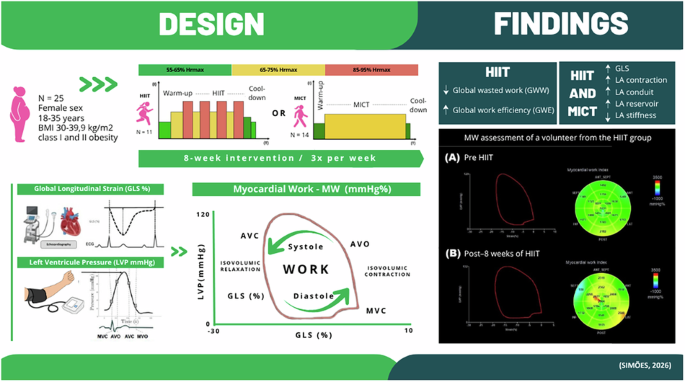
The purpose of this study was to investigate the effects of high-intensity interval training (HIIT) and moderate-intensity continuous training (MICT) on cardiac function and myocardial work (MW) in women with obesity. Twenty-five women with obesity (Age: 28 ± 5 years, BMI: 35.03 ± 3.19 kg/m²) were randomized to 8 weeks of either HIIT (n = 11, 4×4-min at 85–95% of HRmax) or MICT (n = 14, 41-min at 65–75% of HRmax). Cardiac function and strains were evaluated using two-dimensional speckle tracking echocardiography. MW indices were computed via dedicated software. HIIT significantly improved myocardial efficiency, as evidenced by a reduction in global wasted work (GWW [d = 0.64, p = 0.042]) and an increase global work efficiency (GWE [d = 0.83, p = 0.005]). Both HIIT and MICT reduced left ventricular global longitudinal strain (GLS, p = 0.017 for HIIT; p = 0.023 for MICT), left atrial (LA) contraction (p < 0.001 for both), LA conduit (p = 0.002 for both), LA reservoir (p = 0.003 for HIIT; p < 0.001 for MICT), and LA stiffness (p = 0.004 for HIIT, p = 0.031 for MICT). HIIT and MICT both improved myocardial deformation in women with obesity. However, only HIIT improved myocardial efficiency, suggesting a potential superiority for targeting subclinical cardiac dysfunction in this population.
Introduction
Obesity is characterized by metabolic, inflammatory, and vascular disturbances that increase cardiovascular risk [1, 2]. Even in the absence of overt cardiac disease, individuals with obesity often present subclinical myocardial abnormalities, including impaired strain and altered myocardial work (MW) [3, 4]. Obesity is more prevalent among women, in whom obesity-related cardiovascular consequences appear to be more pronounced, increasing the risk of early myocardial dysfunction [5, 6]
Exercise improves cardiometabolic health independently of significant weight loss [7]. High-intensity interval training (HIIT), characterized by brief bouts of vigorous exercise interspersed with recovery periods [8], is a time-efficient modality that induces robust cardiovascular adaptations and often surpasses moderate-intensity continuous training (MICT) in reducing adiposity and systemic inflammation [9,10,11]. Meta-analyses also suggest that HIIT elicits greater improvements in vascular function in individuals with obesity compared with MICT [12]. Supporting this evidence, we previously demonstrated greater reductions in central blood pressure following HIIT versus MICT in women with obesity [13] and in other clinical populations [14].
Given that vascular and hemodynamic adaptations may reduce left ventricular afterload, HIIT could also enhance cardiac function and MW in individuals with obesity. Therefore, we investigated the effects of an 8-week HIIT program compared to MICT on cardiac function and MW in women with obesity
Materials and methods
Forty-four women with obesity were randomly allocated (1:1) to either HIIT or MICT intervention groups using a computer-generated randomization sequence. Inclusion criteria were: female sex; age 18–35 years; obesity grade I or II (BMI 30.0–39.9 kg/m²); stable body weight for ≥12 weeks; no recent participation in weight-loss programs; absence of cardiovascular, metabolic, or endocrine diseases, except for treated hypothyroidism; non-smoking; no use of medications affecting cardiorespiratory or neuromuscular systems; and availability to complete all assessments.
Training protocols
Both interventions were performed outdoors on an official running track, three times a week for eight weeks. HIIT consisted of four 4-min bouts at 85–95% HRmax interspersed with 3-min active recovery at 65–75% HRmax. MICT involved 41 min at 65–75% HRmax followed by a 2-min cool-down. Training protocols are detailed in Supplementary Material and Methods
Assessments
Echocardiography was performed by a single experienced sonographer blinded to group allocation, with a Vivid E95 system (GE Healthcare Vingmed Ultrasound AS, Horten, Norway). Images were analyzed offline using EchoPAC software (GE Healthcare®, Horten, Norway). Systolic blood pressure was measured with an oscillometric device (Omron, Omron Healthcare Brasil, São Paulo, Brazil) immediately before echocardiographic acquisition, and used to compute MW indices. A maximal incremental treadmill test using a ramp protocol assessed VO₂peak and HRmax. Body composition was assessed with a bioelectrical impedance (Maltron® BF-906, Rayleigh, UK). Baseline physical activity levels and dietary intake were assessed using standardized questionnaires. For further details, please see Supplementary Material and Methods.
Participants were instructed to maintain their habitual diet and daily activities and to refrain from initiating any additional exercise programs during the study period. The study was approved by the institutional ethics committee (Protocol: 08935419.5.0000.0104) and registered at the Brazilian Clinical Trials Registry (RBR-3v3dqf). All study procedures were conducted in accordance with the relevant guidelines and regulations. Written informed consent was obtained from all participants prior to their enrollment in the study.
Data analysis
Sample size was calculated with G*Power® (version 3.1) for a repeated-measures ANOVA, with GLS defined a priori as the primary outcome. Assuming an effect size of 0.51 [15], a power of 80%, and a two-sided alpha of 0.05, a minimum of 10 participants was required. Data were analyzed using a per-protocol approach, including only participants who completed the intervention and post-intervention assessments. Data distribution and homogeneity of variances were assessed using the Shapiro–Wilk and Levene’s tests. Continuous data are presented as mean ± standard deviation. Group, time, and group × time effects were analyzed with Bonferroni correction applied when appropriate. Effect sizes were calculated using Cohen’s d. Statistical significance was set at p ≤ 0.05. Data were analyzed using the SPSS (version 23, IBM®, New York, USA).
Results
Twenty-five women completed the intervention (HIIT: n = 11; MICT: n = 14) and were included in the final analysis (Supplementary Table 1). The groups were comparable at baseline for all variables. Mean training attendance was 85%, with no between-group differences
Myocardial work indices
No significant group × time interactions were observed in MW indices. A significant within-group effect of time was observed in the HIIT group, with a reduction in global wasted work (GWW Δ = −15.23 ± 41.49 mmHg·%, p = 0.042) and an increase in global work efficiency (GWE: Δ= 2.39 ± 2.43%, p = 0.005). In contrast, no significant time effects were detected in the MICT group for GWW (Δ= 4.85 ± 62.28 mmHg·%, p = 0.283) or GWE (Δ = 1.21 ± 2.75%, p = 0.101). No significant time effects were observed in global work index (GWI; HIIT p = 0.854; MICT: p = 0.292) or global constructive work (GCW; HIIT p = 0.646; MICT p = 0.396), in any of the groups. Pressure–strain loop (PSL) and MW indices following HIIT and MICT are illustrated in Fig. 1, with a visually larger PSL area observed after HIIT, consistent with improved myocardial efficiency.
Illustration of pressure–strain loop and myocardial work indices following HIIT (A) and MICT (B) in women with obesity. PSL pressure-strain loop, LVP left ventricular pressure, GLS global longitudinal strain, GWI global work index, GCW global constructive work, GWW global wasted work, GWE global work efficiency
Cardiac function and strain
For conventional cardiac structural and functional parameters, no significant group × time interactions were identified. A significant main effect of time was observed in both groups, with increases in left ventricular end-diastolic volume (LVEDV; HIIT p = 0.005; MICT p < 0.001) and stroke volume (LVSV p < 0.001 for both groups), as well as reductions in left atrial stiffness (HIIT: p = 0.004; MICT: p = 0.031)
A significant time effect was observed for left ventricular ejection fraction (LVEF; p = 0.029) and aortic diameter (p = 0.034) only in the HIIT group. No significant time effects were observed in conventional diastolic function parameters in any group (Table 1)
Regarding myocardial deformation, a significant within-group improvement over time was observed in both groups for left ventricular global longitudinal strain (GLS; HIIT p = 0.017; MICT p = 0.023). Similarly, left atrial reservoir (LASr HIIT: p = 0.003; MICT: p < 0.001), conduit (LAScd p = 0.002 for both), and contraction strain (LASct p < 0.001 for both) improved significantly over time in both groups (Table 1)
Body composition, fitness, and hemodynamics
No significant group × time interactions were detected for body composition or fitness variables. A significant time effect was observed only in the HIIT group, which demonstrated reductions in body weight (p = 0.002), BMI (p = 0.002), body fat percentage (p < 0.001), and systolic blood pressure (SBP p = 0.033), along with an increase in VO₂peak (p = 0.024). No significant time effects were observed in the MICT group for these outcomes (Table 1)
Discussion
This study demonstrates that both HIIT and MICT improve cardiac function and LV and LA strain over time in women with obesity. In addition, a significant time effect was observed in MW efficiency, only in the HIIT group, as reflected by a reduction in GWW and an increase in GWE. These findings support the role of aerobic exercise in improving cardiac function and myocardial deformation and suggest that HIIT may confer additional benefits on myocardial efficiency
Obesity impairs LV mechanics through increased afterload, myocardial fibrosis, and altered metabolic signaling [2,3,4]. HIIT may counteract these effects by promoting favorable hemodynamic and myocardial adaptations. While prior studies have shown improvements in conventional systolic and diastolic parameters following HIIT [15, 16], our findings extend this knowledge by incorporating MW indices, which integrate myocardial deformation with non-invasively estimated blood pressure, offering a more sensitive and load-adjusted assessment of cardiac performance [17].
Reduced GWW reflects less non-productive myocardial contraction, representing energy expenditure that does not contribute to effective stroke volume, whereas the increase in GWE indicates a greater proportion of effective MW [17]. These changes are clinically relevant, as myocardial inefficiency is an early marker of subclinical dysfunction in obesity [18]. Importantly, a population-based study has shown that increased GWW and reduced GWE independently predict all-cause mortality in individuals with overweight, even when conventional echocardiographic parameters were preserved [19].
The effects of HIIT on MW indices may relate to its intermittent nature, promoting acute increases in cardiac workload, shear stress, and sympathetic activity, stimuli known to enhance myocardial remodeling and vascular function more than MICT [13, 15, 16]. HIIT has been shown to improve endothelial function and reduce arterial stiffness and central blood pressure [12, 14], key factors that lower cardiac afterload and myocardial wall stress, thereby contributing to greater myocardial efficiency [20].
Both interventions improved GLS and LA strain parameters, which are sensitive markers of early myocardial remodeling [21, 22, 23]. These improvements occurred in parallel with increases in left ventricular end-diastolic volume and stroke volume, suggesting that enhanced preload may have contributed to the observed deformation responses. Increased preload enhances myocardial fiber stretch via the Frank–Starling mechanism, thereby improving longitudinal systolic deformation and atrial reservoir, conduit, and contractile function. Therefore, improvements in GLS may reflect not only enhanced subendocardial contractility but also more favorable loading conditions, which are commonly impaired in obesity due to elevated filling pressures and reduced compliance [21]. Similarly, the improvement in LA strain components indicates greater atrial compliance and booster pump function, facilitating left ventricular filling under increased preload and potentially reducing atrial wall stress. These adaptations are clinically relevant, as impaired LA function and reduced preload reserve are early features of obesity-related cardiomyopathy and precede the development of atrial fibrillation and diastolic dysfunction [22].
The significant time effect in reducing body fat and increasing VO₂peak observed only in the HIIT group underscore its superior effectiveness compared to MICT. HIIT promotes greater energy expenditure and metabolic stimulation through mechanisms such as excess post-exercise oxygen consumption and enhanced lipolysis [10]. Additionally, improvements in VO₂peak reflect HIIT’s capacity to induce central and peripheral cardiovascular adaptations [14]
Collectively, these findings suggest that HIIT may represent a time-efficient strategy to improve cardiorespiratory fitness, body composition, and myocardial function and efficiency in women with obesity. The inclusion of MW assessment provides additional load-adjusted insight into myocardial efficiency, allowing a more sensitive evaluation of exercise-induced cardiac adaptations, beyond conventional echocardiographic measures
Despite the novel findings, this study has important limitations. First, although the sample size was sufficient to detect changes in the primary outcome (GLS), the reduced final sample limits statistical power for secondary outcomes, including MW indices. Second, the high dropout rate (~43%) may have introduced selection bias. However, attrition does not appear to be directly related to the exercise protocols themselves, but may instead reflect sex-specific and contextual barriers to adherence, such as work, family and caregiving demands commonly experienced by women. Third, the inclusion of women only limits generalizability to men, and the absence of a non-exercise control group restricts causal inference. Finally, although physical activity, diet, and medication use were monitored, they were not strictly controlled and may have influenced the outcomes.
Future studies with larger and more diverse cohorts, inclusion of a non-exercise control group, and longer follow-up periods are necessary to confirm these findings and to determine whether improvements in MW translate into clinically meaningful outcomes, such as reduced incidence of heart failure with preserved ejection fraction or atrial fibrillation, in individuals with obesity
Data availability
The data supporting the findings of this study can be made available upon reasonable request from the corresponding author
References
Koskinas KC, Van Craenenbroeck EM, Antoniades C, Blüher M, Gorter TM, Hanssen H, et al. Obesity and cardiovascular disease: an ESC clinical consensus statement. Eur Heart J. 2024;45:4063–98
Ruperez C, Madeo F, de Cabo R, Kroemer G, Abdellatif M. Obesity accelerates cardiovascular ageing. Eur Heart J. 2025;46:2161–85
Alpert MA, Omran J, Bostick BP. Effects of obesity on cardiovascular hemodynamics, cardiac morphology, and ventricular function. Curr Obes Rep. 2016;5:424–34
Zhao H, Jiang M, Wang W, Tao Z, Wang X, Chai Y, et al. Subclinical myocardial work impairment in non-diabetic overweight and obese individuals: Impact of cardiometabolic traits. Int J Cardiol. 2025;433:133321
Manrique-Acevedo C, Chinnakotla B, Padilla J, Martinez-Lemus LA, Gozal D. Obesity and cardiovascular disease in women. Int J Obes. 2020;44:1210–26
Halland H, Lønnebakken MT, Pristaj N, Saeed S, Midtbø H, Einarsen E, et al. Sex differences in subclinical cardiac disease in overweight and obesity (the FATCOR study). Nutr Metab Cardiovasc Dis. 2018;28:1054–60
Oppert JM, Ciangura C, Bellicha A. Health-enhancing physical activity in obesity management: the need to (seriously) go beyond weight loss. Int J Obes. 2025;49:211–13
Coates AM, Joyner MJ, Little JP, Jones AM, Gibala MJ. A perspective on high-intensity interval training for performance and health. Sports Med. 2023;53:85–96
Chang YH, Yang HY, Shun SC. Effect of exercise intervention dosage on reducing visceral adipose tissue: a systematic review and network meta-analysis of randomized controlled trials. Int J Obes. 2021;45:982–97
Poon ET, Li HY, Little JP, Wong SH, Ho RS. Efficacy of interval training in improving body composition and adiposity in apparently healthy adults: an umbrella review with meta-analysis. Sports Med. 2024;54:2817–40
Chen C, Zhang D, Ye M, You Y, Song Y, Chen X. Effects of various exercise types on inflammatory response in individuals with overweight and obesity: a systematic review and network meta-analysis of randomized controlled trials. Int J Obes. 2025;49:214–25
Shishira KB, Vaishali K, Kadavigere R, Sukumar S, Shivashankara KN, Pullinger AS, et al. Effects of high-intensity interval training versus moderate-intensity continuous training on vascular function among individuals with overweight and obesity—a systematic review. Int J Obes. 2024;48:1517–33
Oliveira GH, Boutouyrie P, Simões CF, Locatelli JC, Mendes VHS, Reck HB, et al. The impact of high-intensity interval training (HIIT) and moderate-intensity continuous training (MICT) on arterial stiffness and blood pressure in young obese women: a randomized controlled trial. Hypertens Res. 2020;43:1315–18
Oliveira GH, Okawa RTP, Simões CF, Locatelli JC, Mendes VHS, Reck HB, et al. Effects of high-intensity interval training on central blood pressure: a systematic review and meta-analysis. Arq Bras Cardiol. 2023;120:e20220398
Hollekim-Strand SM, Bjørgaas MR, Albrektsen G, Tjønna AE, Wisløff U, Ingul CB. High-intensity interval exercise effectively improves cardiac function in patients with type 2 diabetes mellitus and diastolic dysfunction: a randomized controlled trial. J Am Coll Cardiol. 2014;64:1758–60
Huang YC, Tsai HH, Fu TC, Hsu CC, Wang JS. High-intensity interval training improves left ventricular contractile function. Med Sci Sports Exerc. 2019;51:1420–28
Trimarchi G, Carerj S, Di Bella G, Manganaro R, Pizzino F, Restelli D, et al. Clinical applications of myocardial work in echocardiography: a comprehensive review. J Cardiovasc Echogr. 2024;34:99–113
Peterson LR, Herrero P, Schechtman KB, Racette SB, Waggoner AD, Kisrieva-Ware Z, et al. Effect of obesity and insulin resistance on myocardial substrate metabolism and efficiency in young women. Circulation. 2004;109:2191–6
Bakija FZ, Tolvaj M, Szijártó Á, Tokodi M, Ferencz A, Károly B, et al. Long-term prognostic value of myocardial work analysis across obesity stages: insights from a community-based study. Int J Obes. 2025;49:2032–41
O’Driscoll JM, Edwards JJ, Wiles JD, Taylor KA, Leeson P, Sharma R. Myocardial work and left ventricular mechanical adaptations following isometric exercise training in hypertensive patients. Eur J Appl Physiol. 2022;122:727–34
Albenque G, Rusinaru D, Bellaiche M, Di Lena C, Gabrion P, Delpierre Q, et al. Resting left ventricular global longitudinal strain to identify silent myocardial ischemia in asymptomatic patients with diabetes mellitus. J Am Soc Echocardiogr. 2022;35:258–66
Galli E, Fournet M, Chabanne C, Lelong B, Leguerrier A, Flecher E, et al. Prognostic value of left atrial reservoir function in patients with severe aortic stenosis: a 2D speckle-tracking echocardiographic study. Eur Heart J Cardiovasc Imaging. 2016;17:533–41
Obert P, Nottin S, Belvisi C, Robert C, Miramont V, de France C, et al. A comprehensive analysis of the impact of high-intensity interval vs. moderate-intensity continuous training on global and regional myocardial function in patients early after acute myocardial infarction – the STRAICT randomized controlled trial. Eur J Prev Cardiol. 2025;33:883–94
Acknowledgements
The current study was funded by Fundação Araucária de Apoio ao Desenvolvimento Científico e Tecnológico do Estado do Paraná (FA) (CP 20/18 PPP), Conselho Nacional de Desenvolvimento Científico e Tecnológico (CNPq) and Coordenação de Aperfeiçoamento de Pessoal de Nível Superior (CAPES). We would like to thank the Avancor employees for their assistance during the assessments
Funding
Fundação Araucária, CP 20/18 PPP. The Article Processing Charge (APC) for the publication of this research was funded by the Coordenação de Aperfeiçoamento de Pessoal de Nível Superior – Brasil (CAPES) (ROR identifier: 00x0ma614)
Author information
Authors and Affiliations
Research Group on Systemic Arterial Hypertension, Arterial Stiffness and Vascular Aging, Maringa, Brazil
Caroline Ferraz Simões, Higor Barbosa Reck, Gustavo Henrique de Oliveira, Victor Hugo de Souza Mendes, Rogério Toshiro Passos Okawa & Wendell Arthur Lopes
Cesumar University, Maringa, Brazil
Caroline Ferraz Simões
Department of Physical Education, State University of Maringa, Maringa, Brazil
Higor Barbosa Reck & Wendell Arthur Lopes
School of Human Sciences (Sport Science, Exercise and Health), The University of Western Australia, Perth, WA, Australia
João Carlos Locatelli
Department of Medicine, State University of Maringa, Maringa, Brazil
Rogério Toshiro Passos Okawa
Diabetes Research Centre, College of Life Sciences, University of Leicester, Leicester, UK
Jamie O’Driscoll
Department of Cardiology, St George’s University Hospitals NHS Foundation Trust,, London, UK
Jamie O’Driscoll
Faculty of Sport, University of Porto, Porto, Portugal
Jorge Mota
Research Centre for Physical Activity, Health and Leisure, Porto, Portugal
Jorge Mota
Authors
- Caroline Ferraz SimõesView author publications
Search author on:PubMed Google Scholar
- Higor Barbosa ReckView author publications
Search author on:PubMed Google Scholar
- João Carlos LocatelliView author publications
Search author on:PubMed Google Scholar
- Gustavo Henrique de OliveiraView author publications
Search author on:PubMed Google Scholar
- Victor Hugo de Souza MendesView author publications
Search author on:PubMed Google Scholar
- Rogério Toshiro Passos OkawaView author publications
Search author on:PubMed Google Scholar
- Jamie O’DriscollView author publications
Search author on:PubMed Google Scholar
- Jorge MotaView author publications
Search author on:PubMed Google Scholar
- Wendell Arthur LopesView author publications
Search author on:PubMed Google Scholar
Contributions
Concept and design (WAL, RTPO); acquisition of data (CFS, GHO, HBR, VHSM); analysis and interpretation of data (CFS, GHO, HBR), drafting of manuscript (CFS, WAL, JCL, JOD), critical revision (WAL, RTPO, JOD, JM). All authors approve the final version of the manuscript
Ethics declarations
Competing interests
The authors declare no competing interests
Additional information
Publisher’s note Springer Nature remains neutral with regard to jurisdictional claims in published maps and institutional affiliations
Supplementary information
Supplemental Material and Methods (download DOCX )
Supplementary Figure 1 (download TIF )
Supplementary Table 1 (download DOCX )
Rights and permissions
Open Access This article is licensed under a Creative Commons Attribution 4.0 International License, which permits use, sharing, adaptation, distribution and reproduction in any medium or format, as long as you give appropriate credit to the original author(s) and the source, provide a link to the Creative Commons licence, and indicate if changes were made. The images or other third party material in this article are included in the article’s Creative Commons licence, unless indicated otherwise in a credit line to the material. If material is not included in the article’s Creative Commons licence and your intended use is not permitted by statutory regulation or exceeds the permitted use, you will need to obtain permission directly from the copyright holder. To view a copy of this licence, visit http://creativecommons.org/licenses/by/4.0/.
About this article
Cite this article
Simões, C.F., Reck, H.B., Locatelli, J.C. et al. High-intensity interval training improves myocardial work and cardiac function in women with obesity: a randomized controlled study.
Int J Obes (2026). https://doi.org/10.1038/s41366-026-02158-4
Received:10 June 2025
Revised:02 June 2026
Accepted:05 July 2026
Published:10 July 2026
Version of record:10 July 2026
DOI
:https://doi.org/10.1038/s41366-026-02158-4
