


Unexpected Structure Detected in The Brains Of Humans And Mice With Alzheimer’s Disease
Health03 August 2026ByIvan Farkas
Add ScienceAlert on Google
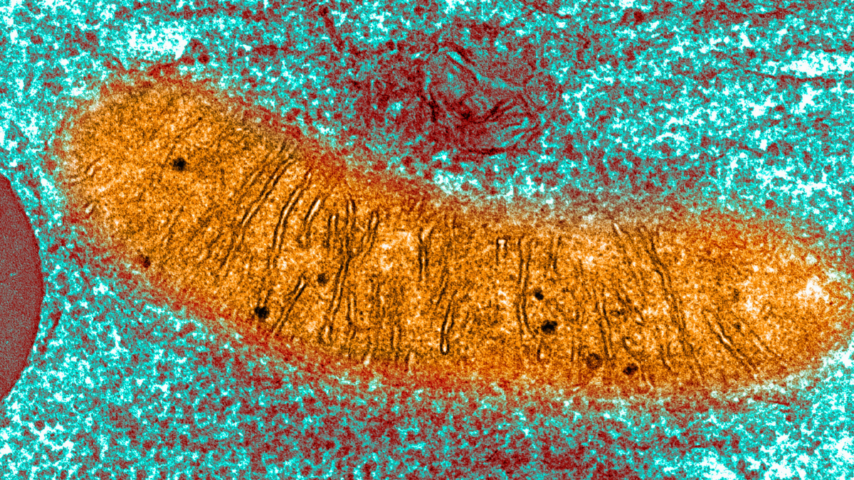
A colored image of mitochondria in a pancreas cell, via transmission electron microscopy.(Callista Images/Connect Images/Getty Images)
Alzheimer’s disease is the most common form of dementia, which affects approximately 57 million people globally – a number that’s projected to rise to more than 150 million by 2050
The incurable neurodegenerative disorder is characterized by the accumulation of beta-amyloid peptides and misfolded tau proteins in the brain, leading to the death of neurons and emerging cognitive difficulties
Recently, attention has shifted toward another potential key component in Alzheimer’s pathology: dysfunction of mitochondria – the energy-generating organelles known as ‘powerhouses of the cells’
In a new study published in NatureNeuroscience, a team of biochemists, geneticists, and gerontologists has pinpointed an entirely new pathological basis for the disease: an accumulation of mitochondrial plaques (MPs)
“This discovery identifies mitochondrial plaques as a previously unrecognized feature of Alzheimer’s disease,” says biochemist Paul Robbins from the University of Minnesota
These MPs result from dysfunctional mitophagy, or the brain’s inability to recycle its failing mitochondria
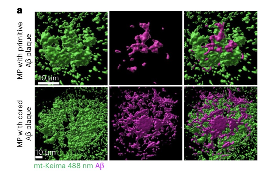
Detected in the brains of genetically engineered mice and postmortem human brains with Alzheimer’s, MPs are aggregations of neutral and acidic mitochondria at different stages of degradation, sometimes still trapped in the acid-imbued ‘cellular trash bags’ that transport or degrade them
“Early ultrastructural investigations showed a prominent presence of degenerating mitochondria in the brain of patients with Alzheimer’s disease, and our recent research indicates impairment of mitophagy in Alzheimer’s models, a process that clears damaged mitochondria,” the researchers say
Previous, indirect measurements in mice engineered with mutations to induce symptoms of Alzheimer’s disease offered only “limited insight” due to inadequate sensitivity
So the researchers directly measured mitophagy in the brains of transgenic mice with a “mitophagy reporter,” called Keima, incorporated into their genomes
Keima is a pH-sensitive fluorescent protein derived from coral, meaning it changes color based on the acidity of its environment. It glows green in the presence of neutral mitochondria and orange in the presence of acidic ones

These two aggregations become more spatially mixed as Alzheimer’s progresses because the MPs harbor abundant amounts of the amyloid precursor protein (APP), from which amyloid-beta peptides originate
In the mouse model, MP aggregation emerged as early as 15 weeks, equivalent to early human adulthood. MP concentrations progressed with age, resulting in abundant acidic mitochondria accumulation by 50–60 weeks, suggesting a significant decline in the brain’s recycling capabilities
And here’s one important bit: the researchers did not find these MPs in the brains of age-matched healthy controls, whether mouse or human
Furthermore, to determine where the MPs were likelier to manifest, the researchers stained the transgenic mice’s brains with a neuronal nuclear protein
“Unlike the amyloid plaques found outside brain cells, these plaques appear to directly affect neurons, which makes them a potential new target for Alzheimer’s disease treatments,” says University of Minnesota cell biologist Xiuli Dan, first author of the study
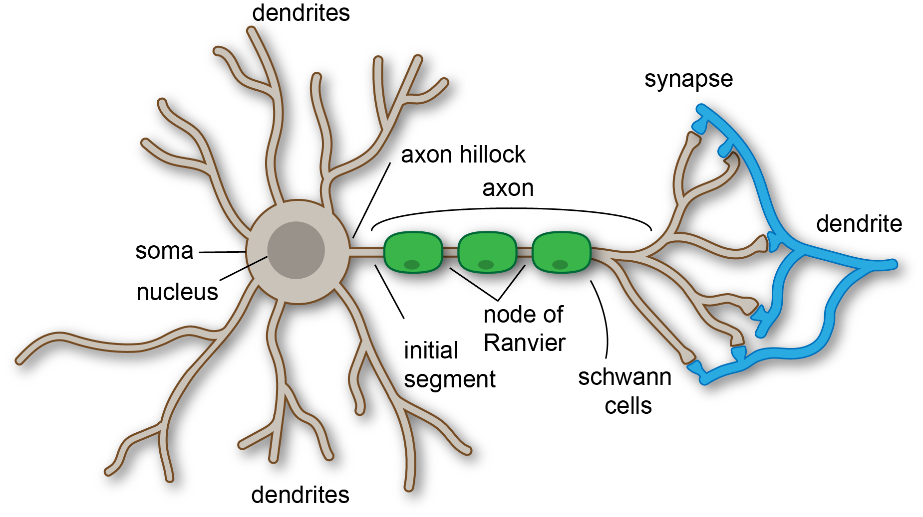
Dan and colleagues found that MPs in neurons were less likely to be associated with the cell bodies. Instead, they accumulated within, and perhaps originated from, neuronal neurites, or the axons and dendrites that extend from the body of neurons
These essential extensions are responsible for sending and receiving the chemical and electrical signals that control basically all brain functions
Such MP accumulation appears to recruit lysosomes, the cellular garbage disposal organelles, which degrade junky old cells using digestive enzymes
However, this vital degradation process cannot keep up with MP accumulation and seems to lead to lysosome dysfunction, or an inability to break down deteriorated mitochondria, resulting in runaway mitochondrial accumulation in the brain
Altogether, this work suggests that Alzheimer’s treatments may need to target numerous
As the researchers note, patients with Alzheimer’s who were immunized to amyloid-beta “revealed successful amyloid plaque clearance yet insufficient protection against ongoing neurodegeneration” – perhaps because MPs affect the parts of neurons that transmit signals
“By understanding how these plaques form and contribute to disease progression, we may be able to develop new strategies to slow or even prevent Alzheimer’s disease,” says Robbins
In addition to a potential new diagnostic feature in MPs, this research hints that future therapies should target mitochondrial accumulation, possibly by finding ways to boost the function of the vacuoles and lysosomes responsible for beneficial degradation
Maybe this could be achieved by inducing an increase in mitophagy processes before they become dysfunctional
Related: Scientists Found a New Alzheimer’s Trigger, And a Drug to Slow It in Mice
In earlier work, inducing mitophagy in ‘Alzheimer’s’ mice has been shown to yield cognitive benefits, supporting its role in healthy brain aging and sidestepping neurodegenerative pathologies
As the researchers conclude, “combination treatments targeting both amyloid-beta plaques and mitochondrial plaques to address the mixed plaques hold great promise to provide better therapeutic outcomes in Alzheimer’s disease.”
This research was published in NatureNeuroscience
This article was fact-checked by Rebecca Dyer and edited by Rebecca Dyer. While we pride ourselves on our process, we are only human. If you spot a mistake, please let us know
