


By Chuck Dinerstein, MD, MBA — Aug 06, 2026
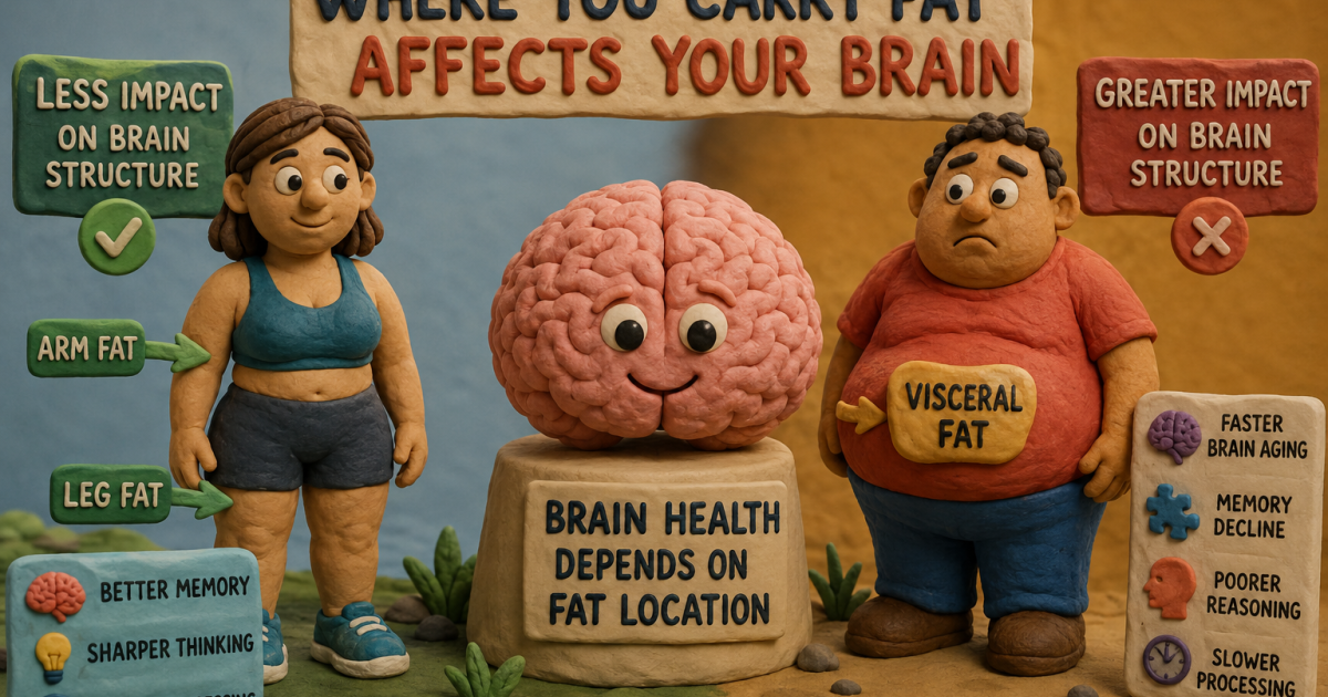
For decades, obesity has been measured largely by how much fat we carry. New research suggests that where fat accumulates may be even more important, showing that regional fat distribution affects brain aging, memory, and cognitive performance in distinct ways.
Image: ACSH
Obesity has beenassociatedwith numerous metabolic and musculoskeletal diseases. It is also linked to structural and functional changes in the brain, including reduced gray matter and disrupted overall functional connectivity. Treating obesity is now part of efforts to mitigate what appears to be age-related cognitive loss we may all face. In short, obesity is a treatment target for preserving “brain health.”
A new studyin Nature’s Mental Health begins with the increasingly denigrated body mass index, or BMI. BMI provides a measure of total body fat, but studies increasingly find that the location of body fat has varying impacts. Fat accumulating along our limbs seems to impair mitochondrial function, reducing energy metabolism and increasing oxidative stress; however, other studies suggest that it may reduce systemic inflammation and have “protective effects.” Visceral fat, which wraps around our organs like a bit of Wagyu beef, is considered a chief culprit in metabolic syndromes, including cardiovascular disease, producing a wealth of inflammatory cytokines that disrupt the blood-brain barrier and lead to neuro-specific inflammation and degeneration.
These distinctions in the impact of fat, depending on where it accumulates, prompted the researchers to look beyond BMI to identify disparities in brain health by where fat was found. They quantified adiposity in the arms, legs, trunk, and viscera and used imaging to measure structural changes in the brain. The researchers had episodic cognitive measures from participants, but without longitudinal cognitive data, they built a model to capture changes. The predictor, termed the brain age gap (BAG), calculated biological age based on structural changes and compared it to the participant’s chronological age. A second analysis showed that the calculated BAGs were a good reflection of, but not the measure of, cognitive decline. The study included roughly 24,000 participants.
Different Fat, Different Damage
Fat accumulation led to loss of brain structure, with reduced overall volume and cortical thinning, but, as suspected, the location of fat accumulation mattered
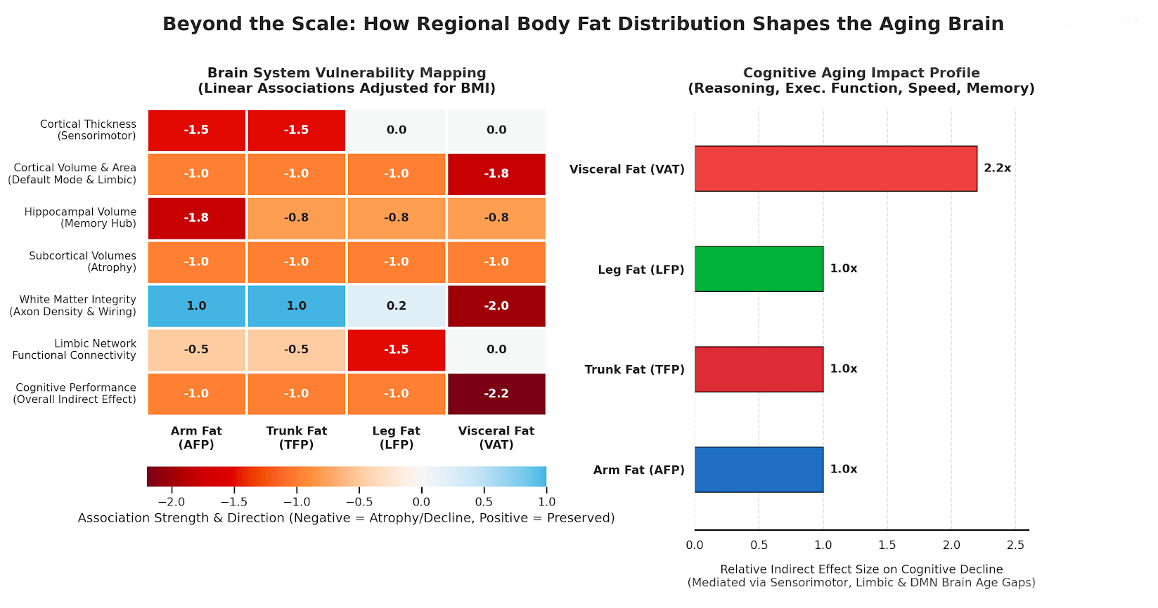
- Arm adiposity was linked to cortical thinning and volume loss across brain structures, including the default mode network (DMN) [1], limbic and sensorimotor networks, and, notably, the hippocampus, a key hub for memory. These changes accelerated brain aging (greater BAG), with measurable declines in memory, reasoning, executive function, and processing speed.
- Trunk fat was associated with similar volume losses in the DMN and limbic systems, as well as in deeper subcortical structures, driving accelerated aging and poorer performance on fluid reasoning, executive function, processing speed, and memory tasks.
- Leg fat caused less structural loss than arm fat, primarily in sparing the sensorimotor areas, but there were similar cognitive declines as seen with the arm.
- Visceral fat was the most destructive of the locales, associated with targeting core DMN hubs and damaging white matter connections, leading to the most accelerated aging and cognitive changes in reasoning, executive function, processing, and memory.
The Brain Pays Attention to Body Geography
The analysis shows that location matters; fat stored in specific body regions targets distinct brain networks independently of overall BMI, driving localized, system-specific brain aging. Arm fat specifically “shrinks” the hippocampus, the memory hub associated with increased Alzheimer’s risk, while leg fat alters functional connectivity within the limbic system, where we experience rewards and emotions. Visceral fat causes the most damage, destroying white matter connectivity through pro-inflammatory signaling. The accelerated aging of these systems directly drives declines in our reasoning, processing, and memory.
This study contributes to the growing understanding that fat is not just a passive energy store but a collection of biologically diverse endocrine tissues capable of altering our physiology in ways we are only beginning to appreciate. Two individuals with identical BMIs may experience markedly different cognitive aging, especially because upper-body fat impairs the hippocampus and visceral fat disrupts white-matter connectivity. The modern rise in obesity, mediated by dysregulation of inflammatory mediators, may be a silent catalyst for the rising rate of late-life cognitive impairment. Cognitive decline may not be a monolithic consequence of our longer aging but a vulnerability shaped by our shape – non-specific weight loss may be insufficient to combat our cognitive decline.
Can You Change Your Brain’s Future?
Perhaps the most empowering takeaway from theNature Mental Healthstudy is that regional fat distribution is a “modifiable health risk factor.” Unlike genetics or chronological age, the “topography” of your body fat can be reshaped. Unfortunately, we lack clearly targeted lifestyle changes to reduce visceral fat. Some studies suggest that aerobic exercise may be more effective at reducing visceral fat than resistance training, but you still need to exercise. A high-quality dietary pattern is still necessary, along with a moderate calorie deficit – so eating right and less remains in the mix. And I cannot end without a word on the elephant in the savannah of obesity care, the GLP-1s. They have been shown to target visceral fat, but their associated muscle loss may lead to other problems. Animal studies have shown improved neuroplasticity and reduced neuroinflammation with the GLP-1s, but oral semaglutide in patients with early Alzheimer’s showed no difference in the rate of cognitive decline compared to a placebo group.
[1] The Default Mode Network is the brain’s interconnected background system that drives inward-focused thoughts, the meditative state we experience when daydreaming, quietly reflecting, or when not focused.
Source: Regional adiposity shapes brain and cognition in adults Nature Mental Health DOI:10.1038s44220-025-00501-8
Tags:
Category
Disease
Chuck Dinerstein, MD, MBA
Director of Medicine
Dr. Charles Dinerstein, M.D., MBA, FACS is Director of Medicine at the American Council on Science and Health. He has over 25 years of experience as a vascular surgeon
Recent articles by this author:
Beyond the scale: where you carry your fat matters
Whole Body MRI has a blind spot
In defense of the Banana Smoothie: Flavanols, food processing and nutritional reductionism
Looking beyond the obvious: three stories to challenge what you think you know
Tick-Tock: The biology behind TikTok’s early dinner trend
All articles by this author
