


Download PDF
Abstract
College students often struggle to regulate emotions under changing academic and social stress, highlighting the need for interventions targeting adaptive emotion regulation (ER). Here, we tested a 5-week online intervention designed to enhance ER and cognitive flexibility in college students. Participants (N = 39) were pseudo-randomly assigned to the experimental (N = 20) or control (N = 19) group, with higher distress individuals preferentially assigned to the experimental condition. Behavioral, self-report, eye-tracking, and brain imaging measures were collected before and after the training. Compared to controls, the experimental group participants showed reduced emotional reactivity to negative stimuli and experiences, along with reduced visual attention to emotionally salient regions. They also showed improvements in the use of attention strategies, positive refocusing, and perceived cognitive control. In the imaging subsample (N = 23; Experimental: N = 15; Control: N = 8), these improvements were accompanied by changes in resting-state functional connectivity, including reduced coupling between affective and higher-order systems and increased efficiency of attentional control and self-referential pathways. These results suggest that training promoted more efficient and flexible interactions among emotion, attention, and cognitive control regions, providing a neural mechanism for the observed behavioral benefits. Overall, the findings support the intervention as a promising approach for improving adaptive ER and resilience during emerging adulthood.
Subjects
- Neuroscience
- Psychology
Introduction
The growing academic, social, and transitional demands of college life have become a persistent source of negative emotions and stress, significantly impacting students’ mental health and psychological well-being1,2. To cope with such challenges, many students rely on strategies that are often ineffective in managing such complex emotional and academic demands3, highlighting the need for intervention programs that help students develop the ability to regulate emotional responses adaptively. Importantly, effective emotion regulation (ER) depends not only on specific strategies but also on the ability to flexibly use them across contexts. This flexibility may be particularly important during young adulthood, including the college years, a period marked by ongoing maturation of prefrontal-limbic circuits that support these ER strategies and their flexible deployment, and by increasing academic, social, and emotional demands4,5. This developmental plasticity may be particularly relevant for resilience — the ability to cope with stressful experiences appropriately or even “better than expected”6— which is increasingly conceptualized as a dynamic process emerging from the interaction between cognitive control and affective systems7. Within this framework, flexibility in cognitive control and ER plays a central role in adaptive responses to stress8. Consistent with these perspectives, we conceptualize resilience as a higher-order capacity to maintain or regain adaptive functioning under challenges, with ER processes serving as proximal mechanisms through which resilience is instantiated, supported, and sustained.
The present intervention targets two core, trainable ER strategies — Focused Attention (FA) and Cognitive Reappraisal (CR) — hypothesized to enhance resilience by improving the flexible modulation of emotional responses across contexts. Building on this integrative framework, the present study examines whether training these ER strategies and their flexible use can improve emotional functioning and the associated neural systems during a developmental period marked by both heightened demands and ongoing neural plasticity. Although prior work, including our own proof-of-principle study in student veterans9,10,11, has shown that the use and effects of these ER strategies can be modified through training, it remains unclear whether such benefits extend to broader college student populations and how they manifest at both behavioral and neural levels, with implications for adaptive functioning and resilience during young adulthood.
Emotion regulation strategies
The ER strategies targeted by the present intervention are seen as complementary mechanisms for strengthening adaptive-related processes. FA is an attentional deployment strategy that involves deliberately directing attention to modify emotional responses (e.g., focusing on the positive parts of a happy memory, or looking away from frightening details of a scary movie)12,13. By targeting early-stage attentional deployment processes, FA is thought to enhance attentional control and perceptual stability, shaping how individuals allocate attention to emotionally salient compared to non-emotional aspects of stimuli [e.g.,14]. Consistent with this, eye-tracking research has shown that ER is reflected in modulations of gaze patterns, with reduced fixation on emotional regions and/or shifts toward neutral/background elements associated with more effective ER15,16,17. However, it remains unclear whether gaze changes directly drive attentional deployment success or instead interact with higher-order regulatory processes involved in reinterpretation and meaning construction18,19. Furthermore, it is also unclear how extended ER training, compared to the brief task-instruction-based ER training commonly used in prior studies, may modulate visual scanning patterns and thereby influence emotional reactivity to possibly confer longer-term protective effects. CR, in contrast, is thought to operate at a later stage of processing, modulating emotional responses through reinterpretation of the meaning of a stimulus or situation (e.g., viewing a difficult experience as an opportunity for growth) and through changes in self-referential evaluation (e.g., shifting from “this says something about my ability or worth” to “this is a specific situation that most people would find challenging”). CR is generally more cognitively demanding than FA12,13, as it engages higher-order appraisal and valuation processes supported by prefrontal systems to reinterpret emotional meaning and regulate responses.
Both attentional deployment/FA and CR have been targeted in previous ER interventions [e.g.,16,20,21] with consistent evidence showing that they down-regulate negative emotional responses. However, prior approaches [reviewed in20,21] have typically trained these strategies in isolation and assessed the effects of training as discrete changes in emotional or cognitive control. For example, attentional deployment/FA training has largely relied on Attention Bias Modification (ABM) paradigms, which showed overall modest effects on clinical outcomes22,23. Similarly, CR training has been associated with reduced stress and negative affect in healthy and clinical populations24,25,26,27,28,29,30, but most of these studies often focused on isolated forms of CR (with reinterpretation10,25 and psychological distancing25being the most typical) and often did not investigate the associated brain mechanisms. Moreover, studies combining multiple ER strategies tend to use simplified training stimuli (such as emotional, scrambled sentences, or non-emotional sounds and numbers [e.g.,31,32]), which may not sufficiently engage the complex perceptual and affective processes involved in everyday emotional experiences. In contrast, our intervention was designed to engage ER processes under ecologically relevant conditions by training FA and CR using emotionally salient stimuli, including affective images and autobiographical memories, across internally and externally driven emotional contexts. Participants also practiced flexibly switching the use of these strategies across temporal perspectives (past, present, and future), engaging attentional, affective, mnemonic, and executive-control processes simultaneously.
Our pilot study in student veterans9, in which participants were trained using the same comprehensive intervention program, showed improved cognitive control and psychological well-being through diminished bottom-up reactivity and normalized connectivity in self-referential and control networks, thus highlighting the intervention’s potential to enhance ER and resilience. Building on this work, the present study extends the intervention to a general college student population. This extension is important, as the two populations face not only common challenges but also experience different types of stressors. Veterans often face chronic, high-intensity stressors involving trauma-related affect and heightened salience processing, whereas typical college students are more commonly exposed to lower-intensity but persistent stressors, such as academic pressure, uncertainty, and sustained cognitive demands. These challenges were further amplified during the COVID-19 pandemic, which introduced additional disruptions, including remote learning, social isolation, and heightened uncertainty about the future33,34. Therefore, adaptive functioning in this academic context relies heavily on attentional control, flexible appraisal, and efficient allocation of cognitive resources. By jointly targeting attentional control and flexible reinterpretation, the present intervention may enable dynamic shifting between attentional stabilization and higher-order reinterpretation35,36, providing a mechanistic basis for enhancing regulatory flexibility — the capacity to adaptively select and switch between ER strategies, based on situational demands.
Emotion regulation and cognitive flexibility
The success of regulation strategies is context-dependent rather than consistently “adaptive” or “maladaptive”37. Thus, outcomes depend not only on the strategy of choice (i.e., FA or CR) but also on a person’s ability to flexibly apply ER strategies based on the contextual demands37,38. This flexibility is increasingly recognized as a core mechanism underlying resilience and adaptive functioning under stress37. For example, flexibility in applying ER strategies has been linked to reduced emotional disturbances and better adjustment38 because it allows individuals to adapt their ER strategies to situational demands, based on actual or perceived controllability of stressors, timing of implementing strategies, or past experiences39,40. Closely related to ER flexibility is cognitive flexibility (CF), the ability to switch perspectives and modify strategies in response to changing circumstances41, emerging from optimal interactions among several cognitive and neural mechanisms to enable flexible adjustment of thoughts and behaviors to changing environmental demands42. The notion of CF reflects both perceived control (i.e., the tendency to believe that difficult situations are controllable) and perceived alternatives (i.e., the capacity to adapt behavior effectively in response to changing environmental demands), which are essential for effective ER, as they both foster a sense of self-efficacy and adaptability in the face of stress. Thus, CF supports flexible ER by allowing individuals to disengage from rigid or maladaptive patterns and to select more successful strategies when confronted with obstacles37,43, to increase distress tolerance and reduce difficulties in emotion ER. This, in turn, leads to increased resilience and quality of life44,45.
Within this framework, our training was designed to enhance CF by teaching participants to flexibly switch between FA and CR strategies across several emotional contexts (i.e., with internal and/or external scenarios, linked to different time perspectives: past, present, and future) using different types of stimuli (i.e., pictures and memories). Together, these strategies target complementary components of flexibility: FA operates at the level of attentional selection, whereas CR operates at the level of meaning construction. By improving both processes, training may enhance students’ capacity to switch adaptively between different perspectives and response options, increasing their sense of control by having a better repertoire of strategies to rely on in different situations. Both components are particularly important in college students, who frequently encounter novel and ambiguous stressors requiring rapid shifts in perspective and adaptive coping. Enhancing these dimensions of cognitive flexibility may therefore reduce reliance on reactive or maladaptive strategies and support more resilient responses across academic and social contexts. Moreover, combining FA and CR within a single intervention may have synergistic benefits by targeting complementary stages of ER, from early attentional selection to later meaning-based reinterpretation46.
Brain mechanisms of emotion regulation and cognitive flexibility
To provide a clear theoretical rationale for the intervention, we situate our approach within neurocognitive models of ER and resilience, which conceptualize adaptive functioning as emerging from dynamic interactions among specific cortical and subcortical brain regions involved in top-down (dorsolateral PFC, dlPFC; ventrolateral PFC, vlPFC; posterior parietal cortex; medial PFC; anterior cingulate cortex, ACC) and bottom-up processing (amygdala, AMY; insula; ventral striatum)47. This system is complemented by CF, which engages frontal (dlPFC; vlPFC; ventromedial PFC, vmPFC; orbitofrontal cortex, OFC), and parietal control networks that enable switching between strategies and mental sets in response to cognitive and emotional demands48. Within this framework, adaptive cognitive and emotional functioning reflects dynamic coordination across large-scale brain networks, not a fixed trait.
As an ER strategy, FA engages frontoparietal attention networks — including the frontal eye fields (FEF) and dlPFC— to support sustained attention and goal maintenance49. In addition, FA also engages the ACC for monitoring attentional conflict and maintaining task focus, along with top-down modulation of sensory cortices (e.g., occipital regions) to bias perceptual processing toward task-relevant inputs50,51. Through these mechanisms, FA biases processing toward task-relevant information and away from emotionally salient stimuli, thereby attenuating emotional responses without altering stimulus meaning through these mechanisms9,52. In contrast, CR relies on distributed prefrontal-limbic networks, including the vmPFC and OFC for subjective valuation and affective meaning, the dlPFC/vlPFC for cognitive control and appraisal manipulation, the ACC for conflict monitoring and regulatory signaling, and the AMY for affective salience and value learning13,53. Together, these systems support the evaluation and reinterpretation of emotional stimuli during regulation.
Bottom-up affective processing, which involves regions such as the AMY, insula, ventral striatum, and posterior sensory cortices, is involved in the automatic responses to salient stimuli47. Top-down processing, mediated by prefrontal regions that include particularly the lateral and medial PFC, is critical for implementing ER strategies (such as FA and CR), by reducing bottom-up AMY activity54, through functional coupling or connectivity between these areas55, and by modulating activity in higher-order executive functions56. Contemporary models of ER move beyond the top-down versus bottom-up dichotomy to emphasize the hierarchical organization of the frontal cortex, in which cognitive control is implemented along a rostro-caudal gradient from abstract, goal-directed representations in anterior prefrontal regions to more concrete, action-oriented processes in posterior regions57,58,59. Within this framework, adaptive ER depends not only on the suppression of bottom-up affective signals but also on the flexible coordination of distributed fronto-limbic networks, including interactions among prefrontal control systems, the AMY, and the ACC. In particular, recent work highlights the AMY–ACC pathway as a critical substrate for efficient emotional appraisal and resilience, supporting rapid evaluation and flexible updating of affective responses under changing demands60. These models further emphasize the role of CF as a critical moderator within this system, enabling the adaptive allocation of cognitive resources across perceptual, attentional, and regulatory domains depending on contextual demands8.
The current approach
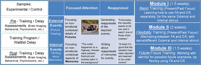
The present study builds directly on these hierarchical and network-based accounts of ER and resilience by extending prior evidence from our group demonstrating that our FA and CR training in student veterans decreased resting-state functional connectivity (rsFC) between emotion and perceptual processing regions, including the AMY and the left occipital pole, consistent with reduced negative emotional bias9. The same training also increased rsFC among cognitive control regions, including the left dlPFC and left dorsal ACC, which support monitoring, attentional control, and cognitive flexibility9. Together, these findings provided proof-of-concept evidence that our training can meaningfully reshape intrinsic functional brain networks and improve ER and well-being. The current study extends this work by testing whether the same training combining FA and CR can modulate hierarchically organized frontal systems and their interactions with limbic and control networks in a broader college student population. Going beyond focusing just on top-down and bottom-up processes, the present study further examines training-related changes in network-level connectivity across multiple neural systems. The training was structured for scalable delivery to college students (either in-person or online), with the goal of improving both accessibility and adaptive functioning. In line with this translational goal, the current intervention was implemented as a structured multi-week training program designed to systematically engage and strengthen these regulatory processes across contexts. Over five weeks, participants practiced FA and CR strategies across external (i.e., emotional pictures) and internal (i.e., about memories) events, past, present, and future scenarios, and stimuli of increasing complexity (Fig. 1). They were also encouraged to use these strategies in daily life. The effectiveness of the training was assessed using behavioral tasks, self-reports, and neuropsychological assessments, along with measures of eye-tracking and rsFC, before and after the training. Collection of eye-tracking data allowed us to examine whether the intervention modulates visual attention patterns linked to subjective ratings of emotional experiences, in the absence of explicit instructions to engage FA during a free viewing task, following training.
Based on our pilot study and prior work implicating limbic, attentional control, and self-referential networks in ER9,53,61, we tested the following sets of hypotheses regarding behavioral, eye-tracking, and rsFC changes following training. To limit the number of analyses relative to sample size, seed regions were selected based on our pilot intervention targeting FA and CR as core regulatory mechanisms in student veterans9, as well as on prior ER literature implicating the involvement of these regions in the specific regulatory processes targeted by our intervention. These regions include AMY-mediated emotional reactivity13,59, frontoparietal attentional and cognitive control49,61, and medial prefrontal/self-referential valuation processes62.
H1: Reduced emotional experience and optimized affective processing
We hypothesized that participants in the experimental group would show reduced emotional reactivity following training, reflected behaviorally in lower ratings of negative emotional images, reduced self-reported negative affect, and increased positive affect. At the neural level, we predicted reduced bottom-up propagation of emotional signals within emotion-processing networks, reflected in decreased connectivity between bilateral AMY seeds and cortical regions implicated in lower-level perception and higher-level cognition. These predictions are grounded in evidence provided by previous ER studies13,59 and by our pilot study in student veterans9.
H2: Enhanced attentional and cognitive control
We further hypothesized that training would strengthen attentional deployment and cognitive control processes. Linked to reduced ratings of the negative images, we explored the possibility that training would modulate visual attention during free viewing of emotional stimuli, such that participants would show reduced dwell time and fixation on emotionally negative aspects of stimuli and increased attention to contextual aspects following training, even in the absence of explicit instructions to do so. We also expected increases in use of FA and CR strategies in everyday life, positive refocusing (a FA strategy involving redirecting attention away from emotional stimuli towards positive content), positive reappraisal (involving reinterpreting situations into a more positive light), and CF (reflected in increased scores on the Perceived Control and Perceived Alternatives subscales of the Cognitive Flexibility Inventory). Neurally, we predicted changes in the rsFC of attentional and executive-control seeds. Specifically, we expected increased connectivity between the FEF and visual processing regions, reflecting enhanced top-down modulation of sensory processing49, along with reduced connectivity within frontoparietal control networks, reflecting more efficient and selective engagement of cognitive control systems9,61.
H3: Reduced influence of valuation and self-referential systems on cognition
Finally, we hypothesized that training would reduce the influence of valuation and self-referential processing systems on cognition and promote more adaptive interpretation of emotional experiences, as well as increases in regulatory flexibility in how these systems are engaged across contexts. Exploratory analyses using mPFC and OFC seeds were expected to identify decreased connectivity between these regions and temporal, parietal, and posterior midline regions associated with self-referential and associative processing, reflecting reduced integration of valuation-related signals with broader cognitive networks. These hypotheses were intended to examine whether training promoted a shift away from internally focused evaluative processing toward more adaptive and flexible ER following training, consistent with prior models of ER and cognitive-emotional training9,13,56.
Methods
Participants and procedures
Fifty-two undergraduate students (ages 18–22; M = 20.13, SD = 1.14; see Table 1 and S1 for full demographic details) from the University of Illinois at Urbana-Champaign were recruited using paper and digital flyers. One participant was excluded due to an incidental finding. The attrition rate was 7.69%, with four participants dropping out before the training. This study was approved by the Institutional Review Board of the University of Illinois at Urbana-Champaign, and all research was performed in accordance with relevant guidelines and regulations applicable when human participants are involved (e.g., Declaration of Helsinki or similar). In accordance with such relevant ethical guidelines, all participants provided informed consent, were debriefed, provided with additional mental health resources, and financially compensated for their time. Participants were screened for factors that might interfere with mood and/or cognition during a phone interview. Participants were asked a series of questions regarding their demographics, academic background, mental health history, substance use, medications, and MRI safety (e.g., surgical history, any medical conditions that would interfere with lying still for long periods, and irremovable piercings). Participants were excluded from the study if they were currently engaged with mental health treatment (i.e., therapy and/or mental health medications, to avoid confounds), using substances regularly, or reported any factors that would risk MRI safety.
Three distress measures were also used to determine study eligibility and group assignment (experimental or control): the Mood and Anxiety Questionnaire – Anhedonic Depression (MASQ-AD, cutoff score = 21)63, the Penn State Worry Questionnaire (PSWQ; moderate cutoff score = 20, high cutoff score = 30)64, and the Posttraumatic Stress Disorder Checklist (PCL-5; cutoff score = 33)65. The study was conducted as a prospective, quasi-experimental training study. Participants were assigned to either the training or waitlist control group using a pseudo-randomized procedure. Participants reporting elevated distress – scoring above the at-risk cutoff scores on at least one of the distress measures – were prioritized for immediate access to training to provide timely support. Given this allocation procedure, we conducted analyses both in a restricted data subset, whose assignment followed the standard pseudo-randomization procedure, and in the full data sample. Reporting both analytic approaches allowed us to evaluate the robustness of findings across allocation conditions. Throughout the manuscript, we use the term “training” to refer to the structured cognitive–emotional program; we avoid clinical terminology, as the study was not designed as a clinical trial, and group allocation was not fully randomized.
The main text reports the subset of the behavioral data (N = 39; Experimental group: N = 20, Control group: N = 19) that does not include those participants (N = 8) marked as high distress and pseudo-randomized into the experimental group. In addition, because the check-in procedure for self-report measures of everyday emotion regulation usage was implemented at a later stage in data collection, responses were unavailable for six participants, yielding an available sample of N = 22 for the corresponding analyses. Analyses for the full dataset (N = 47) are included as supplemental material. Demographic data for the experimental and control groups is shown in Table 1 (and supplemental materials; Table S1).
All participants completed a series of pre- and post-assessments, which included self-report and neuropsychological measures. A subset of participants also completed a behavioral task that involved the collection of eye-tracking (N = 31) and brain imaging data (N = 30). Due to emergency stay-at-home procedures during the COVID-19 pandemic, seven participants were unable to fully complete the eye-tracking or neuroimaging procedures, which needed to be completed in-person. Those who completed both pre- and post- task (N = 24) and neuroimaging (N = 23) sessions were included in the respective analyses. The experimental group participants completed the training over a period of five weeks (two sessions per week, ten sessions total). Before the COVID-19 stay-at-home restrictions, participants had one-on-one meetings with the research team, and during the restrictions, participants completed the training independently by receiving interactive PDF materials over email. The control group participants were waitlisted for training, but they completed the pre- and post-assessment procedures on a timeline similar to that of the experimental group. After the wait period, participants in the control group were offered the cognitive-emotional training before completing their participation in the study.
The cognitive-emotional training
The cognitive-emotional training taught participants how to apply two key emotion regulation strategies (i.e., FA and CR) and associated tactics to both external (emotional pictures) and internal (memories and future worries) stimuli and events. Participants learned a total of 12 tactics
Progressive flexible training
The training promoted the flexible use of FA and CR to external (images) and internal (memories and future worries) stimuli across three progressing modules (Fig. 1). FA emphasized attention to contextual details (people, locations, objects), whereas CR involved reinterpreting aspects of the stimuli (meaning, relevance, consequences). Participants practiced and flexibly applied the learned ER strategies in daily life and reported their usage
Illustration of the training approach. Big-picture details (left-side panel) and specific details regarding the implementation of the training (right-side panel)
External and internal stimuli and multiple time perspectives
FA and CR were applied both to external and internal stimuli, using a picture processing task and a writing task, respectively. In the picture processing task, participants were presented with images composed of an emotional foreground and neutral context in the background and were trained to (1) focus their attention to the context rather than the emotional content (FA), and (2) to reframe the situation depicted in pictures in a positive way (CR). In the writing task, participants wrote about negative personal memories and future worries while applying FA and CR. Namely, participants were trained (1) to write about the contextual details of their identified distressing memories or future worries (FA), such as the time and the place of events, and (2) to focus on reframing the situation in a less negative/more positive way (CR), such as finding positive meanings of personal growth in a setback, by seeing the “silver lining.” For the memory writing task, personal memories were first collected using established procedures in our group66,67,68. Participants were presented with a wide range of life events from which they selected and briefly described the 12 most negative and 12 most positive events that reminded them of specific, unique, and personal situations. For each writing session, participants chose the negative event they wanted to write about.
As illustrated in Fig. 1, the basic and flexibility training linked to present (pictures) and past (memories) events (Modules I and II) also promoted the development of problem-solving skills in preparation for anticipated future challenges (Module III). In this module, the focus shifted to future worrisome events, and participants applied the learned ER skills by mentally simulating possible scenarios of future events (visually cued with a picture processing task) and dealing with personal worries linked to events expected to happen in real life (elaborated upon in a writing task). For this, specific future concerns considered to be in the participants’ control were collected before the final future-focus module of the training (Fig. 1). Participants flexibly applied the learned ER skills in order to find coping solutions to possible future distressing situations, by using a means-ends problem solving procedure69.
Behavioral measures
Participants first completed screening measures to determine eligibility and group assignment. Inferential measures were also employed before and after the program (pre- and post-training) to evaluate the effectiveness of our intervention. We also examined the relationship between results from all questionnaire measures by calculating correlations (Tables S4 and S5). For the measures that included multiple subscales (e.g., Mood and Anxiety Symptom Questionnaire – MASQ; Cognitive Emotion Regulation Questionnaire – CERQ), only the subscales that were theoretically aligned with the aims of the study were included in the analyses.
Screening measures
Self-report measures were used to determine study eligibility and group assignment (experimental or control), such that participants reporting elevated distress (i.e., scoring above the at-risk cutoff scores on at least one distress measure) were prioritized for immediate access to training. These measures are not included in hypothesis testing
Anhedonic Depression was assessed using the 8-item version of the Mood and Anxiety Symptom Questionnaire- Anhedonic Depression Scale (MASQ-AD)63, which assesses anhedonia (i.e., reduced ability to experience pleasure) across positive and negative experiences (e.g., “Felt really bored;” “Felt unattractive”). Participants rated on a 5-point scale (1 = “Not at all;” 5 = “Extremely”) how frequently they experienced various positive and negative experiences during the past week. Items were summed to calculate the final MASQ-AD score, with higher scores indicating increased symptoms of anhedonic depression. Internal consistency at baseline was acceptable (α = 0.774, full sample; α = 0.704, subset without high-distress participants). The anhedonia subscale of the Mood and Anxiety Symptom Questionnaire was used to identify diminished positive affect and reward sensitivity, which are closely linked to adaptive functioning and particularly relevant for our intervention. In contrast, broader depressive symptom subscales were not included in the screening, to ensure that participants were not excluded due to symptoms that are not directly tied to the mechanisms targeted by the intervention (e.g., low mood, fatigue, sleep disturbances).
Worry was measured using the 8-item version of the Penn State Worry Questionnaire (PSWQ)64, which assesses how often people worry (e.g., “I do not tend to worry about things”) using a 5-point scale (1 = “Not at all typical of me;” 5 = “Very typical of me”). Items were summed to calculate the final PSWQ score, with higher scores indicating greater worry. Internal consistency at baseline was good (α = 0.883, full sample; α = 0.872, subset without high-distress participants)
Posttraumatic Stress Disorder (PTSD) Symptoms were assessed using the 20-item Posttraumatic Stress Disorder Checklist (PCL-5)65, which includes challenges associated with a stressful experience (e.g., “Repeated,disturbing dreams of the stressful experience?”). Participants rated on a 5-point scale (0 = “Not at all”; 4 = “Extremely”) how much they were bothered by those challenges. Items were summed to calculate an overall score, with higher scores indicating increased PTSD symptom severity. Internal consistency at baseline was excellent (α = 0.930, full sample; α = 0.912, subset without high-distress participants).
Inferential measures
Inferential analyses focused on a set of measures that were theoretically aligned with the study’s hypotheses and designed to index changes in emotion regulation, affective experience, and flexibility-related processes
Trait Affect was assessed using the Positive and Negative Affective Schedule (PANAS)70, which included descriptors for 10 positive (e.g., “pleased,” “cheerful”) and 10 negative (e.g., “guilty,” “distressed”) affective traits. Three additional positive affect words (“pleased,” “cheerful,” “happy”) and five additional negative affect words (“frustrated,” “down,” “anxious,” “grouchy,” “sad”)71 were also included. Participants rated on a 5-point scale (1 = “Very slightly or not at all;” 5 = “Extremely”) the degree to which they experienced those affective states over a longer period of time. Subscale scores were summed, with higher scores indicating greater negative or positive trait affect. Internal consistency at baseline was good (α = 0.897, full sample; α = 0.871, subset without high-distress participants).
Positive Refocusing and Positive Reappraisal were assessed using the 18-item short-form of the Cognitive Emotion Regulation Questionnaire (CERQ)72, which includes nine strategies (positive refocusing, refocus on planning, positive reappraisal, acceptance, putting into perspective, self-blame, focus on thought/rumination, catastrophizing, and blaming others) that people use in response to threatening or stressful life experiences. Positive refocusing measures the tendency to redirect attention away from negative events and focus on positive or enjoyable experiences (i.e., “I think of pleasant things that have nothing to do with it” and “I think of something nice instead of what has happened”). Positive reappraisal measures the tendency to reinterpret a negative or difficult situation in a way that highlights positive aspects (i.e., “I think I can learn something from the situation” and “I think that I can become a stronger person as a result of what has happened”). Items are rated on a 5-point Likert scale (1 = “I never think like this” to 5 = “I always think like this”) and summed to create a total score, with higher scores reflecting more frequent use of positive refocusing, respectively positive reappraisal. Internal consistency at baseline was acceptable (α = 0.731, full sample; α = 0.725, subset without high-distress participants).
Cognitive Flexibility was measured using the 20-item Cognitive Flexibility Inventory (CFI)73, which provides a total score and two subscale scores: Perceived Control (CFI-Control: i.e., “I am capable of overcoming the difficulties in life that I face”) and Generating Alternatives (CFI-Alternatives: i.e., “I consider multiple options before making a decision). Items were rated on a 7-point scale (1 = “Strongly disagree;” 7 = “Strongly agree”) and summed to calculate the total scores for the two subscales, with higher scores reflecting greater cognitive flexibility. Internal consistency was good at baseline: Control: α = 0.859 (full sample) and α = 0.834 (subset); Alternatives: α = 0.875 and α = 0.861, respectively.
ER use in everyday life. Additional data were collected to assess the use of FA and CR in everyday life in the experimental group only, given that they were actively using the strategies throughout the training. After each training session, experimental group participants reported whether they used FA/CR outside the lab, and provided details about the circumstances (i.e., internal events, external events, or both), frequency (0 = “Never,” 5 = “Multiple times a day”), and effectiveness (1 = “Not at all,” 5 = “Very”). Debriefing similarly assessed frequency and effectiveness, along with qualitative feedback on the circumstances where they found the strategies most helpful and their overall training experience.
Image rating and eye-tracking task
Participants freely viewed and rated a total of 45 composite images in the pre-training task (30 negative and 15 neutral), and 75 composite images in the post-training task (45 negative and 30 neutral), presented using the E-Prime software. Both pre- and post-training tasks were organized in blocks (pre-task = 3 blocks of 15 images each; post-task = 5 blocks of 15 pictures each) matched for human and animal presence, valence, and arousal ratings. Each test was preceded by five image practice trials. Participants were cued (0.5 s) to “look” at each image, displayed for four seconds. After each image presentation, a fixation cross was displayed (2 s), and then participants were prompted to rate their subjective emotional experience triggered by the image (two-second display, five-point scale, 1 = “Not at all negative;” 5 = “Very negative”) using a computer keyboard. Given that a number of these images overlapped with stimuli used in the cognitive-emotional training picture tasks, 30 novel images (15 negative and 15 neutral) were added and balanced across blocks in the post-test to control for potential familiarity effects. Each composite image had a distinguishable foreground and background, developed by overlaying a negative or neutral foreground image upon a visually complex background image. Image components were taken from standardized picture systems, including the International Affective Picture System (IAPS)74 and the Military Affective Picture System (MAPS)75. A validation study confirmed that the negative stimuli used in the pre-training task were negatively valenced (Mvalence = 2.7; SDvalence = 0.64) and arousing (Marousal = 5.97; SDarousal = 0.91), whereas the neutral stimuli were neutral (Mvalence = 4.96; SDvalence = 0.50) and non-arousing (Marousal = 2.16; SDarousal = 0.32). For the additional stimuli included in the post-training task, a validation study confirmed that negative stimuli were negatively valenced (Mvalence = 2.64; SDvalence = 0.67) and arousing (Marousal = 6.07; SDarousal = 0.99), whereas the neutral stimuli were neutral (Mvalence = 4.79; SDvalence = 0.49) and non-arousing (Marousal = 2.17; SDarousal = 0.47).
Eye-tracking data were recorded from a subset of participants while they completed the pre- and post-training tasks (Total: N = 24; Experimental: N = 18; Control: N = 6). Eye positions and movements were recorded from each participant’s left eye using the EyeLink 1000 system at a sampling rate of 1,000 Hz (SR Research, ON, Canada). A pseudorandom 9-point eye-tracking calibration was performed during setup and between every other experimental block
Autobiographical and future events tasks
The Autobiographical Memory Questionnaire (AMQ) was used to acquire and assess emotional memories using established procedures that have been effectively employed in previous research66,67,68. Before the training, participants were prompted with 113 cues of a wide range of life events (e.g., surprise party,looking for an apartment,watching the elections,missing a flight), selected the 12 most negative and 12 most positive events that cued specific, unique, and personal episodes from their life, and provided brief descriptions to serve as reminders for each event (e.g., “Enjoying my first day of school: My grandma picked me up and gave me so many sweets,” or “Crashing my bike on the way to school last spring: I had surgery because my finger was broken”). Participants also rated the phenomenological characteristics for each of the selected events using 7-point Likert scales, including: contextual details (1 = “Not at all;” 7 = “Very Much”), emotional valence (1 = “Very Negative;” 7 = “Very Positive”), emotional intensity (1 = “Not at all;” 7 = “Very Much”), vividness (1 = “Not at all;” 7 = “Very Much”), and personal significance (1 = “Not at all;” 7 = “Very Much”). During the training, experimental group participants chose from their 12 selected negative events for the internal ER writing tasks. At the post-assessment, the same 12 selected events were re-rated. Similarly, worries for expected future events were collected before the training, and participants applied the FA and CR strategies to their own worries during the last phase of the training.
Brain imaging data acquisition
A subset of participants fully completed (i.e., pre- and post-sessions) the neuroimaging procedures (N = 23; Experimental: N = 15, Control: N = 8). MRI scanning was conducted on a 3 T Siemens PRISMA scanner. Sagittal localizer and 3D MPRAGE anatomical images were first obtained (FOV = 230 × 230 mm; TR = 2000 ms; TE = 2.25 ms; volume size = 176 slices; voxel size = 1.0 × 1.0 × 1.0 mm). Resting state functional images consisted of a series of axial images collected using an echoplanar sequence (FOV = 230 × 230 mm; TR = 1500 ms; TE = 30 ms; volume size = 76 slices; voxel size = 1.6 × 1.6 × 1.6 mm; flip angle = 40°; number of volumes = 386), while participants let their mind wander with their eyes open, viewing a fixation cross on the screen. Resting-state functional connectivity (rsFC) was used to index neural changes associated with ER and related adaptive processes, because it captures the intrinsic organization of large-scale brain networks supporting attentional control, valuation, and salience processing, including prefrontal–limbic and default mode–control interactions. Although ER is often examined under conditions of active cognitive or affective challenge, converging evidence indicates that individual differences in rsFC within these networks are associated with regulatory capacity and affective traits, even in the absence of explicit task demands56,76,77,78. Moreover, rsFC is particularly well-suited for intervention studies, as it provides a reliable index of training-related neuroplasticity that is not confounded by task performance, strategy use, or differences in task engagement across sessions14,79,80. This is especially relevant in longitudinal designs, where repeated exposure to ER tasks can introduce practice effects and alter cognitive load. Importantly, rsFC allows us to assess whether the training induces enduring changes in the brain’s intrinsic functional architecture that may generalize across contexts, rather than task-specific adaptations [e.g.,81,82]. Therefore, changes in rsFC may reflect an increased readiness or efficiency of ER-related networks to be engaged under challenge, even if not directly measured during a task.
Data analyses
Behavioral data analyses
The primary goal was to assess potential behavioral changes as a result of the training. This was done by comparing participants’ pre- vs. post-training scores on questionnaires and image rating tasks. To compare pre vs. post differences (for self-reports and ratings) between the two groups, two-way mixed-ANOVAs were performed, with Time (Pre vs. Post) as the within-subjects factor and Group (Experimental vs. Control) as the between-subjects factor; training effects were further examined with pairwise post-hoc comparisons. To examine training effects on emotion ratings, three-way mixed ANOVAs were conducted, with Image Type (Emotionally Negative vs. Neutral) as a within-subjects factor, Condition (Experimental vs. Control) as a between-subjects factor, and Time (Pre vs. Post) as a within-subjects factor. Preliminary analyses were conducted to identify covariates, using ANOVAs for nominal demographic variables and correlations for continuous variables. We expected that certain demographic variables, including cohort, academic year, sex, ethnicity, and age, could influence baseline self-report measures and, therefore, intervention effects. Cohort and age were identified as significant covariates. Specifically, cohort effects were observed such that one of the later cohorts demonstrated higher levels of baseline ER compared to earlier cohorts (see Supplemental Materials). Additionally, age showed significant associations with baseline measures of distress and executive functioning (see Supplemental Materials). Although these effects were not the primary focus of the study, they are not entirely unexpected given developmental and contextual differences (e.g., pandemic-related challenges) that may influence emotion reactivity and ER capacity. Importantly, initial analyses demonstrated significant intervention effects on CF (perceived control subscale, CFI; see Supplemental Materials). However, when cohort and age were included as covariates, these interaction effects were no longer statistically significant, providing a more conservative estimate of intervention effects. Cohort (i.e., Fall 2019 through Spring 2023, with Fall 2019 as reference) was dummy-coded, and age was included as a continuous covariate. Both were included in subsequent mixed ANOVA, with adjusted degrees of freedom. Group-specific effects were examined using paired t-tests within each training group, particularly to assess changes in FA and CR use, which were measured only in the experimental group. Behavioral data were analyzed using the SPSS software (IBM, Version 29). In addition, qualitative data collected through the debriefing survey were used to support interpretations and inform future renditions of the study.
Eye-tracking data analysis
Eye-tracking data were processed using EyeLink Data Viewer (SR Research). Interest-area templates defined foreground and background regions of each image. Visual attention was quantified using dwell-time percentage and fixation percentage within these regions. Emotional ratings collected during the task were averaged across trials. Effects of Image Type (Negative vs. Neutral), Condition (Experimental vs. Control), and Time (Pre vs. Post) were examined using mixed ANOVAs
Brain imaging data analyses
Preprocessing
Functional images from both time points were preprocessed in SPM1283, including: slice timing correction, realignment, segmentation, and coregistration. Data were normalized to the Montreal Neurological Institute (MNI) space and spatially smoothed to enhance the signal-to-noise ratio. Structural and functional data were imported into the CONN toolbox84 for denoising (i.e., removing noise and artifacts), and motion covariates were added to account for head movement during scanning sessions. Denoised data were visually inspected to ensure normal distribution and consistency of functional connectivity values across subjects.
Resting state functional connectivity (rsFC)
Seed-based rsFC analyses were conducted to examine intervention effects on connectivity with regions of interest (ROIs). First, consistent with our pilot study9, the present ROIs included regions involved in emotion processing (i.e., AMY) and cognitive control (i.e., dlFC and mPFC). Second, these ROIs were extended to include additional PFC ROIs associated with higher-order emotion processing and attentional control – i.e., OFC85 and FEF86, respectively. The CONN network atlas (derived from ICA analyses of 497 subjects in a dataset from the Human Connectome Project) and anatomical atlas were used to define the seed masks. For all regions, seeds were identified separately for left and right hemisphere locations, with the exception of the mPFC, which was defined as a single (medial) combined seed including both sides. The corresponding CONN labels are as follows: AMY (atlas.Amygdala R/L); dlPFC (networks.FrontoParietal.PFC L/R); mPFC (networks.DefaultMode.MPFC); OFC (atlas.FOrb L/R); FEF (networks.DorsalAttention.FEF L/R). While significant clusters identified from those seeds were initially labeled in CONN using gyral conventions (e.g., inferior frontal gyrus, IFG; middle frontal gyrus, MFG), visual inspection guided the use of broader cortical labels (e.g., inferior frontal cortex, IFC; middle frontal cortex, MFC) when appropriate. Statistical significance was set at a voxel-level threshold of p < 0.005 (uncorrected) and a cluster-size threshold of p < 0.05 (FDR-corrected). To also account for multiple comparisons in accordance with the number of seed ROIs, the seeds that survived the cluster-level thresholds were additionally subjected to Bonferroni corrections. This step was applied separately to the two distinct categories of ROIs: (1) confirmatory seeds replicating the pilot study (i.e., L/R AMY, L/R dlPFC, and mPFC), and (2) additional seeds selected for their theoretical relevance for higher-level emotion processing and attentional control (i.e., R/L OFC and R/L FEF). Second-level analyses focused on investigating Group (Experimental vs. Control) x Time (Pre vs. Post) interaction effects: [Experimental (Post vs. Pre) vs. Control (Post vs. Pre)]. To confirm that the observed rsFC changes were specific to the training, within-subject main effects contrasts were also tested separately for each group. Interaction effects indicated differences between groups over time, while follow-up main effect analyses further verified whether significant changes in the expected direction occurred within the experimental group alone. Importantly, reporting both interaction and main effects strengthens the interpretation that the observed rsFC changes resulted from the cognitive-emotional training (see Table 3).
Transparency and openness
The study was not preregistered, which we acknowledge as a limitation. All hypotheses were informed by extant literature and guided by patterns identified in an earlier pilot study9. The data can be made available to other scientists upon request, following publication
Results
Emotion regulation: reduced emotional experience and optimized affective processing
Behavioral results
Reduced ratings of negative emotional images in the experimental group
Consistent with H1, the ratings of negative images in the image rating task decreased as a result of training in the experimental group. The three-way interaction [Group (Experimental vs. Control) X Image Type (Emotionally Negative vs. Neutral) X Time (Pre vs. Post)] was nonsignificant, F(1, 22) = 1.359, p = 0.256, ηp2 = 0.058), but pairwise comparisons showed that the experimental group rated negative images less negatively from pre- to post-test (Memo, pre = 3.306, Memo, post = 3.010, p = 0.022, ηp2 = 0.217), with no changes for neutral images (Mneu, pre = 1.122, Mneu, post = 1.129, p = 0.847, ηp2 = 0.002). In the control group, emotional ratings for both the negative (Memo, pre = 3.182, Memo, post = 3.143, p = 0.853, ηp2 = 0.002) and neutral images (Mneu, pre = 1.082, Mneu, post = 1.098, p = 0.779, ηp2 = 0.004) remained unchanged. Similar findings were also found for the full behavioral dataset (n = 47) (Supplementary Materials Table S2).
Reductions in self-reported negative affect
Consistent with H1, results showed decreased negative affect following training. A two-way mixed ANOVA [Time (Pre vs. Post) X Group (Experimental vs. Control)] on PANAS-Negative revealed a significant interaction: F(1, 29) = 9.953, p = 0.004, ηp2 = 0.256 (Fig. 2; Table 2). Post-hoc comparisons indicated significant pre- to post-training reductions only in the experimental group (Experimental group: Mpre = 30.20, Mpost = 24.05, p < 0.001, ηp2 = 0.361; Control group: Mpre = 28.42, Mpost = 28.95, p = 0.499, ηp2 = 0.016). Contrary to our prediction, there were no training-related changes in positive affect (PANAS-Positive). The Time × Condition interaction was nonsignificant:F(1, 29) = 1.26, p = 0.27, ηp2 = 0.042, and there were no significant changes over time in either the Experimental (Mpre = 37.65, Mpost = 32.65, p = 0.735, ηp2 = 0.003) or Control group (Mpre = 36.63, Mpost = 32.65, p = 0.735, ηp2 = 0.007). These results were replicated in the full behavioral dataset (N = 47) (Supplementary Materials Table S2).
Reduction in negative affect (PANAS-Neg) following training. This was confirmed by a significant Time (Pre vs. Post) x Group (Experimental vs. Control) interaction and post-hoc comparisons. *p < 0.05 (two-tailed); ns, non-significant. Error bars represent standard errors of the means
Brain imaging results
Decreased bottom-up propagation of emotional signals
Consistent with H1 and the behavioral results showing lower susceptibility to negative affective influences, the present training reduced bottom-up influences from regions supporting emotional reactivity. These effects were supported both by the interaction effects targeting differences in rsFC as a result of the training and by pre- to post-training comparisons within the experimental group. Specifically, training was associated with decreased rsFC between the left AMY seed and the left hemisphere temporal (lateral, middle temporal gyrus, MTG; superior temporal gyrus, STG); frontal (anterior PFC, rostral mPFC/ACC; frontal pole); and parietal (angular gyrus) regions. The right AMY seed also showed decreased connectivity with a parietal region (SMG, Supramarginal Gyrus). (Fig. 3; Table 3).
Training-related reduced rsFC of regions associated with emotion and sensory processing. The figure illustrates interaction effects identified in a subset of participants who completed both pre- and post-training neuroimaging procedures, which revealed significant changes in the rsFC within groups (Pre vs. Post) and between groups (Experimental vs. Control). Darker blue nodes indicate the seed regions, and light blue nodes indicate regions of their associated rsFC network. ACC, Anterior Cingulate Cortex; AG, Angular Gyrus; AMY, Amygdala; FP, Frontal Pole; MTG, Middle Temporal Gyrus; STG, Superior Temporal Gyrus; SMG, Supramarginal Gyrus.
Attentional and cognitive control: adaptive attention following training
Behavioral results
Increased use of adaptive attention-focused control strategies
Partially confirming our second prediction, self-report data from the experimental group showed increased daily use of FA, but not CR, following training (FA: [t(21) = 2.672, two-sided p = 0.014, d = 0.570]; CR: [t(21) = 0.400, two-sided p = 0.693, d = 0.085]). Consistent with H2, training enhanced the use of positive refocusing (CERQ), a form of FA that involves redirecting attention away from the negative aspects of an event toward more positive or constructive aspects. These findings were supported by a two-way mixed ANOVA, which revealed a significant Time (Pre vs. Post) X Group (Experimental vs. Control) interaction effect: F(1, 29) = 6.009, p = 0.020, ηp2 = 0.172 (Table 2 Fig. 4). Post-hoc comparisons showed significant pre- to post-training increases only in the experimental group (Experimental group: Mpre = 4.25, Mpost = 5.80, p = 0.003, ηp2 = 0.263; Control group: Mpre = 5.68, Mpost = 5.37, p = 0.646, ηp2 = 0.007). However, there were no training-related changes in positive reappraisal (CERQ), and this was also the case for the full behavioral dataset (n = 47; Table S2).
Improvements in positive refocusing (CERQ) as a result of training. This was confirmed by a significant two-way mixed ANOVA Time (Pre vs. Post) x Group (Experimental vs. Control) interaction and post-hoc pairwise comparisons. *significant at p < 0.05 (two-tailed); ns, non-significant. Error bars indicate standard errors of the means
Adaptive shifts in attentional deployment/visual attention
Eye-tracking data further supported training-related changes in attentional deployment. Specifically, the experimental group showed reduced dwell time toward the emotional foreground (FG) regions of negative images and increased attention to neutral background (BG) regions, reflected in a significant Image Type × Condition × Time interaction, F(1, 22) = 4.66, p = 0.042, ηp² = 0.175 (see Figure S1, left-side panel). Post-hoc comparisons showed decreased dwell time toward emotional FGs from pre- to post-training (p = 0.047), with corresponding increases toward BG regions of emotional images (see Figure S1, right-side panel). Similar patterns emerged for fixation percentage, although these effects did not reach significance (all ps > 0.05). No significant changes in dwell time patterns were observed in the control group.
Greater perceived control over thoughts and actions
Consistent with H2, training improved participants’ perceived control over thoughts and actions (CFI-Control). Although the Time × Group interaction was nonsignificant (Table 3), within-group pairwise comparisons showed improvements in the experimental (Mpre = 28.40, Mpost = 32.60, p = 0.011, ηp2 = 0.201), but not the control group (Mpre = 31.53, Mpost = 31.53, p = 0.952, ηp2 < 0.001). Contrary to our predictions, no significant changes were found in the ability to perceive multiple alternative explanations and generate multiple alternative solutions to difficult situations (CFI-Alternatives), as both the interaction (Table 2 and within-group pairwise comparisons were non-significant (Experimental group: Mpre = 75.35, Mpost = 75.25, p = 0.617, ηp2 = 0.009; Control group: Mpre = 70.79, Mpost = 72.42, p = 0.236, ηp2 = 0.048). These findings were also consistent with the full dataset (n = 47; Table S2).
Brain imaging results
Consistent with H2, brain imaging results showed training-related changes in the rsFC of brain areas involved in attentional control (FEF) and top-down processing (dlPFC) with regions involved in visual processing, attention, and cognitive control. These effects were reflected in significant interaction effects and confirmed by within-group analyses in the experimental group (Fig. 5; Table 3)
Enhanced top-down sensory modulation and reduced fronto-parietal connectivity of the left FEF
The left FEF seed showed both decreases and increases in the rsFC following training. For the left FEF seed, decreased connectivity was observed with distributed frontal (including MFG and SFG) and parietal (angular gyrus) regions, involved in cognitive and attention control. Increased connectivity was observed with both early and higher-order visual processing regions (including medial portions of the left occipital lobe – mOC, and lateral portions of the right occipital lobe, covering BA’s 18 and 19 – LOC).
Reduced dlPFC coupling with default mode and premotor networks
The dlPFC seeds were associated with changes in functional connectivity consistent with decreased integration between cognitive control, self-referential processing, and motor planning regions. Specifically, the right dlPFC showed decreased connectivity with a posteromedial parieto-occipital cluster (encompassing the precuneus and lingual gyrus), and the left dlPFC showed decreased connectivity with dorsal premotor cortex regions (Fig. 5; Table 3)
Training-related changes in rsFC of regions associated with attentional control, voluntary eye movements, visual processing, and higher-order cognition. (A) Enhanced top-down sensory modulation and reduced frontoparietal connectivity of the left FEF, and (B) reduced dlPFC coupling with default mode and premotor regions. Interaction effects revealed significant changes in rsFC within groups (Pre vs. Post) and between groups (Experimental vs. Control). Blue edges reflect decreases in functional connectivity, and red edges reflect increases in functional connectivity in the experimental vs. control groups. dlPFC, Dorsolateral Prefrontal Cortex; MFG, Middle Frontal Gyrus; mOC, Medial Occipital Cortex; SFG, Superior Frontal Gyrus; FEF, Frontal Eye Field; LOC, Lateral Occipital Cortex; AG, Angular Gyrus; LG, Lingual Gyrus; postCG, Postcentral Gyrus; preCG, Precentral Gyrus; PC, Precuneus.
Reduced influence of valuation and self-referential systems on cognition
Exploratory connectivity analyses using mPFC and OFC seeds identified decreases in connectivity with temporal, parietal, and occipitotemporal regions (Fig. 6; Table 3), suggesting decreased integration between valuation/self-referential regions and cortical regions involved in cognitive control. The mPFC seed showed decreased connectivity with lateral temporal regions (including the right superior temporal gyrus – STG) and ventral occipitotemporal cortex (fusiform gyrus), consistent with decreased coupling between valuation/self-referential systems and networks supporting social cognition, semantic integration, and affective visual processing. The left OFC seed showed decreased connectivity with clusters in the right inferior parietal cortex and the precuneus (PC) (Fig. 6; Table 3).
Training-related decreases in rsFC of regions associated with valuation (OFC) and self-referential processing (mPFC). Significant interaction effects (Group × Time) were confirmed by within-group analyses in the experimental group. FFG, Fusiform Gyrus; IPC, Inferior Parietal Cortex; mPFC, Medial Prefrontal Cortex; OFC, Orbitofrontal Cortex; PC, Precuneus; SMG, Supramarginal Gyrus; STG, Superior Temporal Gyrus
Discussion
The present findings provide converging behavioral and neural evidence that the combined FA and CR training in a sample of college students promoted a less reactive and more efficient intrinsic functional architecture, primarily through modulation of early-stage attentional and affective processes. Consistent with neurocognitive models of ER and resilience that emphasize CF as a mechanism linking bottom-up and top-down processes [e.g.,8], training reduced bottom-up emotional reactivity, strengthened top-down attentional modulation of sensory processing, and decreased diffuse integration among valuation, self-referential, and control systems, reflecting a shift toward more efficient and adaptive neural regulation. These changes align with behavioral improvements, including reduced negative affect and implicit emotional ratings, increased use of FA, and enhancements in positive refocusing and perceived cognitive control, despite no changes in reappraisal use or flexibility in generating alternative interpretations.
Reduced responses to negative stimuli through diminished bottom-up affective signals
Training revealed reduced bottom-up reactivity to emotional input, providing a mechanistic explanation for the lower subjective ratings of negative images and reduced negative affect observed at the behavioral level. The decreases in AMY connectivity with anterior frontal, lateral temporal, and posterior parietal regions are consistent with a reduction of emotional reactivity, suggesting that bottom-up affective signals are less likely to propagate into systems reflecting semantic elaboration, self-referential processing, and attentional allocation. Prior work shows that AMY-prefrontal connectivity indexes individual differences in affective regulation at rest [e.g.,77], suggesting that reduced coupling may reflect more efficient or less effortful regulation. While traditional bottom-up/top-down frameworks capture aspects of this effect [e.g.,87], our findings are also consistent with models of rostro-caudal organization of the frontal cortex, in which anterior regions support abstract, goal-directed control and posterior regions implement context-specific regulation57,58. Within this framework, reduced AMY connectivity with anterior prefrontal (including the frontal pole) and cingulate regions may reflect a diminished need for higher-order evaluative control, possibly due to more efficient gating of affective input at earlier processing stages. This interpretation is further supported by the observed reductions in AMY-ACC connectivity, suggesting a reorganization of appraisal-related pathways. Consistent with recent work, more efficient AMY-ACC interactions are thought to support rapid and adaptive emotional responding, rather than sustained or effortful regulation60. Decreased coupling with lateral temporal regions (MTG/STG) further suggests reduced integration of emotional signals with semantic elaboration systems13, while reduced connectivity with key nodes of the parietal attentional system (angular and supramarginal) indicates less engagement of attentional reorienting and internally directed processing in response to negative emotional information62. Importantly, these neural changes closely parallel the behavioral findings showing reduced negative affect and lower ratings of negative images following training, suggesting that attenuated AMY coupling with higher-order cortical systems may reflect more efficient gating of emotional input and reduced elaboration of negative emotional experiences.
Enhanced attentional control through strengthened top-down sensory modulation and reduced diffuse coupling within control networks
Training also revealed rsFC changes across frontoparietal and attention networks, consistent with improved control of attention and top-down processing, suggesting a rebalancing of control processes following training. Increases in FEF-occipital cortex connectivity may reflect enhanced top-down modulation of sensory processing, consistent with a shift toward more direct and efficient allocation of visual attentional resources following training. Such patterns have been previously linked to improved attentional stability and reduced reliance on effortful control [e.g.,49]. Moreover, the observed connectivity changes when using the dlPFC as the seed are consistent with a shift toward more efficient and selectively engaged control networks. Specifically, decreased connectivity between the right dlPFC and posterior cingulate/retrosplenial cortex suggests reduced coupling between cognitive control and default mode systems, which may limit interference from self-referential processing and support improved attentional control88 Similarly, reduced connectivity between the left dlPFC seed and premotor regions suggests greater decoupling between cognitive control and motor planning systems, consistent with increased cognitive control efficiency and less impulsive responding51.
This streamlined attention control may help explain the behavioral results showing greater use of FA in daily life and improved capacity to sustain attention on task-relevant external stimuli following training. Consistent with this interpretation, eye-tracking results showed reduced dwell time toward emotionally negative foregrounds along with lower negative emotion ratings following training, suggesting spontaneous deployment of adaptive attentional strategies during free viewing. Together, these findings suggest that the training promoted disengagement from emotionally salient content and greater allocation of attention to non-emotional contextual information, a pattern previously associated with successful emotion regulation and reduced emotional reactivity15,16,89,90,91. These changes provide a clear mechanism for increased attentional stability and enhanced capacity to disengage from negative stimuli, likely reflecting more efficient top-down regulation of attention. In turn, the observed neural changes provide a plausible mechanism for the increased use of FA in everyday life, as individuals may be better able to selectively direct and sustain attention on goal-relevant information, while minimizing interference from internally or externally driven distractions.
Reduced integration among valuation, self-referential, and attentional systems
Decreased connectivity among mPFC, OFC, and dlPFC seeds with temporal, parietal, and default-mode regions suggests reduced integration between valuation, attentional, and self-referential systems. First, reduced mPFC-temporal connectivity suggests less cross-talk between self-referential processing and emotional/semantic elaboration regions, consistent with reduced spontaneous appraisal of the negative content and more effective attentional redirection13,62. Similarly, reduced OFC-parietal connectivity suggests decreased spread of valuation processes into attentional/self-referential systems, supporting a less affectively biased baseline13,88. Together, these patterns are consistent with the behavioral findings showing decreased negative affect, increased positive refocusing, and improved perceived control. Importantly, these patterns of results support the idea that positive refocusing operates as an attentional deployment strategy that does not require the generation of alternative interpretations, consistent with models distinguishing attentional deployment from cognitive change processes52,92,93. Notably, this pattern was not accompanied by changes in CR or flexibility in generating alternative interpretations, indicating a selective modulation of early-stage attentional processes rather than later-stage reinterpretative strategies.
The lack of significant changes in everyday use of CR, positive reappraisal, and flexibility in generating alternatives (CFI-Alternatives) was an interesting null finding. These behavioral patterns align with the rsFC changes showing reduced coupling among valuation (OFC, mPFC), self-referential (default mode), and attentional/control systems, along with decreased coupling within dlPFC and parietal regions, suggesting a shift towards increased network segregation and efficiency rather than enhanced higher-order integrative control. These patterns of neural changes provide important mechanistic insights for our training intervention, suggesting a dissociation between early attentional regulation and higher-order cognitive restructuring. Because reappraisal and generation of alternatives depend on sustained coordination among cognitive control, conflict monitoring, and higher-order abstract evaluative systems53,60,94, the present findings suggest that the intervention preferentially strengthened early attentional regulation processes, whereas higher-order cognitive restructuring processes such as reappraisal and alternative generation likely require more targeted or prolonged training to produce behavioral change.
Considerations regarding developmental, trait vs. state, and resting- vs. task-related connectivity
To contextualize the present findings, it is important to distinguish between trait-like individual differences and state- or experience-dependent plasticity, particularly within a developmental context. Although intrinsic networks can reflect relatively stable individual differences that predict ER and resilience, they also show malleability to learning, development, and environmental demands. Longitudinal and developmental evidence shows that neural features such as baseline network topology and prefrontal-limbic connectivity can predict ER and resilience, while these capacities also continue to change with cortical maturation and experience-dependent reorganization95,96. Current models further suggest that connectivity reflects both stable trait-like characteristics and flexible, state-dependent network configurations97. This distinction is especially relevant in the current sample of college-aged students, which is undergoing maturation of prefrontal and large-scale control networks, potentially increasing both baseline variability and responsiveness to training. Within this framework, our observed pre- to post-changes are best interpreted as experience-dependent reorganization of systems supporting attention and ER, rather than fixed trait changes. Future studies integrating resting-state and task-based approaches98 will help determine how these intrinsic changes relate to ER during explicit challenges, and whether they reflect enduring effects. This interpretation also helps explain differences between the present college sample and prior student veteran sample9. The ongoing maturation of the prefrontal cortex and its connectivity with parietal and temporal association cortices in the college student sample (Mean age = 19.9) may have produced the more distributed pattern of reorganization observed here. In contrast, the student veteran sample (Mean age = 30.9) might be characterized by a more mature neurocognitive system shaped by prior chronic and high-intensity stress exposure, in which intervention effects are more strongly expressed as normalization of dysregulated AMY–Default mode–Control circuitry, consistent with restoration of affective balance rather than developmental optimization. Differences between the typical college student and veteran samples may also be partially influenced by COVID-19-related contextual factors, as pandemic-associated stress and disruptions to academic and data-collection procedures in some of the college sample may have affected baseline affective states, engagement with the intervention, and variability in measured outcomes.
Caveats
A limitation of the present study is the relatively small sample size for the neuroimaging subset, which may increase susceptibility to Type I error and limit the generalizability of the findings. Hence, the neuroimaging findings should be treated with caution, even though our findings survived two levels of corrections for multiple comparisons. Another limitation is that prioritizing higher-distress participants for the experimental condition, for practical and ethical reasons, came at a disadvantage from a methodological standpoint. Namely, the quasi-experimental group assignment, which prioritized participants with higher baseline distress for the intervention, raises the possibility that the observed effects may partially reflect regression to the mean or differences in baseline trajectories rather than training-specific changes. To address this, we conducted analyses on a more restricted subset of participants that did not include data sets from the higher-distress participants, and showed a pattern of results consistent with the full sample, but these analyses cannot fully rule out such concerns. We also note the gender imbalance in our samples, which may further influence the findings. Accordingly, the findings should be interpreted with caution, and future studies using fully randomized and gender-balanced designs will be important to more definitively establish the robustness and generalizability of these effects. Finally, consistent with prior work, we also acknowledge that trait and state mechanisms are likely interdependent. Therefore, future longitudinal designs with well-characterized pre-training baselines and extended follow-up periods will be essential to fully disentangle stable individual differences from training-related changes.
Conclusion
In conclusion, the present findings show that our intervention promotes large-scale reorganization of brain networks characterized by increased network efficiency and more selective engagement of functionally relevant systems. This neural reconfiguration provides a mechanistic explanation for the improvements in emotional responses, attentional control, and flexibility changes observed behaviorally. Importantly, these changes occurred without increased reliance on effortful reappraisal strategies, but rather through more efficient coordination among attentional, affective, and self-referential systems, reflected in altered visual attention to negative information, improved attentional control and positive refocusing, reduced emotional reactivity, coupled with widespread changes in the functional connectivity in the associated networks. Together, these results suggest that enhancing the flexible deployment of FA and CR may support more adaptive engagement with emotionally challenging situations by reducing affective interference and improving coordination between control and perceptual systems. The scalability of the intervention further supports its feasibility and accessibility in college settings. Overall, these findings advance current models of ER by highlighting flexibility and network-level efficiency as potential key mechanisms supporting adaptive functioning during emerging adulthood.
Data availability
The data reported in this manuscript will be made available by the corresponding author upon request after publication
References
Hamaideh, S. H. Stressors and reactions to stressors among university students. Int. J. Soc. Psychiatry. 57, 69–80. https://doi.org/10.1177/0020764009348442 (2011)
Beiter, R. et al. The prevalence and correlates of depression, anxiety, and stress in a sample of college students. J. Affect. Disord. 173, 90–96. https://doi.org/10.1016/j.jad.2014.10.054 (2015)
Gross, J. J. & John, O. P. Individual differences in two emotion regulation processes: Implications for affect, relationships, and well-being. J. Pers. Soc. Psychol.85, 348–362. https://doi.org/10.1037/0022-3514.85.2.348 (2003)
Crone, E. A. & Steinbeis, N. Neural perspectives on cognitive control development during childhood and adolescence. Trends Cogn. Sci.21, 205–215. https://doi.org/10.1016/j.tics.2017.01.003 (2017)
Casey, B. J., Heller, A. S., Gee, D. G. & Cohen, A. O. Development of the emotional brain. Neurosci. Lett.693, 29–34. https://doi.org/10.1016/j.neulet.2017.11.055 (2019)
Parsons, S., Kruijt, A.-W. & Fox, E. A cognitive model of psychological resilience. J. Exp. Psychopathol.7, 296–310 (2016)
Kalisch, R. et al. Deconstructing and reconstructing resilience: A dynamic network approach. Perspect. Psychol. Sci.14, 765–777. https://doi.org/10.1177/1745691619855637 (2019)
Yao, Z. F. & Hsieh, S. Neurocognitive mechanism of human resilience: A conceptual framework and empirical review. Int. J. Environ. Res. Public. Health. 16https://doi.org/10.3390/ijerph16245123 (2019)
Dolcos, S. et al. Cultivating affective resilience: Proof-of-principle evidence of translational benefits from a novel cognitive-emotional training intervention. Front. Psychol.12, 585536. https://doi.org/10.3389/fpsyg.2021.585536 (2021)
McRae, K. et al. The development of emotion regulation: An fMRI study of cognitive reappraisal in children, adolescents and young adults. Soc. Cogn. Affect. Neurosci.7, 11–22. https://doi.org/10.1093/scan/nsr093 (2012)
Cole, W. R. et al. Test-retest reliability of four computerized neurocognitive assessment tools in an active duty military population. Arch. Clin. Neuropsychol.28, 732–742. https://doi.org/10.1093/arclin/act040 (2013)
Gross, J. J. The emerging field of emotion regulation: An integrative review. Rev. Gen. Psychol.2, 271–299 (1998)
Ochsner, K. N., Silvers, J. A. & Buhle, J. T. Functional imaging studies of emotion regulation: A synthetic review and evolving model of the cognitive control of emotion. Ann. N. Y. Acad. Sci.1251, E1–E24. https://doi.org/10.1111/j.1749-6632.2012.06751.x (2012)
Tang, Y. Y., Holzel, B. K. & Posner, M. I. The neuroscience of mindfulness meditation. Nat. Rev. Neurosci.16, 213–225. https://doi.org/10.1038/nrn3916 (2015)
Dolcos, F. et al. The impact of focused attention on emotional evaluation: An eye-tracking investigation. Emotion22, 1088–1099. https://doi.org/10.1037/emo0000895 (2022)
Dolcos, F. et al. The impact of focused attention on emotional experience: A functional mri investigation. Cogn. Affect. Behav. Neurosci.20, 1011–1026. https://doi.org/10.3758/s13415-020-00816-2 (2020)
Dolcos, F. et al. The impact of focused attention on subsequent emotional recollection: A functional MRI investigation. Neuropsychologia138, 107338. https://doi.org/10.1016/j.neuropsychologia.2020.107338 (2020)
Bebko, G. M., Franconeri, S. L., Ochsner, K. N. & Chiao, J. Y. Attentional deployment is not necessary for successful emotion regulation504–512. https://doi.org/10.1037/a0035459 (2014)
Urry, H. L. Seeing, thinking, and feeling: Emotion-regulating effects of gaze-directed cognitive reappraisal. Emotion10, 125–135. https://doi.org/10.1037/a0017434 (2010)
Cohen, N. & Ochsner, K. N. From surviving to thriving in the face of threats: The emerging science of emotion regulation training. Curr. Opin. Behav. Sci.24, 143–155. https://doi.org/10.1016/j.cobeha.2018.08.007 (2018)
Denny, B. T. Getting better over time: A framework for examining the impact of emotion regulation training. Emotion20, 110–114. https://doi.org/10.1037/emo0000641 (2020)
Kuckertz, J. M. & Amir, N. Attention bias modification for anxiety and phobias: Current status and future directions. Curr. Psychiatry Rep.17https://doi.org/10.1007/s11920-014-0545-x (2015)
Mogg, K., Waters, A. M. & Bradley, B. P. Attention bias modification (ABM): Review of effects of multisession ABM training on anxiety and threat-related attention in high-anxious individuals. Clin. Psychol. Sci.5, 698–717. https://doi.org/10.1177/2167702617696359 (2017)
Denny, B. T., Inhoff, M. C., Zerubavel, N., Davachi, L. & Ochsner, K. N. Getting over it: Long-lasting effects of emotion regulation on amygdala response. Psychol. Sci.26, 1377–1388. https://doi.org/10.1177/0956797615578863 (2015)
Denny, B. T. & Ochsner, K. N. Behavioral effects of longitudinal training in cognitive reappraisal. Emotion14, 425–433. https://doi.org/10.1037/a0035276 (2014)
Ng, W. & Diener, E. Daily use of reappraisal decreases negative emotions toward daily unpleasant events. J. Soc. Clin. Psychol.32, 530–545. https://doi.org/10.1521/jscp.2013.32.5.530 (2013)
Kivity, Y. & Huppert, J. D. Emotional reactions to facial expressions in social anxiety: A meta-analysis of self-reports. Emot. Rev.8, 367–375. https://doi.org/10.1177/1754073915594436 (2016)
Doré, B. P., Morris, R. R., Burr, D. A., Picard, R. W. & Ochsner, K. N. Helping others regulate emotion predicts increased regulation of one’s own emotions and decreased symptoms of depression. Pers. Soc. Psychol. Bull.43, 729–739. https://doi.org/10.1177/0146167217695558 (2017)
Finkel, E. J., Slotter, E. B., Luchies, L. B., Walton, G. M. & Gross, J. J. A brief intervention to promote conflict reappraisal preserves marital quality over time. Psychol. Sci.24, 1595–1601. https://doi.org/10.1177/0956797612474938 (2013)
Morris, R. R., Schueller, S. M. & Picard, R. W. Efficacy of a web-based, crowd: Randomized controlled trial. J. Med. Internet Res.17, e72. https://doi.org/10.2196/jmir.4167 (2015)
Sanchez, A., Everaert, J. & Koster, E. H. W. Attention training through gaze-contingent feedback: Effects on reappraisal and negative emotions. Emotion16, 1074–1085. https://doi.org/10.1037/emo0000198 (2016)
Siegle, G. J., Ghinassi, F. & Thase, M. E. Neurobehavioral therapies in the 21st century: Summary of an emerging field and an extended example of cognitive control training for depression. Cogn. Ther. Res.31, 235–262. https://doi.org/10.1007/s10608-006-9118-6 (2007)
Zhang, L. et al. Clinical characteristics of COVID-19-infected cancer patients: A retrospective case study in three hospitals within Wuhan, China. Ann. Oncol.31, 894–901. https://doi.org/10.1016/j.annonc.2020.03.296 (2020)
Aristovnik, A., Keržič, D., Ravšelj, D., Tomaževič, N. & Umek, L. Impacts of the COVID-19 pandemic on life of higher education students: A global perspective. Sustainability12, 8438 (2020)
Article
ADS
CAS
Google ScholarSripada, C. S., Kessler, D. & Angstadt, M. Lag in maturation of the brain’s intrinsic functional architecture in attention-deficit/hyperactivity disorder. Proc. Natl. Acad. Sci. U S A. 111, 14259–14264. https://doi.org/10.1073/pnas.1407787111 (2014)
van Reekum, C. M. et al. Gaze fixations predict brain activation during the voluntary regulation of picture-induced negative affect. Neuroimage36, 1041–1055. https://doi.org/10.1016/j.neuroimage.2007.03.052 (2007)
Bonanno, G. A. & Burton, C. L. Regulatory flexibility: An individual differences perspective on coping and emotion regulation Perspect. Psychol. Sci.8, 591–612. https://doi.org/10.1177/1745691613504116 (2013)
Aldao, A., Sheppes, G. & Gross, J. J. Emotion regulation flexibility. Cogn. Ther. Res.39, 263–278. https://doi.org/10.1007/s10608-014-9662-4 (2015)
Aldao, A. The future of emotion regulation research: Capturing context. Perspect. Psychol. Sci.8, 155–172. https://doi.org/10.1177/1745691612459518 (2013)
Troy, A. S. et al. Psychological resilience: An affect-regulation framework. Ann. Rev. Psychol.74, 547–576. https://doi.org/10.1146/annurev-psych-020122-041854 (2023)
Scott, W. A. Cognitive complexity and cognitive flexibility. Sociometry25, 405–414. https://doi.org/10.2307/2785779 (1962)
Hohl, K. & Dolcos, S. Measuring cognitive flexibility: A brief review of neuropsychological, self-report, and neuroscientific approaches. Front. Hum. Neurosci.18, 1331960. https://doi.org/10.3389/fnhum.2024.1331960 (2024)
Troy, A. S., Shallcross, A. J. & Mauss, I. B. A person-by-situation approach to emotion regulation: Cognitive reappraisal can either help or hurt, depending on the context. Psychol. Sci.24, 2505–2514. https://doi.org/10.1177/0956797613496434 (2013)
Arici-Ozcan, N., Cekici, F. & Arslan, R. The relationship between resilience and distress tolerance in college students: The mediator role of cognitive flexibility and difficulties in emotion regulation. Int. J. Educational Methodol.5, 525–533 (2019)
Genet, J. J. & Siemer, M. Flexible control in processing affective and non-affective material predicts individual differences in trait resilience. Cogn. Emot.25, 380–388. https://doi.org/10.1080/02699931.2010.491647 (2011)
Strauss, G. P., Ossenfort, K. L. & Whearty, K. M. Reappraisal and distraction emotion regulation strategies are associated with distinct patterns of visual attention and differing levels of cognitive demand. PLOS ONE. 11, e0162290. https://doi.org/10.1371/journal.pone.0162290 (2016)
Ochsner, K. N. et al. Bottom-up and top-down processes in emotion generation: Common and distinct neural mechanisms. Psychol. Sci.20, 1322–1331. https://doi.org/10.1111/j.1467-9280.2009.02459.x (2009)
Dajani, D. R. & Uddin, L. Q. Demystifying cognitive flexibility: Implications for clinical and developmental neuroscience. Trends Neurosci.38, 571–578. https://doi.org/10.1016/j.tins.2015.07.003 (2015)
Corbetta, M. & Shulman, G. L. Control of goal-directed and stimulus-driven attention in the brain. Nat. Rev. Neurosci.3, 201–215. https://doi.org/10.1038/nrn755 (2002)
Botvinick, M. M., Braver, T. S., Barch, D. M., Carter, C. S. & Cohen, J. D. Conflict monitoring and cognitive control. Psychol. Rev.108, 624–652. https://doi.org/10.1037/0033-295x.108.3.624 (2001)
Miller, E. K. & Cohen, J. D. An integrative theory of prefrontal cortex function. Annu. Rev. Neurosci.24, 167–202. https://doi.org/10.1146/annurev.neuro.24.1.167 (2001)
Gross, J. Emotion regulation: Current status and future prospects. Psychol. Inq.26, 1–26. https://doi.org/10.1080/1047840X.2014.940781 (2015)
Buhle, J. T. et al. Cognitive reappraisal of emotion: A meta-analysis of human neuroimaging studies. Cereb. Cortex. 24, 2981–2990. https://doi.org/10.1093/cercor/bht154 (2014)
Ochsner, K. N. et al. For better or for worse: Neural systems supporting the cognitive down- and up-regulation of negative emotion. NeuroImage23, 483–499. https://doi.org/10.1016/j.neuroimage.2004.06.030 (2004)
Berboth, S. & Morawetz, C. Amygdala-prefrontal connectivity during emotion regulation: A meta-analysis of psychophysiological interactions. Neuropsychologia153, 107767. https://doi.org/10.1016/j.neuropsychologia.2021.107767 (2021)
Kohn, N. et al. Neural network of cognitive emotion regulation–an ALE meta-analysis and MACM analysis. Neuroimage87, 345–355. https://doi.org/10.1016/j.neuroimage.2013.11.001 (2014)
Koechlin, E. & Summerfield, C. An information theoretical approach to prefrontal executive function. Trends Cogn. Sci.11, 229–235. https://doi.org/10.1016/j.tics.2007.04.005 (2007)
Badre, D. & D’Esposito, M. Is the rostro-caudal axis of the frontal lobe hierarchical? Nat. Rev. Neurosci.10, 659–669. https://doi.org/10.1038/nrn2667 (2009)
Etkin, A., Buchel, C. & Gross, J. J. The neural bases of emotion regulation. Nat. Rev. Neurosci.16, 693–700. https://doi.org/10.1038/nrn4044 (2015)
Yao, Z. F. & Hsieh, S. Amygdala and anterior cingulate cortex activations during emotional stroop and self-negativity appraisal are associated with trait resilience. Pers. Indiv. Differ.246, 1–7. https://doi.org/10.1016/j.paid.2025.113295 (2025)
Dixon, M. L., Thiruchselvam, R., Todd, R. & Christoff, K. Emotion and the prefrontal cortex: An integrative review. Psychol. Bull.143, 1033–1081. https://doi.org/10.1037/bul0000096 (2017)
Andrews-Hanna, J. R., Smallwood, J. & Spreng, R. N. The default network and self-generated thought: Component processes, dynamic control, and clinical relevance. Ann. N Y Acad. Sci.1316, 29–52. https://doi.org/10.1111/nyas.12360 (2014)
Bredemeier, K. et al. Screening for depressive disorders using the mood and anxiety symptoms questionnaire anhedonic depression scale: A receiver-operating characteristic analysis. Psychol. Assess.22, 702–710. https://doi.org/10.1037/a0019915 (2010)
Kertz, S. J., Lee, J. & Björgvinsson, T. Psychometric properties of abbrennaire. Psychol. Assess.26, 1146–1154. https://doi.org/10.1037/a0037251 (2014)
Blevins, C. A., Weathers, F. W., Davis, M. T., Witte, T. K. & Domino, J. L. The posttraumatic stress disorder checklist for DSM-5 (PCL-5): Development and Initial psychometric evaluation. J. Trauma. Stress. 28, 489–498. https://doi.org/10.1002/jts.22059 (2015)
Denkova, E., Dolcos, S. & Dolcos, F. Neural correlates of ‘distracting’ from emotion during autobiographical recollection. Soc. Cognit. Affect. Neurosci.10, 219–230. https://doi.org/10.1093/scan/nsu039 (2015)
Denkova, E., Dolcos, S. & Dolcos, F. The effect of retrieval focus and emotional valence on the medial temporal lobe activity during autobiographical recollection. Front. Behav. Neurosci.https://doi.org/10.3389/fnbeh.2013.00109 (2013)
Iordan, A. D., Dolcos, S. & Dolcos, F. Brain activity and network interactions in the impact of internal emotional distraction. Cereb. Cortex. 29, 2607–2623. https://doi.org/10.1093/cercor/bhy129 (2019)
Platt, J. J. & Spivack, G. Manual for the Means-ends Problem-solving Procedure (MEPS): A Measure of Interpersonal Cognitive Problem-solving Skill (Community Mental Health, Mental Retardation Center, Department of Mental Health Sciences, Hahnemann Medical College and Hospital, 1975)
Watson, D., Clark, L. A. & Tellegen, A. Development and validation of brief measures of positive and negative affect: The PANAS scales. J. Personal. Soc. Psychol.54, 1063–1070. https://doi.org/10.1037/0022-3514.54.6.1063 (1988)
Boden, M. T., Berenbaum, H. & Topper, M. Intuition, affect, and peculiar beliefs. Pers. Indiv. Differ.52, 845–848. https://doi.org/10.1016/j.paid.2012.01.016 (2012)
Garnefski, N. & Kraaij, V. (2012)
Dennis, J. P. & Vander Wal, J. S. The cognitive flexibility inventory: Instrument development and estimates of reliability and validity. Cogn. Ther. Res.34, 241–253. https://doi.org/10.1007/s10608-009-9276-4 (2010)
Lang, P. J., Bradley, M. M. & Cuthbert, B. N. International affective picture system (IAPS): Affective ratings of pictures and instruction manual (NIMH, Center for the Study of Emotion & Attention, 2008)
Goodman, A. M., Katz, J. S. & Dretsch, M. N. Military affective picture system (MAPS): A new emotion-based stimuli set for assessing emotional processing in military populations. J. Behav. Ther. Exp. Psychiatryhttps://doi.org/10.1016/j.jbtep.2015.07.006 (2016)
Morawetz, C., Bode, S., Baudewig, J. & Heekeren, H. R. Effective amygdala-prefrontal connectivity predicts individual differences in successful emotion regulation. Soc. Cogn. Affect. Neurosci.12, 569–585. https://doi.org/10.1093/scan/nsw169 (2017)
Kim, M. J., Gee, D. G., Loucks, R. A., Davis, F. C. & Whalen, P. J. Anxiety dissociates dorsal and ventral medial prefrontal cortex functional connectivity with the amygdala at rest. Cereb. Cortex. 21, 1667–1673. https://doi.org/10.1093/cercor/bhq237 (2011)
McMenamin, B. W., Langeslag, S. J., Sirbu, M., Padmala, S. & Pessoa, L. Network organization unfolds over time during periods of anxious anticipation. J. Neurosci.34, 11261–11273. https://doi.org/10.1523/JNEUROSCI.1579-14.2014 (2014)
Holzel, B. K. et al. Mindfulness practice leads to increases in regional brain gray matter density. Psychiatry Res.191, 36–43. https://doi.org/10.1016/j.pscychresns.2010.08.006 (2011)
Taren, A. A. et al. Mindfulness meditation training alters stress-related amygdala resting state functional connectivity: A randomized controlled trial. Soc. Cogn. Affect. Neurosci.10, 1758–1768. https://doi.org/10.1093/scan/nsv066 (2015)
Cole, M. W., Repovš, G. & Anticevic, A. The frontoparietal control system: A central role in mental health. Neuroscientist20, 652–664. https://doi.org/10.1177/1073858414525995 (2014)
Tavor, I. et al. Task-free MRI predicts individual differences in brain activity during task performance. Science352, 216–220. https://doi.org/10.1126/science.aad8127 (2016)
Ashburner, J. et al. (Wellcome Trust Centre for Neuroimaging, 2014)
Whitfield-Gabrieli, S. & Ford, J. M. Default mode network activity and connectivity in psychopathology. Ann. Rev. Clin. Psychol.8, 49–76. https://doi.org/10.1146/annurev-clinpsy-032511-143049 (2012)
Rolls, E. T. & Grabenhorst, F. The orbitofrontal cortex and beyond: From affect to decision-making. Prog Neurobiol.86, 216–244. https://doi.org/10.1016/j.pneurobio.2008.09.001 (2008)
Japee, S., Holiday, K., Satyshur, M. D., Mukai, I. & Ungerleider, L. G. A role of right middle frontal gyrus in reorienting of attention: A case study. Front. Syst. Neurosci.9, 23. https://doi.org/10.3389/fnsys.2015.00023 (2015)
Volman, I. et al. Reduced serotonin transporter availability decreases prefrontal control of the amygdala. J. Neurosci.33, 8974–8979. https://doi.org/10.1523/JNEUROSCI.5518-12.2013 (2013)
Buckner, R. L., Andrews-Hanna, J. R. & Schacter, D. L. The brain’s default network: Anatomy, function, and relevance to disease. Ann. N Y Acad. Sci.1124, 1–38. https://doi.org/10.1196/annals.1440.011 (2008)
Bebko, G. M., Franconeri, S. L., Ochsner, K. N. & Chiao, J. Y. Look before you regulate: Differential perceptual strategies underlying expressive suppression and cognitive reappraisal. Emotion11, 732–742. https://doi.org/10.1037/a0024009 (2011)
Raila, H. et al. The mindful gaze: Trait mindful people under an instructed emotion regulation goal selectively attend to positive stimuli. Cogn. Emot.38, 256–266. https://doi.org/10.1080/02699931.2023.2270198 (2024)
Sanchez, A., Vazquez, C., Gomez, D. & Joormann, J. Gaze-fixation to happy faces predicts mood repair after a negative mood induction. Emotion14, 85–94. https://doi.org/10.1037/a0034500 (2014)
Ochsner, K. N. & Gross, J. J. The cognitive control of emotion. Trends Cogn. Sci.9, 242–249. https://doi.org/10.1016/j.tics.2005.03.010 (2005)
Sheppes, G., Scheibe, S., Suri, G. & Gross, J. J. Emotion-regulation choice. Psychol. Sci.22, 1391–1396. https://doi.org/10.1177/0956797611418350 (2011)
Botvinick, M. M., Cohen, J. D. & Carter, C. S. Conflict monitoring and anterior cingulate cortex: an update. Trends Cogn. Sci.8, 539–546. https://doi.org/10.1016/j.tics.2004.10.003 (2004)
Roeckner, A. R., Oliver, K. I., Lebois, L. A. M., van Rooij, S. J. H. & Stevens, J. S. Neural contributors to trauma resilience: A review of longitudinal neuroimaging studies. Transl Psychiatry. 11, 508. https://doi.org/10.1038/s41398-021-01633-y (2021)
Hettwer, M. D. et al. Longitudinal variation in resilient psychosocial functioning is associated with ongoing cortical myelination and functional reorganization during adolescence. Nat. Commun.15, 6283. https://doi.org/10.1038/s41467-024-50292-2 (2024)
Lee, K. et al. Human brain state dynamics are highly reproducible and associated with neural and behavioral features. PLoS Biol.22, e3002808. https://doi.org/10.1371/journal.pbio.3002808 (2024)
Clauss, J. A. et al. Neurocircuitry underlying risk and resilience to social anxiety disorder. Depress. Anxiety. 31, 822–833. https://doi.org/10.1002/da.22265 (2014)
Acknowledgements
This work was conducted in part at the Biomedical Imaging Center of the Beckman Institute for Advanced Science and Technology at the University of Illinois at Urbana-Champaign (UIUC-BI-BIC). During the preparation of this manuscript, FD was supported by an Emanuel Donchin Professional Scholarship in Psychology from the University of Illinois. The authors wish to thank members of the Dolcos Lab (Ivy Chen, Autumn Seaton) for assisting with preparation of online-training materials and data collection, as well as Dr. Catharine Fairbairn and and Dr. Thomas Kwapil for providing comments on previous versions of the manuscript. The authors also wish to acknowledge Visiting Professor Fellowships from the Research Institute of the University of Bucharest, to SD and FD.
Author information
Author notes
Kelly Hohl and Sanda Dolcos contributed equally to this work
Authors and Affiliations
Psychology Department, University of Illinois at Urbana-Champaign, Champaign, IL, 61820, USA
Kelly Hohl, Sanda Dolcos, Defne D. Mull, Yagna Reddy, Gillian Ho, Alexa Davis, Jacob S. Faibishenko, Paul C. Bogdan, Zixiao Bian, Simona Buetti, Alejandro Lleras, Howard Berenbaum & Florin Dolcos
University of Illinois at Urbana-Champaign, Beckman Institute for Advanced Science & Technology, Urbana, IL, 61801, USA
Kelly Hohl, Sanda Dolcos, Defne D. Mull, Jacob S. Faibishenko, Paul C. Bogdan, Zixiao Bian & Florin Dolcos
Neuroscience Program, University of Illinois at Urbana-Champaign, Champaign, United States
Sanda Dolcos & Florin Dolcos
Authors
- Kelly HohlView author publications
Search author on:PubMed Google Scholar
- Sanda DolcosView author publications
Search author on:PubMed Google Scholar
- Defne D. MullView author publications
Search author on:PubMed Google Scholar
- Yagna ReddyView author publications
Search author on:PubMed Google Scholar
- Gillian HoView author publications
Search author on:PubMed Google Scholar
- Alexa DavisView author publications
Search author on:PubMed Google Scholar
- Jacob S. FaibishenkoView author publications
Search author on:PubMed Google Scholar
- Paul C. BogdanView author publications
Search author on:PubMed Google Scholar
- Zixiao BianView author publications
Search author on:PubMed Google Scholar
- Simona BuettiView author publications
Search author on:PubMed Google Scholar
- Alejandro LlerasView author publications
Search author on:PubMed Google Scholar
- Howard BerenbaumView author publications
Search author on:PubMed Google Scholar
- Florin DolcosView author publications
Search author on:PubMed Google Scholar
Contributions
S.D., F.D., and H.B. conceived the study; K.H., Y.R, G.H., A.D., and P.B. prepared the study materials and collected the data; K.H., D.M., Y.R., and G.H., conducted the data analyses with support from P.B. and Z.B.; K.H. and S.D. wrote the initial manuscript and revised it based on feedback from F.D.; S.D. and F.D. edited the final version of the manuscript. All authors approved the final submission
Ethics declarations
Competing interests
The authors declare no competing interests
Additional information
Publisher’s note
Springer Nature remains neutral with regard to jurisdictional claims in published maps and institutional affiliations
Supplementary Information
Below is the link to the electronic supplementary material
Supplementary Material 1 (download DOCX )
Rights and permissions
Open Access This article is licensed under a Creative Commons Attribution-NonCommercial-NoDerivatives 4.0 International License, which permits any non-commercial use, sharing, distribution and reproduction in any medium or format, as long as you give appropriate credit to the original author(s) and the source, provide a link to the Creative Commons licence, and indicate if you modified the licensed material. You do not have permission under this licence to share adapted material derived from this article or parts of it. The images or other third party material in this article are included in the article’s Creative Commons licence, unless indicated otherwise in a credit line to the material. If material is not included in the article’s Creative Commons licence and your intended use is not permitted by statutory regulation or exceeds the permitted use, you will need to obtain permission directly from the copyright holder. To view a copy of this licence, visit http://creativecommons.org/licenses/by-nc-nd/4.0/.
About this article
Cite this article
Hohl, K., Dolcos, S., Mull, D.D. et al. Enhancing resilience, flexibility, and well-being through cognitive-emotional training: behavioral and neural evidence.
Sci Rep16, 24914 (2026). https://doi.org/10.1038/s41598-026-63059-0
Received:01 February 2026
Accepted:16 July 2026
Published:18 August 2026
Version of record:18 August 2026
DOI
:https://doi.org/10.1038/s41598-026-63059-0
