


Download PDF
Abstract
With advancing medical capabilities, survival of infants born at 22–23 weeks’ gestation is increasing. However, specific recommendations for nutrition support for these infants are lacking. This review examines the unique characteristics of infants born at 22–23 weeks’ gestation that impact parenteral and enteral nutrition support, highlighting the lack of evidence and knowledge gaps for optimal practices. Current recommendations developed for extremely or very low birth weight infants may be unsuitable for infants born at 22–23 weeks’ gestation due to important differences in anatomy, physiology, body composition, and metabolic capabilities. Key challenges include severely limited intraluminal digestion, profound immune and microbiome immaturity, and reduced metabolic tolerance to parenteral nutrition provision based on current recommendations, none of which are addressed by recommendations developed for larger, more mature preterm populations. Until more specific studies for infants 22–23 weeks gestation are conducted, clinicians must carefully tailor nutritional support for this vulnerable population given their functional and maturational limitations.
Impact
Survival of infants born at 22–23 weeks gestation is increasing, but currently available nutrition support recommendations may be inadequate due to the unique physiological, metabolic, and gastrointestinal characteristics that are not accounted for in current parenteral and enteral nutrition support recommendations
This review synthesizes available evidence and identifies critical knowledge gaps in nutrition support management specific to infants born at 22–23 weeks’ gestation
We provide a framework to guide individualized clinical decision-making and establish research priorities and collaborative efforts in infants born at 22–23 weeks’ gestation
Introduction
With advancing medical capabilities, gestational age viability is decreasing for preterm infants. Survival rates of infants born at 22–23 weeks’ gestation have increased substantially over the past two decades, from 36% in a cohort of infants born in the US between 2013 and 2018 up to 75% in a cohort of infants born in Japan between 2018 and 2020,1,2 creating a growing population of infants whose care presents unique challenges not addressed by existing recommendations.3,4 However, the neurodevelopmental prognosis of many survivors remains poor; a recent cohort reported severe neurodevelopmental impairment in 73% of infants born at 22 weeks and 34% of infants born at 23 weeks.5 A collaborative network has therefore emerged to improve learning and research focused on infants born at 22–23 weeks’ gestation, recognizing their distinct characteristics (Tiny Baby Collaborative).4,6,7,8,9
Many available nutrition support recommendations target infants born with very low birth weight (<1500 g) or extremely low birth weight (<1000 g), but either exclude or inadequately address their application to infants born at 22–23 weeks’ gestation.3
The aims of this review are to: (1) evaluate physiological and metabolic considerations specific to infants born at 22–23 weeks’gestation that impact nutrition support management; (2) assess the appropriateness and limitations of applying current parenteral and enteral nutrition support recommendations for extremely or very preterm infants to this population; (3) discuss the integration of parenteral and enteral nutrition support as it occurs in clinical practice; and (4) identify critical gaps in current knowledge regarding nutritional requirements for this population. Throughout this review, we use the designation “infants born at 22–23 weeks’ gestation” and reserve broader categories such as “extremely preterm” (≤28 weeks’ gestation) and birth weight–based groupings (very low and extremely low birth weight) for the more mature or larger populations from which most existing evidence derives. Because direct data for infants born at 22–23 weeks’ gestation are minimal, this review is primarily intended to describe current knowledge and lack thereof. It also aims to define research priorities rather than to serve as a practice guideline, given that current evidence is mainly extrapolated from more mature or larger infants, we identify uncertainties of its applicability to this targeted population.
Placental support and fetal physiology
Understanding intrauterine fetal metabolism and body composition is necessary when applying nutritional recommendations to infants born at 22–23 weeks’ gestation
Placental transfer and fetal metabolism
Carbohydrates, predominantly glucose and lactate, contribute to approximately 80% of the fetus’ primary energy source.10,11 Maternal glucose is the primary fetal energy source, crossing the placenta via specific glucose transporters. Glucose transporter 1 is the dominant isoform in most fetal tissues and saturates at glucose concentrations of 198–235 mg/dL.10,11 Fetal values are typically lower than maternal levels. Placental glucose transport capacity, fetal glucose utilization, and metabolic demands differ substantially between earlier and later gestational ages.12 As a consequence, it is possible that at 22–23 weeks’ gestation the fetal glucose concentrations are lower than at later stages of development.
Amino acids are necessary for fetal protein synthesis and energy production. Unlike glucose, fetal amino acid concentrations are typically higher than those in maternal blood.13
Fatty acids are needed for fetal cell membranes, hormone synthesis, and energy storage. In early pregnancy, the fetus relies on maternal free fatty acids for energy, which are transported across the placenta bound to plasma proteins.11,14 Lipogenesis and fetal fat accumulation are minimal until the third trimester, when fat accretion and the highest fetal growth occur. This is concurrent with glycogen storage, resulting in limited reserves in infants born at 22–23 weeks’ gestation
Placental hormones and growth factors
Preterm birth causes the cessation of nutrient transfer and disrupts hormonal signals that regulate fetal metabolism and growth, including leptin and insulin-like growth factor
Leptin is produced mainly in the placenta and helps regulate insulin sensitivity and energy metabolism. The absence of leptin after preterm birth could contribute to the poor tolerance to glucose administration observed in extremely preterm infants.15
The placenta modulates insulin-like growth factor availability through binding proteins.16 Insulin-like growth factor has a dual role in insulin-like metabolic effects and growth. Insulin and insulin-like growth factor are anabolic hormones involved in fetal development, and their absence or reduction during this period leads to decreased growth rates.11 Insulin-like growth factor-1 levels are lower at earlier gestations, and their dysregulation could explain both the lower glucose tolerance and altered protein utilization observed after extremely preterm birth.
Body composition at 22–23 weeks’ gestation
The body composition of infants born at 22–23 weeks’ gestation differs substantially from that of more mature infants, with important implications for nutrition support management. A fetus is nearly 95% water in early stages, decreasing to approximately 86% at 27 weeks, and 78% at term.17 At 22–23 weeks’ gestation, body water as a percentage of body weight is therefore expected to be approximately 90%.17 This reduction in total body water throughout gestation is a consequence of the accumulation of fat and body solids, such as proteins and minerals, during fetal growth.
Infants born at 22–23 weeks of gestation have profoundly limited body fat compared to infants at later gestation. At 27 weeks, adipose tissue accounts for only 2% of body mass, compared to 10–15% at term; therefore, earlier gestations, such as 22–23 weeks, can be expected to have even less than 2% total body adipose content. These infants have profoundly limited glycogen stores and minimal subcutaneous tissue. Furthermore, the proportion of visceral to subcutaneous adipose tissue is much higher compared to later gestations. Downstream impacts include the inability to postnatally store and metabolize fat-soluble vitamins.14
Muscle mass in infants at 22–23 weeks’ gestational age is approximately 5% of body weight, compared with 25% at term. This low muscle mass suggests reduced capacity for glucose utilization, as skeletal muscle is a primary site of insulin-mediated glucose uptake.3,18
Gastrointestinal development
Understanding the structure, functional capabilities, enzymatic activity, and microbiome composition of infants born at 22–23 weeks’ gestation is essential for developing appropriate enteral nutrition interventions that address their limitations, maximize nutrient delivery, and minimize adverse events
Anatomical development
Structural and cellular development of the gastrointestinal tract is described here at the tissue level, proceeding from overall changes in intestinal length, to development of the mucosa and its epithelial cell populations, and finally to the enteric neuromuscular apparatus
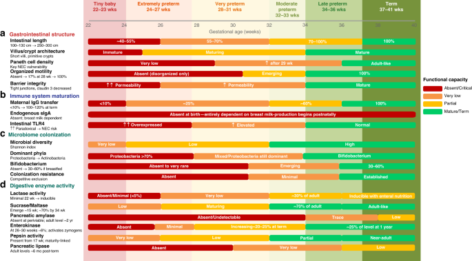
The gross anatomic development of the gastrointestinal system is mostly complete by 20 weeks’ gestation; however, its function continues to mature throughout gestation. The total intestinal length doubles between 22–23 weeks of gestation and term, from approximately 100–130 cm at 22–23 weeks, to ~150–200 cm at 28 weeks, and ~250–300 cm at term.19,20 Length of the large intestine at 22–23 weeks is approximately 15–25 cm, expanding to ~25–40 cm by 28 weeks, and ~40–60 cm at term20 (Fig. 1a).
a Gastrointestinal structure: intestinal length, villus/crypt architecture, Paneth cell density, organized motility, and barrier integrity (tight junctions, permeability).b Immune system maturation: maternal IgG transfer, endogenous sIgA production, and intestinal TLR4/TLR2 expression. c Microbiome colonization: diversity (Shannon index), dominant phyla, Bifidobacterium abundance, and colonization resistance. d Digestive enzyme activity: lactase, sucrase/maltase, pancreatic amylase, enterokinase, pepsin, and pancreatic lipase.
The intestinal absorptive surface area at 22–23 weeks’ gestation is markedly reduced compared to term-age, with well-formed but shorter villi, fewer microvilli, and lower villus-to-crypt ratios.21,22
Goblet cells are present from 9–10 weeks’ gestation, but their density is lower during early development, leading to reduced protection from mucin-2 secretion.23 Paneth cells, which reside in crypt bases, emerge at 13 weeks but remain at low density until 29 weeks’ gestation; adult levels are not achieved until term or postnatally. Paneth cells are crucial components of the gastrointestinal tract’s innate immune system, secreting lysozyme and alpha-defensins.24 The decreased number of Paneth cells is directly associated with susceptibility to necrotizing enterocolitis (NEC)25 (Figs. 1a and 2).
On the left side of the figure are the three interdependent components of the vulnerability triad (gastrointestinal immaturity, immune deficiency, and microbiome dysbiosis) that together predispose the most immature infants to NEC and sepsis. Gastrointestinal immaturity (red circle) includes the structural, motility, and enzymatic deficits that increase intestinal permeability and promote bacterial translocation. Immune deficiency (blue circle) reflects the absence/minimal production of endogenous sIgA, impaired neutrophil function, minimal complement activity, and paradoxical overexpression of TLR4 and TLR2, allowing pathogen proliferation. Microbiome dysbiosis (green circle) is characterized by very low diversity, dominance of Proteobacteria, and the absence of Bifidobacterium, creating an unstable ecosystem that is vulnerable to NICU-acquired pathogens. On the right side of the figure are the four mechanisms by which mother’s own milk (MOM) provides biological protection against each component of the triad: barrier maturation (via EGF, TGF-β, and stem cells), digestive compensation (via BSSL, amylase, and proteases), immune protection (via sIgA, lactoferrin, lysozyme, and live immune cells), and microbiome balance (via >200 HMO species and live bacteria promoting Bifidobacterium growth). A critical distinction is highlighted: fresh MOM functions as active immunotherapy, while pasteurized donor human milk (DHM) provides only partial benefit due to the loss of BSSL, live bacteria, and cellular components during pasteurization. BSSL bile salt-stimulated lipase, CFU colony-forming units, DHM donor human milk, EGF epidermal growth factor, GALT gut-associated lymphoid tissue, HMO human milk oligosaccharides, IgG immunoglobulin G, MMC migrating motor complex, MOM mother’s own milk, NEC necrotizing enterocolitis, NICU neonatal intensive care unit, SCFA short-chain fatty acids, sIgA secretory immunoglobulin A, TGF-β transforming growth factor-beta, TLR toll-like receptor.
The enteric nervous system matures throughout fetal development, with myenteric plexus formation by 9 weeks, submucosal plexus development by 12–14 weeks, and nitrergic neurons appearing at 12 weeks’ gestation.26 Although both plexuses are anatomically present by 12–14 weeks, functional maturity remains limited at 22–23 weeks’ gestation, with reduced neurotransmitter expression and a decrease in interstitial cells of Cajal (pacemaker networks)27,28,29 (Fig. 1a)
Gastrointestinal tract maturation
In utero, amniotic fluid provides bioactive molecules for fetal intestinal maturation, including growth factors and nutrients that support immune system development and barrier formation.30
Gastrointestinal motility is very immature at 22–23 weeks’ gestation. Organized migrating motor complexes are absent before 31 weeks’ gestation, with only disorganized low-amplitude motor activity present31,32 (Fig. 1a). By 28 weeks’ gestation, only approximately 17% of preterm infants demonstrate complete interdigestive cycles compared with term infants,32 and gastric emptying is markedly delayed.33 Coordinated sucking and swallowing does not develop until 32–34 weeks, and intestinal transit time is markedly prolonged at earlier gestational ages.32 These motility limitations provide the physiologic basis for delayed meconium passage, feeding intolerance, risk of bacterial overgrowth, and increased susceptibility to NEC observed in infants born at 22–23 weeks’ gestation.
Digestive enzyme development is also underdeveloped at 22–23 weeks’ gestation, which helps explain some limitations in tolerance and absorption to enteral nutrition
For carbohydrate digestion, sucrase-isomaltase and maltase begin to emerge from approximately 15 weeks’ gestation.34 However, it is very low at 22–23 weeks, approaching term levels in the third trimester. Sucrase reaches roughly 70% of adult activity by 34 weeks, and lactase activity increases with enteral feeding.34,35 Salivary amylases are minimal or absent.36 Pancreatic amylase is detectable in fetal pancreatic acinar cells from approximately 16 weeks’ gestation by immunohistochemistry,37 but functional secretory output at 22–23 weeks is negligible. Amylase activity in amniotic fluid increases substantially only in the third trimester37 (Fig. 1d).
At 22–23 weeks’ gestation, the enzymes for protein digestion are profoundly deficient. Pepsin activity is lower than 50% of term levels reported for very low birth weight infants.38 The high gastric pH further inactivates the limited available peptic activity. Importantly, enterokinase is absent before 26 weeks of gestation.38 Furthermore, at 22–23 weeks’ gestation, trypsin and chymotrypsin are likely non-functional.39 The practical consequence is that intraluminal protein digestion is minimal, so amino acid absorption relies almost entirely on brush border peptidases and intact peptide transporters. Aminopeptidase is among the earliest brush border enzymes to appear and is detectable by 15 weeks’ gestation38 and one of the few relatively functional digestive proteins at 22–23 weeks’ gestation (Fig. 1d).
Enteral fat digestion at 22–23 weeks’ gestation is also decreased. Lingual lipase is present in gastric aspirates collected at birth in infants born at 26–28 weeks’ gestation,36 inferring that the enzyme may appear before 26 weeks based on the pattern of pre-birth accumulation. However, no infants younger than 26 weeks’ gestation were directly studied, and whether meaningful endogenous gastric lipolytic activity exists at 22–23 weeks’ gestation remains unknown.36 Pancreatic lipase is mostly absent at this gestational age,39 and bile acid synthesis is highly reduced.36 Clinically, this profound immaturity of luminal digestion implies that a substantial proportion of the macronutrients delivered may be incompletely hydrolyzed and therefore malabsorbed at 22–23 weeks’ gestation. This provides a physiologic rationale for prioritizing feedings that supply their own digestive enzymes and bioactive support (such as fresh mother’s own milk), for cautious feeding volumes and advancement. Whether enteral enzyme or bile salt supplementation could augment macronutrient absorption in this population has not been studied and remains a research question.
In the near-complete absence of functional enzymes, fresh mother’s own milk plays an exceptional role in this population. Beyond its growth factors and immunologic components, it provides exogenous bile salt-stimulated lipase to help hydrolyze triglycerides along with milk amylase and proteases that partially compensate for the infant’s endogenous deficiencies.40,41 Bile salt-stimulated lipase is destroyed by pasteurization,42 meaning that pasteurized donor human milk cannot provide this compensatory function (Fig. 2). At 22–23 weeks’ gestation, fresh mother’s own milk is not merely preferred, it is physiologically irreplaceable.
Intestinal barrier function is also immature at 22–23 weeks’ gestation. Tight junction protein expression, particularly claudin 3, is reduced, resulting in increased paracellular permeability43 (Fig. 1a). Direct measurement of intestinal permeability using the lactulose-to-mannitol ratio has not been performed in infants born at 22–23 weeks’ gestation, but available data at 25–26 weeks’ showed markedly elevated ratios compared to term infants.44 The combination of a very thin intestinal mucus layer also facilitates bacterial translocation and increases the risk of NEC (Fig. 2).
Gastrointestinal Immunity and Microbiome
Peyer’s patches are identifiable as early as 24 weeks’ gestation but lack germinal centers, which form only postnatally in response to microbial exposure.45 Endogenous secretory immunoglobulin A production is absent.46 Placental transfer of maternal IgG happens predominantly in the late-gestation; less than 10% of maternal IgG has been transferred by 24 weeks’ gestation (Fig. 1b).47 Innate immune signaling via Toll-like receptors (TLR) is critically dysregulated at this gestational age (Fig. 1b). TLR-4 is paradoxically overexpressed on immature enterocytes, amplifying inflammatory responses to luminal bacterial ligands and increasing NEC susceptibility.47 TLR-2 is co-expressed at elevated levels at 21–28 weeks’ gestation and acts synergistically with TLR-4 to modulate the Th1/Th2 and Treg/Th17 immune axes playing an important role in intestinal immune programming, tolerance, and NEC pathogenesis.48 Clinically, this paradoxical innate-immune dysregulation implies that even small stimuli can provoke a disproportionate proinflammatory response in the immature intestine, which is central to the heightened NEC susceptibility of these infants and underscores why feeding choices matter: mother’s own milk dampens TLR4-mediated signaling and promotes immune tolerance, whereas non-human substrates and dysbiotic colonization amplify it. Human milk contains bioactive components that support intestinal maturation and function, including sIgA and human milk oligosaccharides, which act as selective prebiotics to establish a healthy intestinal microbiome and immunoglobulin A49 (Fig. 1b).
Intestinal barrier maturation is associated with increased fecal microbiota biodiversity; early, exclusive human milk feeding promotes both microbial diversity and intestinal barrier maturation.50,51 The intestinal microbiome of infants born at 22–23 weeks’ gestation is affected by extreme immaturity, high rates of cesarean delivery, prolonged antibiotic exposure, delayed initiation of enteral feeding, and exposure to the NICU environment.49,52 At 22–23 weeks’ gestation, microbial diversity is extremely low. Initial colonization is delayed, and the dominant phyla are Proteobacteria (>70–80% of total), particularly Enterobacteriaceae (Klebsiella, Escherichia coli), with Firmicutes and Actinobacteria nearly absent.53,54Bifidobacterium species, which dominate the intestinal tract of breastfed term infants, are probably absent or very rare in infants born at 22–23 weeks’ gestation and may not appear for many weeks postnatally, even with human milk feeding. By 28 weeks’ gestation, diversity expands, with increasing Firmicutes (Staphylococcus, Enterococcus) and emerging Bifidobacterium, though Proteobacteria remain predominant55 (Fig. 1c). Compared with the more diverse, Bifidobacterium-predominant community of more mature and term human milk-fed infants, this profile lacks colonization resistance, facilitating the colonization of NICU-derived or pathogenic organisms, a key reason these infants are predisposed to the dysbiosis-associated complications described below.
Fresh mother’s own milk is the single most impactful intervention for microbiome establishment,49 providing live bacteria (~105–10⁷ CFU/mL), >200 human milk oligosaccharide species that selectively promote Bifidobacterium, secretory immunoglobulin A, lactoferrin, and immune cells.56,57,58 Heat-processed donor human milk retains human milk oligosaccharides and some immunoglobulins but lacks live bacteria, providing reduced benefit.49,58
Dysbiosis patterns are strongly associated with NEC and late-onset sepsis, and infants born at 22–23 weeks’ gestation are at high risk (Fig. 2). The NEC incidence at 22–23 weeks’ gestation in a recent analysis from 11 international neonatal networks was 6%–27%.59 A characteristic Proteobacteria bloom (especially Gammaproteobacteria) precedes NEC onset by 7–14 days.53 Intestinal translocation is a major mechanism of late-onset sepsis, with the intestinal microbiome often identical to the blood isolate. The microbiome remains unbalanced for 2–4 months, with its composition fluctuating in response to antibiotic use, type of enteral feeding, and clinical events (Fig. 2).60
Metabolic transition at birth
Abnormalities in metabolic adaptations after birth are common among infants born at 22–23 weeks’ gestation. These infants have very limited glycogen stores and delayed maturation of gluconeogenic pathways, making them dependent on exogenous glucose for energy supply within minutes after birth.3,11,14
Limited data suggest these infants have both insulin deficiency and insulin resistance.61 The reduced concentration of circulating cortisol in preterm infants, combined with immaturity of their adrenal glands, contributes to lower lipolysis and gluconeogenesis.14,62,63 These factors make them particularly vulnerable to developing hypoglycemia if inadequate glucose is supplied. At the same time, and arguably more frequently, the combination of insulin deficiency, insulin resistance, and immature glucose metabolism predisposes these infants to hyperglycemia. Both hypoglycemia and hyperglycemia therefore occur in this population. In the first hours after birth, hypoglycemia is the most common, which later transitions to hyperglycemia. Glucose provision must be titrated to avoid each, as discussed under Carbohydrate/Glucose Management.
The concept of an “adaptive approach” to nutrition support interventions in these infants warrants careful consideration.3 Perhaps the answer is not to replicate what they would receive in utero, but to support maturation without disruption, adapting recommendations for more mature infants depending on their response. This requires acknowledging that clinicians are providing external nutrition that a fetus at 22–23 weeks of gestation was never physiologically intended to receive, so additional research specifically designed for this population is urgently needed to determine whether current nutrition interventions are physiologically appropriate.
Parenteral nutrition management
Given their gastrointestinal immaturity, infants born at 22–23 weeks’ gestation rely on parenteral nutrition during the early postnatal period. However, current parenteral nutrition recommendations developed for extremely low birth weight infants may require significant modification for infants born at 22–23 weeks’ gestation.3
Carbohydrate/glucose management
Infants born at 22–23 weeks’ gestation exhibit distinct physiological characteristics that affect glucose uptake in insulin-sensitive tissues.3,14,61 Current recommendations for glucose administration in preterm infants by Koletzko et al. in the stable phase for infants weighing 500–1000 g are to provide glucose at 3–6 mg/kg/min during the initial phase, adjust by 1–2 mg/kg/min daily toward the goal of ~7–10 mg/kg/min.18 The European Society for Paediatric Gastroenterology, Hepatology and Nutrition (ESPGHAN) recommends glucose infusion rates of 4–8 mg/kg/min on day 1, advancing over 2–3 days to a target of 8–10 mg/kg/min (minimum 4, maximum 12 mg/kg/min) for preterm newborns, with dose adjustments guided by blood glucose levels and illness phase.64 Notably, the American Society for Parenteral and Enteral Nutrition(ASPEN) guidelines for preterm infants do not provide specific glucose infusion rate targets but recommend against the routine use of insulin, given associated risks of hypoglycemia and higher mortality,65 increased lactate, acidosis, and reduced protein synthesis, and explicitly identify infants born at 22–23 weeks’ gestation as a priority population for future research.66 For infants at 22–23 weeks’ gestation, these recommendations are likely to require modification. Their lower muscle and adipose tissue mass suggests potentially lower glucose requirements, while their immature insulin response impairs glucose tolerance, increasing the risk of hyperglycemia.3 Clinical management requires careful consideration of individual factors, as persistent hyperglycemia inhibits amino acid incorporation, and maintaining euglycemia is needed for optimal nutrient utilization and development.3,61,67
Based on these physiological differences, initial glucose infusion rates may need to be lower than those recommended for the general population (3–4 mg/kg/min rather than 4–6 mg/kg/min) if the infant is not hypoglycemic, with more frequent monitoring of glucose response. Advancement of glucose infusion should be individualized based on metabolic capacity (with close blood glucose monitoring and adjustment guided by the glycemic response), and the use of insulin administration remains a priority research question.
Protein/amino acid provision
Amino acids are necessary for cell signaling and protein synthesis.13 In the fetus, protein may be catabolized for energy when substrate availability is insufficient.13
For preterm infants born at 22–23 weeks’ gestation, it is critical to provide an amino acid solution to achieve a positive nitrogen balance and prevent cumulative protein deficits. Preterm infants rapidly accrue energy and protein deficits after birth, underscoring the rationale for prompt initiation of parenteral nutrition.66,68 Studies have reported tolerance to amino acid doses of up to 3.5 g/kg/day in preterm infants weighing 401–1500 g, with some evidence that higher early protein intake is associated with improved neurodevelopmental outcomes at 18 months corrected age.67,69,70 However, these studies primarily enrolled more mature preterm populations. The ESPGHAN recommends initiating parenteral amino acids at a minimum of 1.5 g/kg/day on the first postnatal day, advancing to 2.5–3.5 g/kg/day from day 2 with non-protein energy intakes exceeding 65 kcal/kg/day.71 The ASPEN recommends initiating amino acids promptly after birth, targeting a minimum of 3 g/kg/day and not exceeding 3.5 g/kg/day.66 Importantly, across combined analyses of multiple randomized controlled trials, higher versus lower parenteral amino acid doses did not significantly improve growth outcomes at 36 weeks’ postmenstrual age or at 2 years of age.66
The ProVIDE trial randomized infants <1000 g to receive an additional 1 g/day of amino acids for the first 5 postnatal days.72 Higher amino acid provision significantly increased the risk of moderate-to-severe neurodevelopmental impairment at 2 years’ corrected age (RR 1.95, 95% CI 1.09–3.48). However, the protocol did not standardize co-nutrient administration, had heterogeneous baseline amino acid provision across centers, had few infants born at 22–23 weeks’ gestation, and lacked individualized weight-based dosing.3 A likely contributor to this harm is an inadequate non-protein energy-to-protein ratio: when amino acids are increased without an adequate increase in non-protein energy, the protein cannot be used for growth and must instead be oxidized and excreted at a metabolic cost, justifying why amino acid provision should always be matched to sufficient concurrent non-protein energy.
For infants born at 22–23 weeks of gestation, available evidence supports initiating parenteral amino acids at 2–2.5 g/kg/day immediately after birth, with cautious advancement to 3–3.5 g/kg/day as clinical stability permits.3 It is likewise essential to maintain an adequate non-protein energy-to-protein ratio (non-protein energy >25 kcal per gram of protein) to ensure that the provided amino acids are directed toward protein synthesis rather than oxidized for energy.18,71,73
Lipid/triglyceride management
Fatty acids are essential for cell membrane integrity, hormonal synthesis, and energy storage.14 Lipids are particularly relevant to brain and retinal development, with linoleic acid, alpha-linolenic acid, and docosahexaenoic acid serving as essential components for proper myelination and neurocognitive maturation.3,74 However, infants born at 22–23 weeks’ gestation have very limited fat stores and an impaired capacity to metabolize large quantities of fatty acids, increasing their susceptibility to hypertriglyceridemia.3
The ESPGHAN guidelines recommend initiating lipid injectable emulsions in preterm infants immediately after birth and no later than the second postnatal day, with a maximum parenteral lipid intake of 4 g/kg/day.75 For critically ill neonates, phase-specific lipid dosing has been proposed: 1–2 g/kg/day during the early acute phase, 2–3 g/kg/day during the late acute phase, and 3–4 g/kg/day during the recovery phase.76
For infants born at 500–1000 g, a starting dose of ≤2 g/kg/day on the first day of initiation, with subsequent advancement toward 3 g/kg/day as tolerated, is recommended by Koletzko et al.18 These birth weight–based thresholds warrant caution, because many infants born at 22–23 weeks’ gestation weigh at or below the 500 g threshold, and also because weight alone is not equivalent to maturity in lipid metabolism. ASPEN also recommends daily advancement of lipid injectable emulsion to a target of 3 g/kg/day to prevent essential fatty acid deficiency; however, the evidence is of very low quality.66 Importantly, ASPEN also recommends against routinely reducing lipid injectable emulsion doses to prevent sepsis or parenteral nutrition–associated liver disease, as available randomized control trial data do not support lipid restriction as an effective preventive strategy.66
Regarding lipid injectable emulsion composition, ESPGHAN recommends composite emulsions, with or without fish oil, over pure soybean oil, given their more balanced fatty acid profile, higher alpha-tocopherol content, and lower phytosterol load;75 ASPEN provides no specific composition recommendation.66 However, in a secondary analysis, the use of a multicomponent lipid injectable emulsions containing fish oil was associated with a significant reduction in severe retinopathy of prematurity (ROP stage ≥3) compared to other lipid injectable emulsions (risk difference −0.04; 95% CI −0.08 to −0.01; p = 0.02), a finding of potential relevance given the high risk of ROP in 22–23 weeks’ gestational age infants.66 ESPGHAN further recommends that 20% formulations be run continuously over 24 h and protected from light using validated tubing, given the morbidity and mortality risks associated with photooxidation products.75
Limited information exists on the metabolic capacity of infants at 22–23 weeks’ gestational age to tolerate the lipid doses outlined in current recommendations.3,66,75 For infants born at 22–23 weeks’ gestation, a lower initial dose of lipid injectable emulsion of 0.5–1 g/kg/day during the immediate postnatal period may be more appropriate, with careful upward titration contingent on triglyceride tolerance. Given the small weight of these infants, the maximum tolerated dose may not be sufficient to sustain a continuous 24-h infusion. ESPGHAN recommends reducing the infusion rate of lipid injectable emulsion if plasma triglyceride concentrations during infusion exceed 3 mmol/L (265 mg/dL).75 However, further research must identify acceptable threshold values for fatty acids to guide management of lipid injectable emulsions to likewise prevent essential fatty acid deficiency.
Energy requirements
Research suggests that 55 kcal/kg/day is needed to meet the fetal basal energy expenditure, which may translate to an acceptable initial goal for parenteral energy provision in infants born at 22–23 weeks’ gestation.18 The ESPGHAN acknowledges the metabolic disturbances during critical illness and advocates a phased approach, starting at 40–55 kcal/kg/day and gradually increasing to 90–120 kcal/kg/day as the infant stabilizes.77 This, combined with lower muscle and adipose tissue mass at 22–23 weeks’ gestation, likely further affects basal energy requirements.3 Thus, the challenge lies in providing sufficient energy to support growth and development while avoiding metabolic complications from excessive provision or metabolic intolerance. Regular monitoring of growth, metabolic tolerance, and clinical status is fundamental for guiding energy provision.
Micronutrients: vitamins and minerals
The extremely low body fat percentage (<2%) of infants born at 22–23 weeks’ gestation significantly impacts the storage and metabolism of fat-soluble vitamins.3,78 These infants also experience higher oxidative stress, potentially increasing their requirements for antioxidant micronutrients. Current micronutrient recommendations for preterm infants are based mainly on expert opinion rather than definitive evidence from randomized clinical trials and therefore may not be appropriate for this population.78
Mineral homeostasis presents additional challenges. Calcium accumulation increases proportionally with total body weight and gestational age, as bone makes up an increasingly large portion of fetal weight, with approximately 80% of total body calcium and phosphorus accumulating during the third trimester.79 Thus, infants born at 22–23 weeks’ gestation have minimal accrual by birth.74
The complex interplay between protein provision and mineral utilization requires careful consideration, as high protein provision without adequate phosphorus can lead to mineral mobilization from bone.3,79 High amino acid provision may induce early hypophosphatemia via a refeeding-like syndrome when co-administration of phosphorus is insufficient. In this clinical scenario, available phosphorus is diverted to cellular energy metabolism and ATP-generation, reducing phosphorus available in serum to potentially dangerous levels and diverting from bone mineralization.79 This risk is compounded by the minimal skeletal mineral reserve characteristic of infants at 22–23 weeks’ gestation. For early postnatal parenteral nutrition management, recommended provisions include 0.8–2 mMol/kg/day for calcium, 1–2 mMol/kg/day for phosphorus, and 0.1–0.2 mMol/kg/day for magnesium, increasing as the infant grows and stabilizes.79 A molar calcium-to-phosphorus ratio 0.8–1.0 is recommended to reduce the incidence of early postnatal hypercalcemia and hypophosphatemia.79 However, these targets were derived from broader preterm populations, so their adequacy at 22–23 weeks of gestation remains unknown. Frequent monitoring of alkaline phosphatase, calcium, phosphorus, and magnesium concentrations is recommended to guide individualized adjustments.79
Enteral nutrition management
Delayed initiation of enteral nutrition primarily leads to progressive intestinal villous atrophy, impairs development of functional peristalsis and impedes postnatal maturation of digestive enzymes, compromising subsequent tolerance and potentially increasing clinical risk.80 The challenge for infants born at 22–23 weeks is balancing the benefits of enteral feeding against the risks of gastrointestinal complications.81
Initiation of enteral feeding
Mother’s own milk remains the preferred enteral substrate for infants born at 22–23 weeks’ gestation.81 Human milk contains bioactive components that support intestinal maturation and function and provide developmental signals.49 A dose-dependent relationship exists between mother’s own milk volume and reduced rates of NEC, late-onset sepsis, chronic lung disease, retinopathy of prematurity, and neurodevelopmental impairment.81 Oral immunotherapy using small amounts of colostrum or human milk promotes immune function and gastrointestinal development, and neonates receiving this intervention achieve full enteral nutrition in fewer days, have reduced sepsis rates, and experience shortened hospital stays.82,83,84,85,86 However, no studies are reported specifically for the 22–23 weeks’ gestation population.
Donor human milk is the recommended substitute when mother’s own milk is not sufficiently available.81 The American Academy of Pediatrics (AAP) and ESPGHAN conditionally recommend pasteurized donor human milk over formula for infants born before 32 weeks’ gestation or weighing less than 1500 g when the mother’s own milk is unavailable or insufficient.80,81 Long-term outcomes comparing donor milk to formula are conflicting across sepsis, neurodevelopment, and mortality, and donor milk has been associated with impaired growth in more mature preterm infants.87,88,89 Donor milk storage time may affect outcomes, with longer storage times associated with an increased risk of complications, especially in infants <750 g.90,91 If a substitute for mother’s own milk is needed, the AAP recommends transitioning to preterm formula at approximately 34–36 weeks’ postmenstrual age, when NEC risk is lower.81
The timing of enteral nutrition initiation in infants born at 22–23 weeks’ gestation is debated, given the higher risk for NEC, spontaneous intestinal perforation, slow intestinal motility, and delayed stooling. A 2022 Cochrane review found no benefit in reducing NEC or mortality by delaying enteral nutrition initiation beyond 4 days of age in extremely preterm infants.92 Delayed initiation (>3 days) after birth has been associated with increased bronchopulmonary dysplasia, retinopathy of prematurity, and multiple morbidities.93 Current recommendations for stable extremely preterm infants suggest that minimal enteral nutrition should be started as soon as possible and within 48 h of age, though the optimal approach for infants born at 22–23 weeks’ gestation remains uncertain.18,80
Reported protocols for this population vary widely.3 Initial volumes of around 10 mL/kg/day, with individualized advancement, appear reasonable based on available evidence.94
Enteral advancement
No consensus exists regarding the optimal rate of enteral nutrition advancement in infants born at 22–23 weeks’ gestation. ESPGHAN recommends advancing enteral feeds by 18–30 mL/kg/day in stable extremely preterm infants.80 The landmark Speed of Increasing Milk Feeds Trial compared slow feeding progression at 18 mL/kg/day with faster progression at 30 mL/kg/day in 2804 infants across the United Kingdom and Ireland and found no significant difference in NEC, late-onset sepsis, or mortality between groups, supporting the safety of faster advancement in stable extremely preterm infants.95 However, it included very few infants at 23 weeks’ gestation and fewer <1% with birth weight <500 g. Again, these data derive mainly from infants more mature than 22–23 weeks’ gestation, so their applicability to the most immature infants remains uncertain. Further, feeding intolerance affects up to 40–50% of extremely preterm infants, with dysmotility being the main cause.80 Importantly, ESPGHAN advises against routine monitoring of gastric residuals in clinically stable infants, as this practice has not been shown to reduce feeding complications and may unnecessarily delay the advancement of enteral nutrition.80 Given the lack of specific data for this population, individualized advancement and monitoring appear most appropriate, and more research is needed.
Probiotic supplementation
Multiple meta-analyses have demonstrated that probiotic supplementation reduces the incidence of NEC and late-onset sepsis in preterm infants, with the most consistent evidence supporting multispecies regimens containing Lactobacillus and Bifidobacterium strains.96,97,98 Whether these findings extend to infants born at 22–23 weeks’ gestation remains uncertain. Notably, subgroup analysis of the current Cochrane review for extremely preterm or extremely low birth weight infants did not demonstrate a clear benefit in NEC (RR 0.92, 95% CI [0.69–1.22]), all-cause mortality (RR 0.92, 95% CI [0.72 – 1.18]), or late-onset invasive infection (RR 0.93, 95% CI [0.78–1.09]).98 The extreme immune immaturity at this gestational age raises theoretical concerns about probiotic bacteremia or fungemia, which may have greater consequences than in more mature infants.99 Until adequately powered trials specifically enroll infants at 22–23 weeks’ gestational age, clinicians must weigh the plausible benefit against the uncharacterized safety profile in this population.
Human milk fortification
Human milk alone is inadequate to meet estimated micronutrient and mineral requirements for infants born at 22–23 weeks’ gestation.3
Multi-component human milk fortifiers are preferred, given globally elevated requirements. ESPGHAN recommends fortifying human milk “early with phosphate followed by early introduction of multicomponent breast milk fortifiers” and states that fortifiers “do not seem to be associated with feeding intolerance and are safe even when introduced early with enteral feeds.” 80 Initiation at an enteral volume of 40–100 mL/kg/day is suggested.100 In a randomized trial of infants born at less than 28 weeks’ gestation, early fortification initiated on day 2, compared with day 14, was associated with improved linear growth without increased adverse events.101 Individualized fortification strategies have shown improved growth outcomes compared to standard fortification.102
Whether human milk-based fortifiers offer advantages over bovine-based fortifiers in this population remains uncertain. ESPGHAN concludes there is currently “insufficient data from adequately powered studies to determine the optimal strategy” and “insufficient evidence to recommend the routine use of human milk-derived fortifiers until further high-quality data is available.” 80 The AAP similarly concludes there is no evidence to recommend human milk-derived over hydrolyzed bovine-derived fortifiers in very low birth weight infants receiving human milk as the base diet, citing two randomized trials that found no differences in NEC, sepsis, growth, or neurodevelopmental outcomes.81,103,104
Enteral energy, micronutrient and mineral requirements
Energy requirements for infants born at 22–23 weeks’ gestation as they transition to enteral nutrition warrant careful consideration. Koletzko et al. recommend 110–130 kcal/kg/day enterally for stable preterm infants weighing up to 2000 g, while ESPGHAN recommends 115–140 kcal/kg/day for “healthy, growing preterm infants” without clarifying weight or gestational age.18,80 Higher provision (e.g., minimum 120–125 kcal/kg/day and 4 g protein/kg/day) may be needed to support adequate growth or compensate for reduced absorption.105 Infants born at 22–23 weeks’ gestation may experience early postnatal metabolic disturbances and critical illness, limiting their ability to initially tolerate increased energy provision. Therefore, higher provision during less-acute illness phases may require catch-up nutrition, with enteral recommendations of 110–160 kcal/kg/day and 4.5 g protein/kg/day for “recovery in critically ill neonates.” 76
Micronutrient requirements for extremely preterm infants in general include vitamin D (400–700 IU/kg/day, maximum 1000 IU/day), iron (2–3 mg/kg/day starting by 2 weeks of age), calcium (approaching 200 mg/kg/day), phosphorus (68–115 mg/kg/day), and sodium (up to 8 mEq/kg/day).80,106,107 However, these recommendations have not been specifically validated for infants at 22–23 weeks’ gestation. Protein targets during full enteral nutrition range from 3.5–4 g/kg/day (up to 4.5 g/kg/day) per ESPGHAN recommendations, without special acknowledgment for infants born at 22–23 weeks’ gestation.80 Urinary sodium losses in infants at 23–25 weeks range from 6 to 7 mEq/kg/day during the first month after birth, often requiring supplementation beyond what is provided by fortified enteral feeding.106 However, requirements may be altered for infants born at 22–23 weeks’ gestation based on stage of development and postnatal interventions and exposures.
The transition: integrating parenteral and enteral nutrition
Infants born at 22–23 weeks’ gestation usually have a prolonged period of receiving both parenteral and enteral nutrition support. Maintaining adequate nutrition during the transition from parenteral to enteral nutrition requires careful attention. The transition typically progresses through three phases: an initial phase with minimal enteral nutrition, an intermediate phase with advancing enteral feeding while weaning off parenteral nutrition, and a final phase with full enteral nutrition.80
During the transition period, parenteral nutrition should be adjusted to complement enteral provision. This requires careful calculation of combined parenteral and enteral protein, energy, and micronutrient provision. Nutritional deficits that occur during the transition explain not only declines in growth but also increased risk of adverse neurodevelopmental outcomes, late-onset sepsis, and other neonatal morbidities.69,108
Infants born at 22–23 weeks’ gestation often have prolonged parenteral nutrition dependency with associated complications (cholestasis, catheter-related infections), high rates of enteral feeding intolerance or gastrointestinal complications, and difficulty achieving adequate growth velocity, so special attention to each phase is warranted.80,94
Special considerations
It is important to acknowledge that in many low- and middle-income countries, the decision to resuscitate infants born 22–23 weeks’ gestation is also influenced by the availability of appropriate life-sustaining equipment, trained personnel, or intensive care capacity. When available, oropharyngeal immunotherapy with colostrum should be considered a low-cost intervention with potential immune benefits, and available enteral multivitamin or other singular nutrient (e.g., vitamin D, calcium, phosphorus, etc.) supplementation should be incorporated into nutritional protocols whenever feasible to address common micronutrient deficiencies.
Heat-processed donor human milk is often unavailable in low- and middle-income countries due to the absence of human milk banks and regulatory bodies. In these settings, informal milk sharing among mothers may be a common and, at times, necessary practice. Increasing the total enteral volume of mother’s own milk to increase total nutrient intake, or using calorie-dense hindmilk for feedings, are mechanisms that could also be employed in the absence of designated multicomponent human milk fortification options.
Kangaroo Mother Care is among the most evidence-based, cost-effective interventions in reduction, increases the volume and bioactive content of expressed milk, and supports maternal-infant bonding, which is critical for sustaining breastfeeding during the prolonged NICU course typical of infants at 22–23 weeks’ gestational age.109
Research priorities
Almost every aspect of currently available nutrition support recommendations lacks evidence specific to infants born at 22–23 weeks’ gestation. We propose the following research priorities: gastrointestinal structure and motility; digestive enzyme production and nutrient absorption; metabolic tolerance to parenteral nutrition and targeted nutrient provision; overall macronutrient and micronutrient requirements to promote optimal development; mother’s own milk and immune-microbiome interactions; and methodological improvements. Until research provides data specific to infants born at 22–23 weeks’ gestation, clinicians must carefully extrapolate and individualize nutrition support interventions to these infants. Research priorities are summarized in Table 1.
Conclusions
Infants born at 22–23 weeks’ gestation represent a population whose nutritional requirements cannot be addressed using recommendations provided for more mature preterm infants, given their immature gastrointestinal structure and function and significant limitations of their metabolic capacity
The lack of evidence specific to this population represents both an urgent knowledge gap and a call to action. Until dedicated trials and collaborative multicenter research generate evidence for infants born at 22–23 weeks’ gestation, clinicians must carefully individualize and actively manage all aspects of nutrition support, recognizing that strategies effective in larger preterm infants may require substantial modification in infants born at 22–23 weeks’ gestation
References
Bell, E. F. et al. Mortality, in-hospital morbidity, care practices, and 2-year outcomes for extremely preterm infants in the US, 2013–2018. JAMA327, 248–263 (2022)
Article
PubMed
PubMed Central
<a href="http://scholar.google.com/scholar_lookup?&title=Mortality%2C%20in-hospital%20morbidity%2C%20care%20practices%2C%20and%202-year%20outcomes%20for%20extremely%20preterm%20infants%20in%20the%20US%2C%202013%E2%80%932018&journal=JAMA&doi=10.1001%2Fjama.2021.23580&volume=327&pages=248-263&publication_year=2022&author=Bell%2CEF” rel=”nofollow noopener” target=”_blank”>Google ScholarIsayama, T. et al. Survival and unique clinical practices of extremely preterm infants born at 22–23 weeks’ gestation in Japan: a national survey. Arch. Dis. Child. Fetal Neonatal Ed.110, 17–22 (2024)
Romero-Lopez, M., Naik, M., Hartman, T., Anderson-Berry, A. & Thoene, M. Evidence and unknowns for the relevancy of applying current parenteral nutrition support recommendations among infants born less than 750 g or younger than 25 weeks’ gestation: a narrative review. JPEN J. Parenter. Enteral Nutr.49, 560–571 (2025)
Rysavy, M. A. et al. An immature science: intensive care for infants born at ≤23 weeks of gestation. J. Pediatr.233, 16–25.e1 (2021)
Haga, M. et al. Prevalence and risk factors for neurodevelopmental impairment in very preterm infants without severe intraventricular hemorrhage or periventricular leukomalacia. Early Hum. Dev.206, 106286 (2025)
Pitcher, G. J. et al. Meconium obstruction of prematurity in tiny babies – towards developing an international consensus. J. Perinatol. https://doi.org/10.1038/s41372-025-02395-x (2025)
Stoll, C. M. et al. Approaches to incubator humidification at <25 weeks’ gestation and potential impacts on infants. J. Perinatol. https://doi.org/10.1038/s41372-025-02294-1 (2025)
Ågren, J., Naseh, N., Bell, E. F. & Rysavy, M. A. Tiny baby math: supralinear implications for management of infants born at less than 24 weeks gestation. J. Perinatol. https://doi.org/10.1038/s41372-025-02273-6 (2025)
Bernardini, L. B. et al. It’s the little things. A framework and guidance for programs to care for infants 22-23 weeks’ gestational age. J. Perinatol. https://doi.org/10.1038/s41372-025-02252-x (2025)
Hay, W. W. Jr. Placental-fetal glucose exchange and fetal glucose metabolism. Trans. Am. Clin. Climatol. Assoc.117, 321–340 (2006)
Rao, P. N., Shashidhar, A. & Ashok, C. In utero fuel homeostasis: lessons for a clinician. Indian J. Endocrinol. Metab.17, 60–68 (2013)
Bozzetti, P. et al. The relationship of maternal and fetal glucose concentrations in the human from midgestation until term. Metabolism37, 358–363 (1988)
Brown, L. D. et al. Placental transfer and fetal requirements of amino acids. in Fetal and Neonatal Physiology, 6th edn. 408–422 (Elsevier, 2022)
Herrera, E. & Amusquivar, E. Lipid metabolism in the fetus and the newborn. Diabetes Metab. Res. Rev.16, 202–210 (2000)
de Knegt, V. E. et al. The role of leptin in fetal growth during pre-eclampsia. Int. J. Mol. Sci.22, 4569 (2021)
Han, V. K., Bassett, N., Walton, J. & Challis, J. R. The expression of insulin-like growth factor (IGF) and IGF-binding protein (IGFBP) genes in the human placenta and membranes: evidence for IGF-IGFBP interactions at the feto-maternal interface. J. Clin. Endocrinol. Metab.81, 2680–2693 (1996)
CAS
PubMed
Google ScholarFriis-Hansen, B. Body water compartments in children: changes during growth and related changes in body composition. Pediatrics28, 169–181 (1961)
Koletzko, B. et al. Scientific basis and practical application of nutritional care for preterm infants. World Rev. Nutr. Diet.122, XIII–XIV (2021)
Touloukian, R. J. & Smith, G. J. Normal intestinal length in preterm infants. J. Pediatr. Surg.18, 720–723 (1983)
Bardwell, C. et al. Establishing normal ranges for fetal and neonatal small and large intestinal lengths: results from a prospective postmortem study. World J. Pediatr. Surg.5, e000397 (2022)
Grand, R. J., Watkins, J. B. & Torti, F. M. Development of the human gastrointestinal tract. A review. Gastroenterology70, 790–810 (1976)
Shulman, R. J., Wong, W. W. & Smith, E. O. Influence of changes in lactase activity and small-intestinal mucosal growth on lactose digestion and absorption in preterm infants. Am. J. Clin. Nutr.81, 472–479 (2005)
Kim, Y. S. & Ho, S. B. Intestinal goblet cells and mucins in health and disease: recent insights and progress. Curr. Gastroenterol. Rep.12, 319–330 (2010)
Lueschow, S. R. & McElroy, S. J. The Paneth cell: the curator and defender of the immature small intestine. Front. Immunol.11, 587 (2020)
McElroy, S. J., Underwood, M. A. & Sherman, M. P. Paneth cells and necrotizing enterocolitis: a novel hypothesis for disease pathogenesis. Neonatology103, 10–20 (2013)
Indrio, F. et al. Development of the gastrointestinal tract in newborns as a challenge for an appropriate nutrition: a narrative review. Nutrients14, 1405 (2022)
Wallace, A. S. & Burns, A. J. Development of the enteric nervous system, smooth muscle and interstitial cells of Cajal in the human gastrointestinal tract. Cell Tissue Res. 319, 367–382 (2005)
Kenny, S. E. et al. Delayed maturation of the interstitial cells of Cajal: a new diagnosis for transient neonatal pseudoobstruction. Report of two cases. J. Pediatr. Surg.33, 94–98 (1998)
Lenfestey, M. W. & Neu, J. Gastrointestinal development: implications for management of preterm and term infants. Gastroenterol. Clin. North Am.47, 773–791 (2018)
Chaaban, H., Burge, K. & McElroy, S. J. Evolutionary bridges: how factors present in amniotic fluid and human milk help mature the gut. J. Perinatol.44, 1552–1559 (2024)
Bisset, W. M., Watt, J. B., Rivers, R. P. & Milla, P. J. Ontogeny of fasting small intestinal motor activity in the human infant. Gut29, 483–488 (1988)
Berseth, C. L. Gestational evolution of small intestine motility in preterm and term infants. J. Pediatr.115, 646–651 (1989)
Riezzo, G. et al. Gastric electrical activity and gastric emptying in term and preterm newborns. Neurogastroenterol. Motil.12, 223–229 (2000)
Antonowicz, I. & Lebenthal, E. Developmental pattern of small intestinal enterokinase and disaccharidase activities in the human fetus. Gastroenterology72, 1299–1303 (1977)
Raul, F., Lacroix, B. & Aprahamian, M. Longitudinal distribution of brush border hydrolases and morphological maturation in the intestine of the preterm infant. Early Hum. Dev.13, 225–234 (1986)
Hamosh, M. et al. Fat digestion in the newborn. Characterization of lipase in gastric aspirates of premature and term infants. J. Clin. Investig.67, 838–846 (1981)
Davis, M. M., Hodes, M. E., Munsick, R. A., Ulbright, T. M. & Goldstein, D. J. Pancreatic amylase expression in human pancreatic development. Hybridoma5, 137–145 (1986)
Commare, C. E. & Tappenden, K. A. Development of the infant intestine: implications for nutrition support. Nutr. Clin. Pract.22, 159–173 (2007)
Zoppi, G., Andreotti, G., Pajno-Ferrara, F., Njai, D. M. & Gaburro, D. Exocrine pancreas function in premature and full term neonates. Pediatr. Res.6, 880–886 (1972)
Hernell, O. & Bläckberg, L. Human milk bile salt-stimulated lipase: functional and molecular aspects. J. Pediatr.125, S56–S61 (1994)
Heitlinger, L. A., Lee, P. C., Dillon, W. P. & Lebenthal, E. Mammary amylase: a possible alternate pathway of carbohydrate digestion in infancy. Pediatr. Res.17, 15–18 (1983)
Wardell, J. M., Wright, A. J., Bardsley, W. G. & D’Souza, S. W. Bile salt-stimulated lipase and esterase activity in human milk after collection, storage, and heating: nutritional implications. Pediatr. Res.18, 382–386 (1984)
Patel, R. M. et al. Probiotic bacteria induce maturation of intestinal claudin 3 expression and barrier function. Am. J. Pathol.180, 626–635 (2012)
van Elburg, R. M., Fetter, W. P., Bunkers, C. M. & Heymans, H. S. Intestinal permeability in relation to birth weight and gestational and postnatal age. Arch. Dis. Child. Fetal Neonatal Ed.88, F52–F55 (2003)
Spencer, J., MacDonald, T. T., Finn, T. & Isaacson, P. G. The development of gut associated lymphoid tissue in the terminal ileum of fetal human intestine. Clin. Exp. Immunol.64, 536–543 (1986)
Granger, C. L. et al. Secretory immunoglobulin A in preterm infants: determination of normal values in breast milk and stool. Pediatr. Res.92, 979–986 (2022)
Leaphart, C. L. et al. A critical role for TLR4 in the pathogenesis of necrotizing enterocolitis by modulating intestinal injury and repair. J. Immunol.179, 4808–4820 (2007)
Gribar, S. C., Richardson, W. M., Sodhi, C. P. & Hackam, D. J. No longer an innocent bystander: epithelial toll-like receptor signaling in the development of mucosal inflammation. Mol. Med.14, 645–659 (2008)
Underwood, M. A. & Sohn, K. The microbiota of the extremely preterm infant. Clin. Perinatol.44, 407–427 (2017)
Saleem, B. et al. Intestinal barrier maturation in very low birthweight infants: relationship to feeding and antibiotic exposure. J. Pediatr.183, 31–36.e1 (2017)
Ma, B. et al. Microbial biomarkers of intestinal barrier maturation in preterm infants. Front. Microbiol.9, 2755 (2018)
Henderickx, J. G. E., Zwittink, R. D., van Lingen, R. A., Knol, J. & Belzer, C. The preterm gut microbiota: an inconspicuous challenge in nutritional neonatal care. Front. Cell. Infect. Microbiol.9, 85 (2019)
Warner, B. B. et al. Gut bacteria dysbiosis and necrotising enterocolitis in very low birthweight infants: a prospective case-control study. Lancet387, 1928–1936 (2016)
Torrazza, R. M. et al. Intestinal microbial ecology and environmental factors affecting necrotizing enterocolitis. PLoS ONE8, e83304 (2013)
Hartz, L. E., Bradshaw, W. & Brandon, D. H. Potential NICU environmental influences on the neonate’s microbiome: a systematic review. Adv. Neonatal Care15, 324–335 (2015)
Bode, L. Human milk oligosaccharides: every baby needs a sugar mama. Glycobiology22, 1147–1162 (2012)
Fernández, L. et al. The human milk microbiota: origin and potential roles in health and disease. Pharmacol. Res.69, 1–10 (2013)
Ballard, O. & Morrow, A. L. Human milk composition: nutrients and bioactive factors. Pediatr. Clin. North Am.60, 49–74 (2013)
Isayama, T. et al. Outcomes of preterm infants born at 22 to 23 weeks’ gestation in 11 international neonatal networks. JAMA Pediatr179, 1183–1193 (2025)
Patel, R. M. & Denning, P. W. Intestinal microbiota and its relationship with necrotizing enterocolitis. Pediatr. Res.78, 232–238 (2015)
Mitanchez-Mokhtari, D. et al. Both relative insulin resistance and defective islet beta-cell processing of proinsulin are responsible for transient hyperglycemia in extremely preterm infants. Pediatrics113, 537–541 (2004)
Heckmann, M., Wudy, S. A., Haack, D. & Pohlandt, F. Serum cortisol concentrations in ill preterm infants less than 30 weeks gestational age. Acta Paediatr89, 1098–1103 (2000)
Fernandez, E. F. & Watterberg, K. L. Relative adrenal insufficiency in the preterm and term infant. J. Perinatol.29, S44–S49 (2009)
Mesotten, D., Joosten, K., van Kempen, A. & Verbruggen, S. ESPGHAN/ESPEN/ESPR/CSPEN guidelines on pediatric parenteral nutrition: carbohydrates. Clin. Nutr.37, 2337–2343 (2018)
Beardsall, K. et al. Early insulin therapy in very-low-birth-weight infants. N. Engl. J. Med.359, 1873–1884 (2008)
Robinson, D. T. et al. Guidelines for parenteral nutrition in preterm infants: the American Society for Parenteral and Enteral Nutrition. JPEN J. Parenter. Enteral Nutr.47, 830–858 (2023)
Poindexter, B. B., Langer, J. C., Dusick, A. M. & Ehrenkranz, R. A. Early provision of parenteral amino acids in extremely low birth weight infants: relation to growth and neurodevelopmental outcome. J. Pediatr.148, 300–305 (2006)
Embleton, N. E., Pang, N. & Cooke, R. J. Postnatal malnutrition and growth retardation: an inevitable consequence of current recommendations in preterm infants? Pediatrics107, 270–273 (2001)
Stephens, B. E. et al. First-week protein and energy intakes are associated with 18-month developmental outcomes in extremely low birth weight infants. Pediatrics123, 1337–1343 (2009)
Ibrahim, H. M., Jeroudi, M. A., Baier, R. J., Dhanireddy, R. & Krouskop, R. W. Aggressive early total parental nutrition in low-birth-weight infants. J. Perinatol.24, 482–486 (2004)
van Goudoever, J. B., Carnielli, V., Darmaun, D. & Sainz de Pipaon, M. ESPGHAN/ESPEN/ESPR/CSPEN guidelines on pediatric parenteral nutrition: amino acids. Clin. Nutr.37, 2315–2323 (2018)
Bloomfield, F. H., Jiang, Y., Harding, J. E., Crowther, C. A. & Cormack, B. E. Early amino acids in extremely preterm infants and neurodisability at 2 years. N. Engl. J. Med.387, 1661–1672 (2022)
te Braake, F. W., van den Akker, C. H., Wattimena, D. J., Huijmans, J. G. & van Goudoever, J. B. Amino acid administration to premature infants directly after birth. J. Pediatr.147, 457–461 (2005)
Lapillonne, A., Groh-Wargo, S., Gonzalez, C. H. & Uauy, R. Lipid needs of preterm infants: updated recommendations. J. Pediatr.162, S37–S47 (2013)
Lapillonne, A. et al. ESPGHAN/ESPEN/ESPR/CSPEN guidelines on pediatric parenteral nutrition: lipids. Clin. Nutr.37, 2324–2336 (2018)
Moltu, S. J. et al. Nutritional management of the critically ill neonate: a position paper of the ESPGHAN committee on nutrition. J. Pediatr. Gastroenterol. Nutr.73, 274–289 (2021)
Joosten, K., Embleton, N., Yan, W. & Senterre, T. ESPGHAN/ESPEN/ESPR/CSPEN guidelines on pediatric parenteral nutrition: energy. Clin. Nutr.37, 2309–2314 (2018)
Bronsky, J., Campoy, C. & Braegger, C. ESPGHAN/ESPEN/ESPR/CSPEN guidelines on pediatric parenteral nutrition: vitamins. Clin. Nutr.37, 2366–2378 (2018)
Mihatsch, W. et al. ESPGHAN/ESPEN/ESPR/CSPEN guidelines on pediatric parenteral nutrition: calcium, phosphorus and magnesium. Clin. Nutr.37, 2360–2365 (2018)
Embleton, N. D. et al. Enteral nutrition in preterm infants (2022): a position paper from the ESPGHAN Committee on Nutrition and invited experts. J. Pediatr. Gastroenterol. Nutr.76, 248–268 (2023)
Parker, M. G. et al. Promoting human milk and breastfeeding for the very low birth weight infant: clinical report. Pediatrics157, e2025073625 (2026)
Rodriguez, N. A., Meier, P. P., Groer, M. W. & Zeller, J. M. Oropharyngeal administration of colostrum to extremely low birth weight infants: theoretical perspectives. J. Perinatol.29, 1–7 (2009)
Xavier Ramos, M. S. et al. Oropharyngeal colostrum immunotherapy and nutrition in preterm newborns: meta-analysis. Rev. Saude Publica55, 59 (2021)
Lee, J. et al. Oropharyngeal colostrum administration in extremely premature infants: an RCT. Pediatrics135, e357–e366 (2015)
Romano-Keeler, J. et al. Oral colostrum priming shortens hospitalization without changing the immunomicrobial milieu. J. Perinatol.37, 36–41 (2017)
Martins, C. D. C. et al. Oropharyngeal colostrum immunotherapy and risk reduction of mortality in very low birth weight premature newborns: a clinical trial. J. Pediatr.100, 32–39 (2024)
Quigley, M., Embleton, N. D. & McGuire, W. Formula versus donor breast milk for feeding preterm or low birth weight infants. Cochrane Database Syst. Rev.7, CD002971 (2019)
O’Connor, D. L. et al. Effect of supplemental donor human milk compared with preterm formula on neurodevelopment of very low-birth-weight infants at 18 months: a randomized clinical trial. JAMA316, 1897–1905 (2016)
Colaizy, T. T. et al. Neurodevelopmental outcomes of extremely preterm infants fed donor milk or preterm infant formula: a randomized clinical trial. JAMA331, 582–591 (2024)
Wagner, J., Hanson, C. & Anderson Berry, A. Donor human milk for premature infants: a review of current evidence. ICAN Infant Child Adolesc. Nutr.5, 71–77 (2013)
Camilon, T. J. M. et al. Donor human milk storage and gastrointestinal morbidities in preterm infants: a case-control study. JPEN J. Parenter. Enteral Nutr.49, 717–723 (2025)
Young, L., Oddie, S. J. & McGuire, W. Delayed introduction of progressive enteral feeds to prevent necrotising enterocolitis in very low birth weight infants. Cochrane Database Syst. Rev.1, CD001970 (2022)
Konnikova, Y. et al. Late enteral feedings are associated with intestinal inflammation and adverse neonatal outcomes. PLoS ONE10, e0132924 (2015)
Viswanathan, S., Merheb, R., Wen, X., Collin, M. & Groh-Wargo, S. Standardized slow enteral feeding protocol reduces necrotizing enterocolitis in micropremies. J. Neonatal Perinatal Med.10, 171–180 (2017)
Dorling, J. et al. Controlled trial of two incremental milk-feeding rates in preterm infants. N. Engl. J. Med.381, 1434–1443 (2019)
Dermyshi, E. et al. The “golden age” of probiotics: a systematic review and meta-analysis of randomized and observational studies in preterm infants. Neonatology112, 9–23 (2017)
Patel, R. M. & Underwood, M. A. Probiotics and necrotizing enterocolitis. Semin. Pediatr. Surg.27, 39–46 (2018)
Sharif, S., Meader, N., Oddie, S. J., Rojas-Reyes, M. X. & McGuire, W. Probiotics to prevent necrotising enterocolitis in very preterm or very low birth weight infants. Cochrane Database Syst. Rev.7, CD005496 (2023)
Poindexter, B. Use of probiotics in preterm infants. Pediatrics147, e2021051485 (2021)
Thanigainathan, S. & Abiramalatha, T. Early fortification of human milk versus late fortification to promote growth in preterm infants. Cochrane Database Syst. Rev.7, CD013392 (2020)
Salas, A. A. et al. Early human milk fortification in infants born extremely preterm: a randomized trial. Pediatrics152, e2023061603 (2023)
Rochow, N. et al. Individualized target fortification of breast milk with protein, carbohydrates, and fat for preterm infants: a double-blind randomized controlled trial. Clin. Nutr.40, 54–63 (2021)
Jensen, G. B. et al. Effect of human milk-based fortification in extremely preterm infants fed exclusively with breast milk: a randomised controlled trial. EClinicalMedicine68, 102375 (2024)
O’Connor, D. L. et al. Nutrient enrichment of human milk with human and bovine milk-based fortifiers for infants born weighing <1250 g: a randomized clinical trial. Am. J. Clin. Nutr.108, 108–116 (2018)
Whyte, R. K. et al. Energy balance and nitrogen balance in growing low birthweight infants fed human milk or formula. Pediatr. Res.17, 891–898 (1983)
Segar, D. E. et al. Physiological approach to sodium supplementation in preterm infants. Am. J. Perinatol.35, 994–1000 (2018)
Baker, R. D. & Greer, F. R. Diagnosis and prevention of iron deficiency and iron-deficiency anemia in infants and young children (0-3 years of age). Pediatrics126, 1040–1050 (2010)
Ehrenkranz, R. A. et al. Growth in the neonatal intensive care unit influences neurodevelopmental and growth outcomes of extremely low birth weight infants. Pediatrics117, 1253–1261 (2006)
Conde-Agudelo, A. & Díaz-Rossello, J. L. Kangaroo mother care to reduce morbidity and mortality in low birthweight infants. Cochrane Database Syst. Rev.2016, CD002771 (2016)
Acknowledgements
We thank Dr. Cydney Meyer for her help and advice during manuscript editing. The authors usedChatGPT (OpenAI, GPT-5 mini) to improve readability and language in selected sections; all content, interpretation, andconclusions were developed independently by the authors, who reviewed and take full responsibility for the final tex
Funding
Gerber Foundation National Research Grants, Project #10287; National Center for Advancing Translational Sciences R03TR005545 (MR-L). National Institutes of Health, National Institute of General Medical Sciences, GM115458 (AA-B)
Author information
Authors and Affiliations
Division of Neonatal-Perinatal Medicine, Department of Pediatrics, McGovern Medical School at UTHealth Houston, Houston, TX, USA
Mar Romero-Lopez
Institute for Clinical Research and Learning Health Care, McGovern Medical School at UTHealth Houston, Houston, TX, USA
Mar Romero-Lopez
Department of Pharmacy Services, Children’s Memorial Hermann Hospital, Texas Medical Center, Houston, TX, USA
Mamta Naik
Division of Neonatology, Department of Pediatrics, University of Nebraska Medical Center, Omaha, NE, USA
Melissa Thoene & Ann Anderson-Berry
Child Health Research Institute, University of Nebraska Medical Center and Children’s Nebraska, Omaha, NE, USA
Melissa Thoene & Ann Anderson-Berry
Authors
- Mar Romero-LopezView author publications
Search author on:PubMed Google Scholar
- Mamta NaikView author publications
Search author on:PubMed Google Scholar
- Melissa ThoeneView author publications
Search author on:PubMed Google Scholar
- Ann Anderson-BerryView author publications
Search author on:PubMed Google Scholar
Contributions
All the authors contributed to the study’s conceptualization. Mar Romero-Lopez wrote the original draft. All authors contributed to edits and approved the final manuscript. We greatly appreciate Dr. Cydney Meyer for her help and advice during the manuscript editing process
Ethics declarations
Competing interests
The authors declare no competing interests The authors report no proprietary or commercial interest in this article.
Additional information
Publisher’s note Springer Nature remains neutral with regard to jurisdictional claims in published maps and institutional affiliations
Rights and permissions
Open Access This article is licensed under a Creative Commons Attribution 4.0 International License, which permits use, sharing, adaptation, distribution and reproduction in any medium or format, as long as you give appropriate credit to the original author(s) and the source, provide a link to the Creative Commons licence, and indicate if changes were made. The images or other third party material in this article are included in the article’s Creative Commons licence, unless indicated otherwise in a credit line to the material. If material is not included in the article’s Creative Commons licence and your intended use is not permitted by statutory regulation or exceeds the permitted use, you will need to obtain permission directly from the copyright holder. To view a copy of this licence, visit http://creativecommons.org/licenses/by/4.0/.
About this article
Cite this article
Romero-Lopez, M., Naik, M., Thoene, M. et al. Nutritional management at 22–23 weeks gestational age: evidence and knowledge gaps.
Pediatr Res (2026). https://doi.org/10.1038/s41390-026-05370-0
Received:31 March 2026
Revised:18 June 2026
Accepted:13 July 2026
Published:17 August 2026
Version of record:17 August 2026
DOI
:https://doi.org/10.1038/s41390-026-05370-0
